

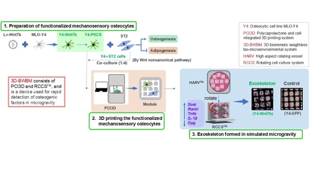
3D-bioprinted osteocytes expressing Wnt7b protect osteoblast differentiation from microgravity

Maintaining bone formation in microgravity/weightless environments remains a major challenge. Under weightless conditions, osteocytes act as mechanosensors to inhibit Wnt canonical signaling and bone formation by secreting sclerostin. This study explores whether osteocytic Wnt7b can counteract microgravity-induced bone loss through Wnt non-canonical signaling. Unlike previous bioprinting studies that focused on structural scaffolds or generic cell types, a novel bioprinted scaffold consisting of polycaprolactone (supportive) and osteocyte (functional) hydrogels was constructed in this study. Osteocytes overexpressing Wnt7b were co-cultured with bone marrow stromal cells (ST2) in a 3D biomimetic weightless biomicroenvironmental system (3D-BWBM) to assess osteogenic and lipogenic differentiation. The results indicated that osteocytic Wnt7b enhanced osteogenic differentiation and mineralization of ST2 cells via the Wnt non-canonical pathway PKCδ, while suppressing the expression of lipogenic markers (Pparg, Cebpa) and adipogenesis. Reverse transcription quantitative polymerase chain reaction (RT-qPCR) analysis revealed elevated expression of Sost and Mef2c, downregulation of the Wnt target gene Opg, and elevated expression of pro-osteoclastogenic cytokine Rankl and pro-inflammatory cytokines Tnfa and Il1b, thus validating the microgravity effect. Unlike conventional 2D culture of RCCS™ cells, the 3D hydrogels were printed with tunnels (500 μm) for efficient nutrient/ metabolite exchange, resulting in good cell growth, high cell viability (97%), and a six-fold increase in proliferative activity within 7 days. Wnt7b osteocytes were still able to maintain the osteogenic differentiation of ST2 cells, as evidenced by elevated alkaline phosphatase activity, mineralization (1.8-fold increase), and a decrease in osteoblast marker genes (Alpl, Runx2, Col1a1). In conclusion, Wnt7b-PCKδ signaling counteracts microgravity-induced bone loss, and further in vivo studies on osteocytic Wnt7b are warranted to confirm this causal relationship.
- McCarthy ID. Fluid shifts due to microgravity and their effects on bone: a review of current knowledge. Ann Biomed Eng. 2005;33:95-103. doi: 10.1007/s10439-005-8967-6
- Lang T, LeBlanc A, Evans H, Lu Y, Genant H, Yu A. Cortical and trabecular bone mineral loss from the spine and hip in long-duration spaceflight. J Bone Miner Res. 2004;19: 1006-1012. doi: 10.1359/jbmr.040307
- Durnova G, Kaplansky A, Morey-Holton E. Histomorphometric study of tibia of rats exposed aboard American spacelab life sciences 2 shuttle mission. J Gravit Physiol. 1996;3:80-81.
- Carmeliet G, Bouillon R. The effect of microgravity on morphology and gene expression of osteoblasts in vitro. Faseb J. 1999; 13(Suppl):S129-S134. doi: 10.1096/fasebj.13.9001.s129
- Smith SM, Wastney ME, O’Brien KO, et al. Bone markers, calcium metabolism, and calcium kinetics during extended-duration space flight on the mir space station. J Bone Miner Res. 2005; 20:208-218. doi: 10.1359/jbmr.041105
- Vico L, Hargens A. Skeletal changes during and after spaceflight. Nat Rev Rheumatol. 2018;14: 229-245. doi: 10.1038/nrrheum.2018.37
- Robling AG, Bonewald LF. The osteocyte: new insights. Annu Rev Physiol. 2020;82:485-506. doi: 10.1146/annurev-physiol-021119-034332
- Delgado-Calle J, Bellido T. The osteocyte as a signaling cell. Physiol Rev. 2022;102:379-410. doi: 10.1152/physrev.00043.2020
- Spatz JM, Wein MN, Gooi JH, et al. The Wnt inhibitor sclerostin is up-regulated by mechanical unloading in osteocytes in vitro. J Biol Chem. 2015;290:16744-16758. doi: 10.1074/jbc.M114.628313
- Lin C, Jiang X, Dai Z, et al. Sclerostin mediates bone response to mechanical unloading through antagonizing Wnt/beta-catenin signaling. J Bone Miner Res. 2009;24:1651-1661. doi: 10.1359/jbmr.090411
- Tu X, Rhee Y, Condon KW, et al. Sost downregulation and local Wnt signaling are required for the osteogenic response to mechanical loading. Bone. 2012;50:209-217. doi: 10.1016/j.bone.2011.10.025
- Bloomfield SA, Martinez DA, Boudreaux RD, Mantri AV. Microgravity stress: bone and connective tissue. Compr Physiol. 2016;6:645-686. doi: 10.1002/cphy.c130027
- Tu X, Joeng KS, Nakayama KI, et al. Noncanonical Wnt signaling through G protein-linked PKCdelta activation promotes bone formation. Dev Cell. 2007;12:113-127. doi: 10.1016/j.devcel.2006.11.003
- Chen J, Tu X, Esen E, et al. WNT7B promotes bone formation in part through mTORC1. PLoS Genet. 2014;10:e1004145. doi: 10.1371/journal.pgen.1004145
- Cui Y, Liu W, Zhao S, Zhao Y, Dai J. Advances in microgravity directed tissue engineering. Adv Healthc Mater. 2023;12:e2202768. doi: 10.1002/adhm.202202768
- Bradbury P, Wu H, Choi JU, et al. Modeling the impact of microgravity at the cellular level: implications for human disease. Front Cell Dev Biol. 2020;8;96. doi: 10.3389/fcell.2020.00096
- Silvani G, Basirun C, Wu H, et al. A 3D‐bioprinted vascularized glioblastoma‐on‐a‐chip for studying the impact of simulated microgravity as a novel pre‐clinical approach in brain tumor therapy. Adv Ther. 2021;4:2100106. doi: 10.1002/adtp.202100106
- Van Ombergen A, Chalupa-Gantner F, Chansoria P, et al. 3D bioprinting in microgravity: opportunities, challenges, and possible applications in space. Adv Healthc Mater. 2023;12:e2300443. doi: 10.1002/adhm.202300443
- Kang HW, Lee SJ, Ko IK, et al. A 3D bioprinting system to produce human-scale tissue constructs with structural integrity. Nat Biotechnol. 2016;34:312-319. doi: 10.1038/nbt.3413
- Mochi F, Scatena E, Rodriguez D, Ginebra M-P, Del Gaudio C. Scaffold-based bone tissue engineering in microgravity: potential, concerns and implications. NPJ Microgravity. 2022;8:45. doi: 10.1038/s41526-022-00236-1
- Tu X, Delgado-Calle J, Condon KW, et al. Osteocytes mediate the anabolic actions of canonical Wnt/β-catenin signaling in bone. Proc Natl Acad Sci USA. 2015;112:E478-E486. doi: 10.1073/pnas.1409857112
- Morey-Holton ER, Globus RK. Hindlimb unloading of growing rats: a model for predicting skeletal changes during space flight. Bone. 1998;22:83S-88S. doi: 10.1016/S8756-3282(98)00019-2
- Wang P, Wang X, Wang B, Li X. 3D printing of osteocytic Dll4 integrated with PCL for cell fate determination towards osteoblasts in vitro. Bio-Design Manuf. 2022;5: 497-511. doi: 10.1007/s42242-022-00196-1
- Gong W, Li M, Zhao L, et al. Sustained release of a highly specific GSK3β inhibitor SB216763 in the PCL scaffold creates an osteogenic niche for osteogenesis, anti-adipogenesis, and potential angiogenesis. Front Bioeng Biotechnol. 2023;11:1215233. doi: 10.3389/fbioe.2023.1215233
- Liu Y, Ruan X, Li J, et al. The osteocyte stimulated by Wnt agonist SKL2001 is a safe osteogenic niche improving bioactivities in a polycaprolactone and cell integrated 3d module. Cells. 2022;11:831. doi: 10.3390/cells11050831
- Yuste I, Luciano FC, González-Burgos E, Lalatsa A, Serrano DR. Mimicking bone microenvironment: 2D and 3D in vitro models of human osteoblasts. Pharmacol Res. 2021;169:105626. doi: 10.1016/j.phrs.2021.105626
- Zhou Z, Pang Y, Ji J, et al. Harnessing 3D in vitro systems to model immune responses to solid tumours: a step towards improving and creating personalized immunotherapies. Nat Rev Immunol. 2024;24:18-32.doi: 10.1038/s41577-023-00896-4
- Hammond TG, Hammond JM. Optimized suspension culture: the rotating-wall vessel. Am J Physiol Renal Physiol. 2001;281:F12-F25. doi: 10.1152/ajprenal.2001.281.1.F12
- Wang X, Tu X, Ma Y, et al. Wnt3a-induced ST2 decellularized matrix ornamented PCL scaffold for bone tissue engineering. Biocell. 2022;46:2089-2099. doi: 10.32604/biocell.2022.020069
- Luo Y, Liu Y, Wang B, Tu X. CHIR99021-treated osteocytes with Wnt activation in 3D-printed module form an osteogenic microenvironment for enhanced osteogenesis and vasculogenesis. Int J Mol Sci. 2023;24:6008. doi: 10.3390/ijms24066008
- Zhang J, Zhang Y, Chen J, Gong W, Tu X. The osteocyte with SB216763-activated canonical Wnt signaling constructs a multifunctional 4D intelligent osteogenic module. Biomolecules. 2024;14:354. doi: 10.3390/biom14030354
- Liu G, Chen J, Wang X, Liu Y, Ma Y, Tu X. Functionalized 3D-printed ST2/gelatin methacryloyl/polcaprolactone scaffolds for enhancing bone regeneration with vascularization. Int J Mol Sci. 2022;23:8347. doi: 10.3390/ijms23158347
- Abraham RT. Identification of TOR signaling complexes: more TORC for the cell growth engine. Cell. 2002;111:9-12. doi: 10.1016/s0092-8674(02)01009-7
- Gschwendt M, Muller HJ, Kielbassa K, et al. Rottlerin, a novel protein kinase inhibitor. Biochem Biophys Res Commun. 1994;199:93-98. doi: 10.1006/bbrc.1994.1199
- Aderem A. The MARCKS brothers: a family of protein kinase C substrates. Cell. 1992;71:713-716. doi: 10.1016/0092-8674(92)90546-o
- Leupin O, Kramer I, Collette NM, et al. Control of the SOST bone enhancer by PTH using MEF2 transcription factors. J Bone Miner Res. 2007;22:1957-1967. doi: 10.1359/jbmr.070804
- Robling AG, Niziolek PJ, Baldridge LA, et al. Mechanical stimulation of bone in vivo reduces osteocyte expression of Sost/sclerostin. J Biol Chem. 2008;283:5866-5875. doi: 10.1074/jbc.M705092200
- Spatz JM, Fields EE, Yu EW, et al. Serum sclerostin increases in healthy adult men during bed rest. J Clin Endocrinol Metab. 2012;97:E1736-E1740. doi: 10.1210/jc.2012-1579
- Uda Y, Azab E, Sun N, Shi C, Pajevic PD. Osteocyte mechanobiology. Curr Osteoporos Rep. 2017;15: 318-325. doi: 10.1007/s11914-017-0373-0
- Metzger CE, Anand Narayanan S, Phan PH, Bloomfield SA. Hindlimb unloading causes regional loading-dependent changes in osteocyte inflammatory cytokines that are modulated by exogenous irisin treatment. NPJ Microgravity. 2020;6:28. doi: 10.1038/s41526-020-00118-4
- Lau P, Vico L, Rittweger J. Dissociation of bone resorption and formation in spaceflight and simulated microgravity: potential role of myokines and osteokines? Biomedicines. 2022;10:342. doi: 10.3390/biomedicines10020342.
- Zhang Y, Zhao Y, Xie Z, Li M, Liu Y, Tu X. Activating Wnt/β-catenin signaling in osteocytes promotes osteogenic differentiation of BMSCs through BMP-7. Int J Mol Sci. 2022;23:16045. doi: 10.3390/ijms232416045
- Sapir-Koren R, Livshits G. Osteocyte control of bone remodeling: is sclerostin a key molecular coordinator of the balanced bone resorption-formation cycles? Osteoporos Int. 2014;25:2685-2700. doi: 10.1007/s00198-014-2808-0
- Yuan Z, Li Q, Luo S, et al. PPARγ and Wnt signaling in adipogenic and osteogenic differentiation of mesenchymal stem cells. Curr Stem Cell Res Ther. 2016;11:216-225. doi: 10.2174/1574888x10666150519093429
- Borner C, Guadagno SN, Fabbro D, et al. Expression of four protein kinase C isoforms in rat fibroblasts. Differential alterations in ras-, src-, and fos-transformed cells. J Biol Chem. 1992; 267:12900-12910. doi: 10.1016/S0021-9258(18)42360-5
- Ohno S, Akita Y, Hata A, et al. Structural and functional diversities of a family of signal transducing protein kinases, protein kinase C family; two distinct classes of PKC, conventional cPKC and novel nPKC. Adv Enzyme Regul. 1991;31:287-303. doi: 10.1016/0065-2571(91)90018-h
- Hug H, Sarre TF. Protein kinase C isoenzymes: divergence in signal transduction? Biochem J. 1993;291(Pt 2): 329-343. doi: 10.1042/bj2910329
- Fleming I, MacKenzie SJ, Vernon RG, et al. Protein kinase C isoforms play differential roles in the regulation of adipocyte differentiation. Biochem J. 1998;333(Pt 3):719-727. doi: 10.1042/bj3330719
- Tang Y, Wu X, Lei W, et al. TGF-beta1-induced migration of bone mesenchymal stem cells couples bone resorption with formation. Nat Med. 2009;15:757-765. doi: 10.1038/nm.1979
- Smith SM, Heer MA, Shackelford LC, et al. Benefits for bone from resistance exercise and nutrition in long-duration spaceflight: evidence from biochemistry and densitometry. J Bone Miner Res. 2012;27:1896-1906. doi: 10.1002/jbmr.1647
- Halloran BP, Bikle DD, Harris J, et al. Regional responsiveness of the tibia to intermittent administration of parathyroid hormone as affected by skeletal unloading. J Bone Miner Res. 1997;12:1068-1074. doi: 10.1359/jbmr.1997.12.7.1068
- Spatz JM, Ellman R, Cloutier AM, et al. Sclerostin antibody inhibits skeletal deterioration in mice exposed to partial weight-bearing. Life Sci Space Res (Amst). 2017;12: 32-38. doi: 10.1016/j.lssr.2017.01.001
- Barbehenn EK, Lurie P, Wolfe SM. Osteosarcoma risk in rats using PTH 1-34. Trends Endocrinol Metab. 2001; 12:383. doi: 10.1016/s1043-2760(01)00489-1
- Hildreth BE, 3rd, Werbeck JL, Thudi NK, et al. PTHrP 1-141 and 1-86 increase in vitro bone formation. J Surg Res. 2010;162:e9-e17. doi: 10.1016/j.jss.2010.02.023
- Costa-Almeida R, Granja PL, Gomes ME. Gravity, tissue engineering, and the missing link. Trends Biotechnol. 2018;36:343-347. doi: 10.1016/j.tibtech.2017.10.017
- Artegiani B, Clevers H. Use and application of 3D-organoid technology. Hum Mol Genet. 2018;27:R99-R107. doi: 10.1093/hmg/ddy187
- He J, Zhang X, Xia X, et al. Organoid technology for tissue engineering. J Mol Cell Biol. 2020;12:569-579. doi: 10.1093/jmcb/mjaa012
- Yi SA, Zhang Y, Rathnam C, Pongkulapa T, Lee KB. Bioengineering approaches for the advanced organoid research. Adv Mater. 2021;33:e2007949. doi: 10.1002/adma.202007949
- Hwang YS, Cho J, Tay F, et al. The use of murine embryonic stem cells, alginate encapsulation, and rotary microgravity bioreactor in bone tissue engineering. Biomaterials. 2009;30:499-507. doi: 10.1016/j.biomaterials.2008.07.028
- Avitabile E, Fusco L, Minardi S, et al. Bioinspired scaffold action under the extreme physiological conditions of simulated space flights: osteogenesis enhancing under microgravity. Front Bioeng Biotechnol. 2020;8:722. doi: 10.3389/fbioe.2020.00722
- Hann SY, Cui H, Esworthy T, et al. Dual 3D printing for vascularized bone tissue regeneration. Acta Biomater. 2021;123:263-274. doi: 10.1016/j.actbio.2021.01.012
- Zhou F, Hong Y, Liang R, et al. Rapid printing of bio-inspired 3D tissue constructs for skin regeneration. Biomaterials. 2020;258:120287. doi: 10.1016/j.biomaterials.2020.120287
- Zhang W, Shi W, Wu S, et al. 3D printed composite scaffolds with dual small molecule delivery for mandibular bone regeneration. Biofabrication. 2020;12:035020. doi: 10.1088/1758-5090/ab906e
- Chen S, Shi Y, Zhang X, Ma J. Evaluation of BMP-2 and VEGF loaded 3D printed hydroxyapatite composite scaffolds with enhanced osteogenic capacity in vitro and in vivo. Mater Sci Eng C Mater Biol Appl. 2020;112:110893. doi: 10.1016/j.msec.2020.110893
- West-Livingston LN, Park J, Lee SJ, Atala A, Yoo JJ. The role of the microenvironment in controlling the fate of bioprinted stem cells. Chem Rev. 2020; 120:11056-11092. doi: 10.1021/acs.chemrev.0c00126