

Construction of gelatin–alginate scaffolds containing chondrocytes using 3D bioprinting technology for the study of in vitro cartilage senescence

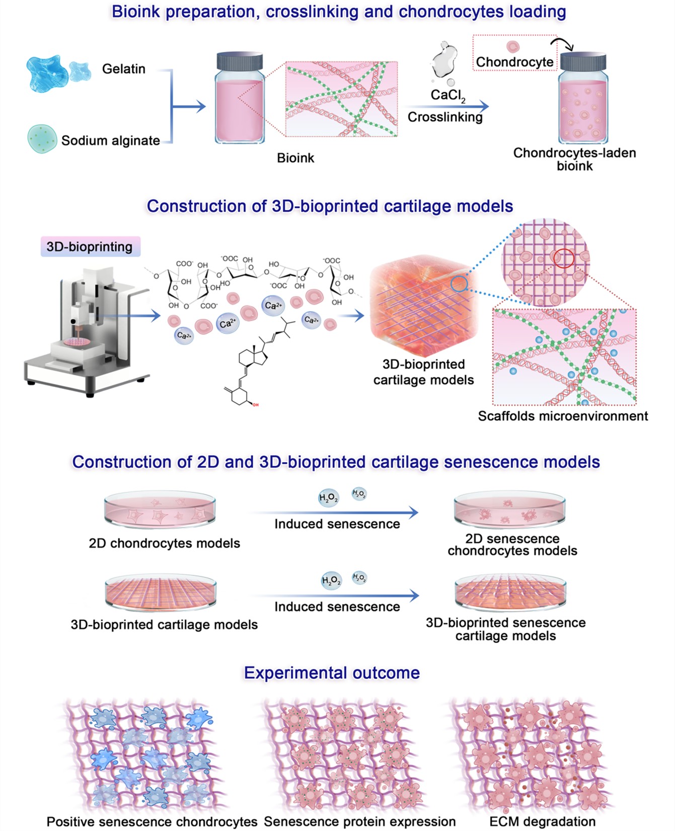
Osteoarthritis (OA) is an age-related degenerative joint disease characterized by progressive cartilage deterioration. Chondrocyte senescence is recognized as a key contributor to the onset and progression of OA. Establishing reliable cartilage senescence models is, therefore, essential for elucidating the underlying mechanisms and developing preventive strategies for OA. 3D-bioprinted models offer significant advantages in precisely controlling tissue architecture, enabling spatial delivery of bioactive molecules, and supporting dynamic cell culture. In this study, we employed 3D bioprinting technology to construct cartilage models and subsequently established cartilage senescence models using hydrogen peroxide (H₂O₂). Firstly, gelatin–sodium alginate hydrogel scaffolds provided favorable mechanical strength and porosity, creating a supportive microenvironment for chondrocyte proliferation. Secondly, these scaffolds exhibited excellent biocompatibility and effectively promoted extracellular matrix synthesis and secretion. By comparing H₂O₂-induced 2D chondrocyte senescence models with 3D-bioprinted cartilage senescence models, our results demonstrated that the 3D models more closely mimicked the molecular characteristics of naturally aged human cartilage. Therefore, the 3D-bioprinted cartilage senescence models represent a promising experimental platform for investigating the pathogenesis and prevention of age-related OA.
- Li Y, Li L, Wang M, et al. O-alg-THAM/gel hydrogels functionalized with engineered microspheres based on mesenchymal stem cell secretion recruit endogenous stem cells for cartilage repair. Bioact Mater. 2023;28(5):255-272. doi: 10.1016/j.bioactmat.2023.05.003.
- Guo X, Xi L, Yu M, et al. Regeneration of articular cartilage defects: therapeutic strategies and perspectives. Tissue Eng Rev. 2023;14(3):20417314231164765. doi: 10.1177/20417314231164765.
- Liu Z, Lu T, Ma L, et al. DNA demethylation of promoter region orchestrates SPI-1-induced ADAMTS-5 expression in articular cartilage of osteoarthritis mice. J Cell Physiol. 2024;239(2):e31170. doi: 10.1002/jcp.31170.
- Bank NC, Sanghvi P, Hecht CJ, et al. The epidemiology of posttraumatic osteoarthritis of the knee in the United States: an analysis of 948,853 patients from 2000 to 2022. J Am Acad Orthop Surg. 2024;32(7):313-320. doi: 10.5435/JAAOS-D-23-00662.
- Zhang XX, He SH, Liang X, et al. Aging, cell senescence, the pathogenesis and targeted therapies of osteoarthritis. Front Pharmacol. 2021;12:728100. doi: 10.3389/fphar.2021.728100.
- Loeser RF. Aging processes and the development of osteoarthritis. Curr Opin Rheumatol. 2013;25(1):108-113. doi: 10.1097/BOR.0b013e32835a9428.
- June RK, Liu-Bryan R, Long F, et al. Emerging role of metabolic signaling in synovial joint remodeling and osteoarthritis. J Orthop Res. 2016;34(12):2048-2058. doi: 10.1002/jor.23420.
- Ray A, Ray BK. An inflammation-responsive transcription factor in the pathophysiology of osteoarthritis. Biorheology. 2008;45(3-4):399-409.
- Yang Y, Zhao X, Wang S, et al. Ultra-durable cell-free bioactive hydrogel with fast shape memory and on-demand drug release for cartilage regeneration. Nat Commun. 2023;14(1):7771. doi: 10.1038/s41467-023-43334-8.
- Hu Y, Lyu C, Teng L, et al. Glycopolypeptide hydrogels with adjustable enzyme-triggered degradation: a novel proteoglycans analogue to repair articular-cartilage defects. Mater Today Bio. 2023;20(5):100659. doi: 10.1016/j.mtbio.2023.100659.
- Zeng Q, Gong Y, Zhu N, et al. Lipids and lipid metabolism in cellular senescence: emerging targets for age-related diseases. Ageing Res Rev. 2024;97(6):102294. doi: 10.1016/j.arr.2024.102294.
- Jimenez PA, Glasson SS, Trubetskoy OV, et al. Spontaneous osteoarthritis in Dunkin Hartley guinea pigs: histologic, radiologic, and biochemical changes. Lab Anim Sci. 1997;47(6):598-601.
- Säämänen AK, Salminen HJ, Dean PB, et al. Osteoarthritis-like lesions in transgenic mice harboring a small deletion mutation in type II collagen gene. Osteoarthritis Cartilage. 2000;8(4):248-257. doi: 10.1053/joca.2000.0298.
- Murakami T, Ishida T, Tanaka S, et al. Inflammation and subsequent nociceptor sensitization in the bone marrow are involved in an animal model of osteoarthritis pain. Life Sci. 2023;324(7):121736. doi: 10.1016/j.lfs.2023.121736.
- Seo JE, Le Y, Revollo J, et al. Evaluating the mutagenicity of N-nitrosodimethylamine in 2D and 3D HepaRG cell cultures using error-corrected next generation sequencing. Arch Toxicol. 2024;98(6):1919-1935. doi: 10.1007/s00204-024-03731-4.
- Yagi M, Endo K, Komori K, et al. Comparison of the effects of oxidative and inflammatory stresses on rat chondrocyte senescence. Sci Rep. 2023;13(1):7697. doi: 10.1038/s41598-023-34825-1.
- Shboul SA, DeLuca VA, Dweiri YA, et al. Can 3D bioprinting solve the mystery of senescence in cancer therapy? Ageing Res Rev. 2022;81(11):101732. doi: 10.1016/j.arr.2022.101732.
- Żuchowska A, Baranowska P, Flont M, et al. Review: 3D cell models for organ-on-a-chip applications. Anal Chim Acta. 2024;1301(5):342413. doi: 10.1016/j.aca.2024.342413.
- Ziadlou R, Barbero A, Martin I, et al. Anti-inflammatory and chondroprotective effects of vanillic acid and epimedin C in human osteoarthritic chondrocytes. Biomolecules. 2020;10(6):932. doi: 10.3390/biom10060932.
- Huang S, Song X, Li T, et al. Pellet coculture of osteoarthritic chondrocytes and infrapatellar fat pad-derived mesenchymal stem cells with chitosan/hyaluronic acid nanoparticles promotes chondrogenic differentiation. Stem Cell Res Ther. 2017;8(1):264. doi: 10.1186/s13287-017-0719-7.
- Stüdle C, Vallmajó-Martín Q, Haumer A, et al. Spatially confined induction of endochondral ossification by functionalized hydrogels for ectopic engineering of osteochondral tissues. Biomaterials. 2018;171(7):219-229. doi: 10.1016/j.biomaterials.2018.04.025.
- Castro-Viñuelas R, Viudes-Sarrión N, Rojo-García AV, et al. Mechanical loading rescues mechanoresponsiveness in a human osteoarthritis explant model despite Wnt activation. Osteoarthritis Cartilage. 2025;33(6):692-702. doi: 10.1016/j.joca.2024.02.945.
- Byron CR, Trahan RA. Comparison of the effects of interleukin-1 on equine articular cartilage explants and cocultures of osteochondral and synovial explants. Front Vet Sci. 2017;4(9):152. doi: 10.3389/fvets.2017.00152.
- Lin H, Lozito TP, Alexander PG, et al. Stem cell-based microphysiological osteochondral system to model tissue response to interleukin-1beta. Mol Pharm. 2014;11(7):2203-2212. doi: 10.1021/mp500136b.
- Dönges L, Damle A, Mainardi A, et al. Engineered human osteoarthritic cartilage organoids. Biomaterials. 2024;308(7):122549. doi: 10.1016/j.biomaterials.2024.122549.
- Hall GN, Mendes LF, Gklava C, et al. Developmentally engineered callus organoid bioassemblies exhibit predictive in vivo long bone healing. Adv Sci (Weinh). 2020;7(2):1902295. doi: 10.1002/advs.201902295.
- SunY, You Y, Wu Q, et al. Genetically inspired organoids prevent joint degeneration and alleviate chondrocyte senescence via Col11a1-HIF1alpha-mediated glycolysis- OXPHOS metabolism shift. Clin Transl Med. 2024;14(2):e1574. doi: 10.1002/ctm2.1574.
- Shen C, Wang J, Li G, et al. Boosting cartilage repair with silk fibroin-DNA hydrogel-based cartilage organoid precursor. Bioact Mater. 2024;35(2):429-444. doi: 10.1016/j.bioactmat.2024.02.016.
- Thompson CL, Hopkins T, Bevan C, et al. Human vascularised synovium-on-a-chip: a mechanically stimulated, microfluidic model to investigate synovial inflammation and monocyte recruitment. Biomed Mater. 2023;18(6):065013. doi: 10.1088/1748-605X/acf976.
- Lin Z, Li Z, Li EN, et al. Osteochondral tissue chip derived from iPSCs: modeling OA pathologies and testing drugs. Front Bioeng Biotechnol. 2019;7(12):411. doi: 10.3389/fbioe.2019.00411.
- Ong LJY, Fan X, Sun AR, et al. Controlling microenvironments with organs-on-chips for osteoarthritis modelling. Cells. 2023;12(4):579. doi: 10.3390/cells12040579.
- Rothbauer M, Reihs EI, Fischer A, et al. A progress report and roadmap for microphysiological systems and organ-on-a-chip technologies to be more predictive models in human (knee) osteoarthritis. Front Bioeng Biotechnol. 2022;10(6):886360. doi: 10.3389/fbioe.2022.886360.
- Nu Aye KT, Ferreira JN, Chaweewannakorn C, et al. Advances in the application of iron oxide nanoparticles (IONs and SPIONs) in three-dimensional cell culture systems. SLAS Technol. 2024;29(3):100132. doi: 10.1016/j.slast.2024.100132.
- Wu KY, Tabari A, Mazerolle E, et al. Towards precision ophthalmology: the role of 3D printing and bioprinting in oculoplastic surgery, retinal, corneal, and glaucoma treatment. Biomimetics (Basel). 2024;9(3):145. doi: 10.3390/biomimetics9030145.
- Jiu J, Liu H, Li D, et al. 3D bioprinting approaches for spinal cord injury repair. Biofabrication. 2024;16(3):032003. doi: 10.1088/1758-5090.
- Das S, Valoor R, Ratnayake P, et al. Bioprinting complex cartilaginous structures with clinically compliant biomaterials. Adv Funct Mater. 2015;25(48):7406-7417. doi: 10.1021/acsabm.3c01194.
- Schuurman W, Levett PA, Pot MW, et al. Gelatin–methacrylamide hydrogels as potential biomaterials for fabrication of tissue-engineered cartilage constructs. Macromol Biosci. 2013; 13(5):551-561. doi: 10.1002/mabi.201200471.
- Ávila HM, Schwarz S, Rotter N, et al. 3D bioprinting of human chondrocyte-laden nanocellulose hydrogels for patient-specific auricular cartilage regeneration. Bioprinting. 2016;1–2:22-35. doi: 10.1016/j.bprint.2016.08.003.
- Markstedt K, Mantas A, Tournier I, et al. 3D Bioprinting human chondrocytes with nanocellulose–alginate bioink for cartilage tissue engineering applications. Biomacromolecules. 2015;16(5):1489-1496. doi: 10.1021/acs.biomac.5b00188.
- Rastogi P, Kandasubramanian B Review of alginate-based hydrogel bioprinting for application in tissue engineering. Biofabrication. 2019;11(4):042001. doi: 10.1088/1758-5090/ab331e.
- Zhang X, Liu K, Qin M, et al. Abundant tannic acid modified gelatin/sodium alginate biocomposite hydrogels with high toughness, antifreezing, antioxidant and antibacterial properties. Carbohydr Polym. 2023;309(6):120702. doi: 10.1016/j.carbpol.2023.120702.
- Lai Y, Cao H, Wang X, et al. Porous composite scaffold incorporating osteogenic phytomolecule icariin for promoting skeletal regeneration in challenging osteonecrotic bone in rabbits. Biomaterials. 2018;153(1):1-13. doi: 10.1016/j.biomaterials.2017.10.025.
- Liu Z, Fang R, Meng Q The method of type II collagenase digestion can quickly obtain a large number of highly purified rat articular cartilage cells. J Clin Rehabil Tissue Eng Res. 2011;15(10):9323-9326. doi: 10.3969/j.issn.1673-8225.2011.50.004.
- Chen W, Xu Y, Li Y, et al. 3D printing electrospinning fiber-reinforced decellularized extracellular matrix for cartilage regeneration. Chem Eng J. 2020;15(2):122986. doi: 10.1016/j.cej.2019.122986.
- Wu Y, (William) Lin ZY, Wenger AC, et al. 3D bioprinting of liver-mimetic construct with alginate/cellulose nanocrystal hybrid bioink. Bioprinting. 2018;9(3):1-6. doi: 10.1016/j.bprint.2017.12.001.
- Chung JHY, Naficy S, Yue Z, et al. Bio-ink properties and printability for extrusion printing living cells. Biomater Sci. 2013;1(7):763-773. doi: 10.1039/c3bm00012e.
- Duan B, Hockaday LA, Kang KH, et al. 3D bioprinting of heterogeneous aortic valve conduits with alginate/gelatin hydrogels. Biomed Mater Res. 2013;101(5):1255-1264. doi: 10.1002/jbm.a.34420.
- Ferris CJ, Gilmore KJ, Beirne S, et al. Bio-ink for on-demand printing of living cells. Biomater Sci. 2013;1(2):224-230. doi: 10.1039/c2bm00114d.
- Skardal A, Atala A. Biomaterials for integration with 3-D bioprinting. Ann Biomed Eng. 2015;43(3):730-746. doi: 10.1007/s10439-014-1207-1.
- Hu W, Wang Z, Xiao Y, et al. Advances in crosslinking strategies of biomedical hydrogels. Biomater Sci. 2019;7(3):843-855. doi: 10.1039/c8bm01246f.
- Shanto PC, Al Fahad Md A, Jung HI, et al. Multi-functional dual-layer nanofibrous membrane for prevention of postoperative pancreatic leakage. Biomaterials. 2024;307(6):122508. doi: 10.1016/j.biomaterials.2024.122508.
- Mohamadhoseini M, Mohamadnia Z. Alginate-based self-healing hydrogels assembled by dual cross-linking strategy: fabrication and evaluation of mechanical properties. Int J Biol Macromol. 2021;191(11):139-151. doi: 10.1016/j.ijbiomac.2021.09.062.
- Miri AK, Khalilpour A, Cecen B, et al. Multiscale bioprinting of vascularized models. Biomaterials. 2019;198(4):204-216. doi: 10.1016/j.biomaterials.2018.08.006.
- Bhaladhare S, Bhattacharjee S. Chemical, physical, and biological stimuli responsive nanogels for biomedical applications (mechanisms, concepts, and advancements): a review. Int J Biol Macromol. 2023;226(1):535-553. doi: 10.1016/j.ijbiomac.2022.12.076.
- Giuseppe MD, Law N, Webb B, et al. Mechanical behaviour of alginate-gelatin hydrogels for 3D bioprinting. J Mech Behav Biomed Mater. 2018;79(3):150-157. doi: 10.1016/j.jmbbm.2017.12.018.
- Caliari SR, Burdick JA. A practical guide to hydrogels for cell culture. Nat Methods. 2016;13(5):405-414. doi: 10.1038/nmeth.3839.
- Mahmoudi C, Douma NT, Mahmoudi H, et al. Developing and characterizing a biocompatible hydrogel obtained by cross-linking gelatin with oxidized sodium alginate for potential biomedical applications. Polymers (Basel). 2024;16(22):3143. doi: 10.3390/polym16223143.
- Schwarz S, Kuth S, Distler T, et al. 3D printing and characterization of human nasoseptal chondrocytes laden dual crosslinked oxidized alginate-gelatin hydrogels for cartilage repair approaches. Mater Sci Eng. 2020;116(11):111189. doi: 10.1016/j.msec.2020.111189.
- Zhao P, Deng C, Xu H, et al. Fabrication of photo-crosslinked chitosan gelatin scaffold in sodium alginate hydrogel for chondrocyte culture. Bio Med Mater Eng. 2014;24(1):633-641. doi: 10.3233/bme-130851.
- Xu HQ, Liu JC, Zhang ZY, et al. A review on cell damage, viability, and functionality during 3D bioprinting. Mil Med Res. 2022;9(1):70. doi: 10.1186/s40779-022-00429-5.
- Shi P, Laude A, Yeong WY. Investigation of cell viability and morphology in 3D bio-printed alginate constructs with tunable stiffness. J Biomed Mater Res. 2017;105(4):1009-1018. doi: 10.1002/jbm.a.35971.
- Soltan N, Ning L, Mohabatpour F, et al. Printability and cell viability in bioprinting alginate dialdehydegelatin scaffolds. ACS Biomater Sci Eng. 2019;5(6):2976-2987. doi: 10.1021/acsbiomaterials.9b00167.
- Coryell PR, Diekman BO, Loeser RF. Mechanisms and therapeutic implications of cellular senescence in osteoarthritis. Nat Rev Rheumatol. 2021;17(1):47-57. doi: 10.1038/s41584-020-00533-7.
- Lou C, Deng A, Zheng H, et al. Pinitol suppresses TNF-α-induced chondrocyte senescence. Cytokine. 2020;130:155047. doi: 10.1016/j.cyto.2020.155047.
- Arra M, Swarnkar G, Ke K, et al. Generation in chondrocytes is a potential therapeutic target for osteoarthritis. Nat Commun. 2020;11(1):3427. doi: 10.1038/s41467-020-17242-0.
- Ma MW, Wang J, Zhang Q, et al. NADPH oxidase in brain injury and neurodegenerative disorders. Mol Neurodegener. 2017;12(1):7. doi: 10.1186/s13024-017-0150-7.
- Liang H, Luo R, Li G, et al. The proteolysis of ECM in intervertebral disc degeneration. Int J Mol Sci. 2022;23(3):1715. doi: 10.3390/ijms23031715.
- Coppé JP, Desprez PY, Krtolica A, et al. The senescence-associated secretory phenotype: the dark side of tumor suppression. Annu Rev Pathol. 2010;5:99-118. doi: 10.1146/annurev-pathol-121808-102144.
- Sun Y, Wu Q, Dai K, et al. Generating 3D-cultured organoids for preclinical modeling and treatment of degenerative joint disease. Signal Transduct Target Ther. 2021;6(1):380. doi: 10.1038/s41392-021-00675-4.
- Gao F, Xu Z, Liang Q, et al. Osteochondral regeneration with 3D-printed biodegradable high-strength supramolecular polymer reinforced-gelatin hydrogel scaffolds. Adv Sci. 2019;6(15):1900867. doi: 10.1002/advs.201900867.
- Gao F, Xu Z, Liang Q, et al. Direct 3D printing of high strength biohybrid gradient hydrogel scaffolds for efficient repair of osteochondral defect. Adv Funct Mater. 2018;28(13): 1706644. doi: 10.1002/adfm.201706644.
- Chen D, Wu JY, Kennedy KM, et al. Tissue engineered autologous cartilage-bone grafts for temporomandibular joint regeneration. Sci Transl Med. 2020;12(565):eabb6683. doi: 10.1126/scitranslmed.abb6683.