

Collagen bioinks redefined: Optimizing ionic strength and growth factor delivery for cartilage tissue engineering

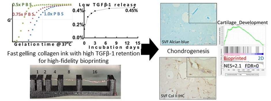
Tissue engineering of hyaline cartilage for regenerative medicine and the treatment of osteoarthritis has advanced significantly over the past decade, driven by developments in 3D bioprinting and biomaterials science. Despite these advances, standardized biofabrication protocols approved for clinical applications remain elusive, underscoring the need for research into widely accessible, non-immunogenic, and biocompatible bioinks that support chondrogenesis. This study proposes a strategy to improve the gelation kinetics of collagen bioinks by fine-tuning their ionic strength and reports a highly efficient sequestration of TGF-β1 within them, alongside their compatibility with bioprinting live chondrocytes and adipose-derived stem cells for cartilage tissue engineering. By adjusting sodium chloride and phosphate-buffered saline (PBS) concentrations, we demonstrate that reduced ionic strengths accelerate gelation, facilitating high-fidelity bioprinting while supporting high cell viability and proliferation. Furthermore, at 1% collagen concentration, the hydrogel effectively immobilized TGF-β1, with less than 0.5% released over two weeks, indicating potent sequestration capability. Using adipose-derived mesenchymal stromal cells, histomorphological and transcriptomic analyses reveal that the presence of TGF-β1 significantly enhances chondrogenesis. These results underscore the neglected role of ionic strength in optimizing collagen ink properties for advanced bioprinting applications and highlight the potential of collagen hydrogels as effective carriers for sustained growth factor delivery, paving the way for successful cartilage tissue engineering strategies.
- Dhavalikar P, Lan Z, Kar R, Salhadar K, Gaharwar AK, Cosgriff-Hernandez E. Biomedical applications of additive manufacturing. In: Biomaterials Science. 4th ed. San Diego, USA: Elsevier; 2020:623-639. doi: 10.1016/B978-0-12-816137-1.00040-4
- Sagi I, Afratis NA. Collagen. Vol 1944. In: Sagi I, Afratis NA, eds. New York, NY: Springer; 2019. doi: 10.1007/978-1-4939-9095-5
- Osidak EO, Kozhukhov VI, Osidak MS, Domogatsky SP. Collagen as bioink for bioprinting: a comprehensive review. Int J Bioprint. 2024;6(3):270. doi: 10.18063/ijb.v6i3.270
- Li Z, Ruan C, Niu X. Collagen-based bioinks for regenerative medicine: fabrication, application and prospective. Med Nov Technol Devices. 2023;17(37):100211. doi: 10.1016/j.medntd.2023.100211
- Stepanovska J, Supova M, Hanzalek K, Broz A, Matejka R. Collagen bioinks for bioprinting: a systematic review of hydrogel properties, bioprinting parameters, protocols, and bioprinted structure characteristics. Biomedicines. 2021;9(9):1137. doi: 10.3390/biomedicines9091137
- Marques CF, Diogo GS, Pina S, Oliveira JM, Silva TH, Reis RL. Collagen-based bioinks for hard tissue engineering applications: a comprehensive review. J Mater Sci Mater Med. 2019;30(3):32. doi: 10.1007/s10856-019-6234-x
- Li Y, Asadi A, Monroe MR, Douglas EP. pH effects on collagen fibrillogenesis in vitro: electrostatic interactions and phosphate binding. Mater Sci Eng C. 2009;29(5):1643-1649. doi: 10.1016/j.msec.2009.01.001
- Rezabeigi E, Griffanti G, Nazhat SN. Effect of fibrillization ph on gelation viscoelasticity and properties of biofabricated dense collagen matrices via gel aspiration-ejection. Int J Mol Sci. 2023;24(4):3889. doi: 10.3390/ijms24043889
- Kalbitzer L, Pompe T. Fibril growth kinetics link buffer conditions and topology of 3D collagen I networks. Acta Biomater. 2018;67:206-214. doi: 10.1016/j.actbio.2017.11.051
- Li Y, Douglas EP. Effects of various salts on structural polymorphism of reconstituted type I collagen fibrils. Colloids Surf B Biointerfaces. 2013;112:42-50. doi: 10.1016/j.colsurfb.2013.07.037
- Achilli M, Mantovani D. Tailoring mechanical properties of collagen-based scaffolds for vascular tissue engineering: the effects of pH, temperature and ionic strength on gelation. Polymers (Basel). 2010;2(4):664-680. doi: 10.3390/polym2040664
- Stamov DR, Stock E, Franz CM, Jähnke T, Haschke H. Imaging collagen type I fibrillogenesis with high spatiotemporal resolution. Ultramicroscopy. 2015;149:86-94. doi: 10.1016/j.ultramic.2014.10.003
- Gobeaux F, Mosser G, Anglo A, et al. Fibrillogenesis in dense collagen solutions: a physicochemical study. J Mol Biol. 2008;376(5):1509-1522. doi: 10.1016/j.jmb.2007.12.047
- Zhang J, Wei B, He L, et al. Systematic modulation of gelation dynamics of snakehead (Channa argus) skin collagen by environmental parameters. Macromol Res. 2017;25(11):1105-1114. doi: 10.1007/s13233-017-5149-y
- Harris JR, Reiber A. Influence of saline and pH on collagen type I fibrillogenesis in vitro: fibril polymorphism and colloidal gold labelling. Micron. 2007;38(5):513-521. doi: 10.1016/j.micron.2006.07.026
- Harris JR, Soliakov A, Lewis RJ. In vitro fibrillogenesis of collagen type I in varying ionic and pH conditions. Micron. 2013;49:60-68. doi: 10.1016/j.micron.2013.03.004
- Salinas-Fernandez S, Garcia O, Kelly DJ, Buckley CT. The influence of pH and salt concentration on the microstructure and mechanical properties of meniscus extracellular matrix-derived implants. J Biomed Mater Res A. 2024;112(3):359-372. doi: 10.1002/jbm.a.37634
- Zhu S, Yuan Q, Yin T, et al. Self-assembly of collagen-based biomaterials: preparation, characterizations and biomedical applications. J Mater Chem B. 2018;6(18):2650-2676. doi: 10.1039/c7tb02999c
- Cooke ME, Rosenzweig DH. The rheology of direct and suspended extrusion bioprinting. APL Bioeng. 2021;5(1):011502. doi: 10.1063/5.0031475
- Xing JY, Yang L, Li YL. Effect of anions on type I collagen fibrillogenesis in aqueous solution. ISWREP 2011 – Proc 2011 Int Symp Water Resour Environ Prot. 2011;4: 2975-2978. doi: 10.1109/ISWREP.2011.5893502
- Zhu J, Kaufman LJ. Collagen I self-assembly: revealing the developing structures that generate turbidity. Biophys J. 2014;106(8):1822-1831. doi: 10.1016/j.bpj.2014.03.011
- Diamantides N, Wang L, Pruiksma T, et al. Correlating rheological properties and printability of collagen bioinks: the effects of riboflavin photocrosslinking and pH. Biofabrication. 2017;9(3):034102. doi: 10.1088/1758-5090/aa780f
- Yang YL, Motte S, Kaufman LJ. Pore size variable type I collagen gels and their interaction with glioma cells. Biomaterials. 2010;31(21):5678-5688. doi: 10.1016/j.biomaterials.2010.03.039
- Lai G, Li Y, Li G. Effect of concentration and temperature on the rheological behavior of collagen solution. Int J Biol Macromol. 2008;42(3):285-291. doi: 10.1016/j.ijbiomac.2007.12.010
- Xie J, Bao M, Bruekers SMC, Huck WTS. Collagen gels with different fibrillar microarchitectures elicit different cellular responses. ACS Appl Mater Interfaces. 2017;9(23):19630-19637. doi: 10.1021/acsami.7b03883
- Tran-Ba KH, Lee DJ, Zhu J, Paeng K, Kaufman LJ. Confocal rheology probes the structure and mechanics of collagen through the sol-gel transition. Biophys J. 2017;113(8): 1882-1892. doi: 10.1016/j.bpj.2017.08.025
- Sarrigiannidis SO, Rey JM, Dobre O, González-García C, Dalby MJ, Salmeron-Sanchez M. A tough act to follow: collagen hydrogel modifications to improve mechanical and growth factor loading capabilities. Mater Today Bio. 2021;10(January):100098. doi: 10.1016/j.mtbio.2021.100098
- Goebel EJ, Hart KN, McCoy JC, Thompson TB. Structural biology of the TGFβ family. Exp Biol Med. 2019;244(17): 1530-1546. doi: 10.1177/1535370219880894
- Shah SS, Mithoefer K. Current applications of growth factors for knee cartilage repair and osteoarthritis treatment. Curr Rev Musculoskelet Med. 2020;13(6):641-650. doi: 10.1007/s12178-020-09664-6
- Johnstone B, Hering TM, Caplan AI, Goldberg VM, Yoo JU. In vitro chondrogenesis of bone marrow-derived mesenchymal progenitor cells. Exp Cell Res. 1998;238(1):265-272. doi: 10.1006/excr.1997.3858
- Kundu J, Shim JH, Jang J, Kim SW, Cho DW. An additive manufacturing-based PCL-alginate-chondrocyte bioprinted scaffold for cartilage tissue engineering. J Tissue Eng Regen Med. 2015;9(11):1286-1297. doi: 10.1002/term.1682
- Zhu W, Cui H, Boualam B, et al. 3D bioprinting mesenchymal stem cell-laden construct with core–shell nanospheres for cartilage tissue engineering. Nanotechnology. 2018;29(18):185101. doi: 10.1088/1361-6528/aaafa1
- Hauptstein J, Forster L, Nadernezhad A, Groll J, Teßmar J, Blunk T. Tethered TGF-β1 in a hyaluronic acid-based bioink for bioprinting cartilaginous tissues. Int J Mol Sci. 2022;23(2):924. doi: 10.3390/ijms23020924
- Komsa-Penkova R, Stavreva G, Belemezova K, Kyurkchiev S, Todinova S, Altankov G. Mesenchymal stem-cell remodeling of adsorbed type-I collagen – the effect of collagen oxidation. Int J Mol Sci. 2022;23(6):3058. doi: 10.3390/ijms23063058
- Ge SX, Jung D, Jung D, Yao R. ShinyGO: a graphical gene-set enrichment tool for animals and plants. Bioinformatics. 2020;36(8):2628-2629. doi: 10.1093/bioinformatics/btz931
- Szklarczyk D, Kirsch R, Koutrouli M, et al. The STRING database in 2023: protein-protein association networks and functional enrichment analyses for any sequenced genome of interest. Nucleic Acids Res. 2023;51(1D):D638-D646. doi: 10.1093/nar/gkac1000
- Deyl Z, Mikšík I, Eckhardt A. Preparative procedures and purity assessment of collagen proteins. J Chromatogr B Anal Technol Biomed Life Sci. 2003;790(1-2):245-275. doi: 10.1016/S1570-0232(03)00158-2
- Drzewiecki KE, Grisham DR, Parmar AS, Nanda V, Shreiber DI. Circular dichroism spectroscopy of collagen fibrillogenesis: a new use for an old technique. Biophys J. 2016;111(11):2377-2386. doi: 10.1016/j.bpj.2016.10.023
- Mertz EL, Leikin S. Interactions of inorganic phosphate and sulfate anions with collagen. Biochemistry. 2004;43(47):14901-14912. doi: 10.1021/bi048788b
- Hayashi T, Nagai Y. Factors affecting the interactions of collagen molecules as observed by in vitro fibril formation: III. non-helical regions of the collagen molecules. J Biochem. 1974;76(1):177-186. doi: 10.1093/oxfordjournals.jbchem.a130543
- Weinstock A, King PC, Wuthier RE. The ion-binding characteristics of reconstituted collagen. Biochem J. 1967;104(3):705_b1-705_b1. doi: 10.1042/bj1040705_b1a
- Freudenberg U, Behrens SH, Welzel PB, et al. Electrostatic interactions modulate the conformation of collagen I. Biophys J. 2007;92(6):2108-2119. doi: 10.1529/biophysj.106.094284
- Oh S, Nguyen QD, Chung KH, Lee H. Bundling of collagen fibrils using sodium sulfate for biomimetic cell culturing. ACS Omega. 2020;5(7):3444-3452. doi: 10.1021/acsomega.9b03704
- Lien YH, Stern R, Fu JCC, Siegel RC. Inhibition of collagen fibril formation in vitro and subsequent cross-linking by glucose. Science. 1984;225(4669):1489-1491. doi: 10.1126/science.6147899
- Sbirkov Y, Molander D, Milet C, et al. A colorectal cancer 3D bioprinting workflow as a platform for disease modeling and chemotherapeuticsScreening. Front Bioeng Biotechnol. 2021;9(November):1-12. doi: 10.3389/fbioe.2021.755563
- Chaudhuri O, Cooper-White J, Janmey PA, Mooney DJ, Shenoy VB. Effects of extracellular matrix viscoelasticity on cellular behaviour. Nature. 2020;584(7822):535-546. doi: 10.1038/s41586-020-2612-2
- Schinagl RM, Gurskis D, Chen AC, Sah RL. Depth-dependent confined compression modulus of full-thickness bovine articular cartilage. J Orthop Res. 1997;15(4):499-506. doi: 10.1002/jor.1100150404
- Beketov EE, Isaeva EV, Yakovleva ND, et al. Bioprinting of cartilage with bioink based on high-concentration collagen and chondrocytes. Int J Mol Sci. 2021;22(21):11351. doi: 10.3390/ijms222111351
- Isaeva EV, Beketov EE, Demyashkin GA, et al. Cartilage formation in vivo using high concentration collagen-based bioink with MSC and decellularized ECM granules. Int J Mol Sci. 2022;23(5):2703. doi: 10.3390/ijms23052703
- Gospodinova A, Nankov V, Tomov S, Redzheb M, Petrov PD. Extrusion bioprinting of hydroxyethylcellulose-based bioink for cervical tumor model. Carbohydr Polym. 2021;260(February):117793. doi: 10.1016/j.carbpol.2021.117793
- Sbirkov Y, Redzheb M, Forraz N, McGuckin C, Sarafian V. High hopes for the biofabrication of articular cartilage – what lies beyond the horizon of tissue engineering and 3D bioprinting? Biomedicines. 2024;12(3):665. doi: 10.3390/biomedicines12030665
- Cui X, Gao G, Yonezawa T, Dai G. Human cartilage tissue fabrication using three-dimensional inkjet printing technology. J Vis Exp. 2014;(88):1-5. doi: 10.3791/51294
- Bourin P, Bunnell BA, Casteilla L, et al. Stromal cells from the adipose tissue-derived stromal vascular fraction and culture expanded adipose tissue-derived stromal/stem cells: a joint statement of the International Federation for Adipose Therapeutics and Science (IFATS) and the International So. Cytotherapy. 2013;15(6):641-648. doi: 10.1016/j.jcyt.2013.02.006
- Sang S, Mao X, Cao Y, et al. 3D bioprinting using synovium-derived MSC-laden photo-cross-linked ECM bioink for cartilage regeneration. ACS Appl Mater Interfaces. 2023;15(7):8895-8913. doi: 10.1021/acsami.2c19058
- Grafe I, Alexander S, Peterson JR, et al. TGF-β family signaling in mesenchymal differentiation. Cold Spring Harb Perspect Biol. 2018;10(5):1-50. doi: 10.1101/cshperspect.a022202
- Baba AB, Rah B, Bhat GR, et al. Transforming growth factor-beta (TGF-β) signaling in cancer-A betrayal within. Front Pharmacol. 2022;13(February):1-16. doi: 10.3389/fphar.2022.791272
- Stromps JP, Paul NE, Rath B, Nourbakhsh M, Bernhagen J, Pallua N. Chondrogenic differentiation of human adipose-derived stem cells: a new path in articular cartilage defect management? Biomed Res Int. 2014;2014:740926. doi: 10.1155/2014/740926