

Electrospun polylactic acid-glycolic acid composite hydrogel scaffold loaded with 3D extracellular vesicles for nasal septal cartilage defect repair

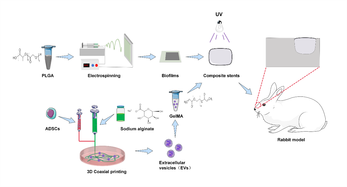
The nasal septum plays an important role in the growth and support of the human nose, and defects can cause nasal deformities. Extracellular vesicles (EVs) have demonstrated great potential in tissue repair. Stem cell EVs are widely used in the repair of articular cartilage defects, but their use for nasal septal cartilage defects has not been reported. Due to the low yield and loss of EVs during in situ injection, improved preparation methods and better carriers are needed for the effective sustained release of EVs in wounds. In this study, swelling and degradation experiments were initially conducted on the scaffold, along with mechanical performance testing, including observation of the scaffold morphology using scanning electron microscopy (SEM). Subsequently, in vitro cell experiments were conducted to evaluate the ability of 3D EVs to promote chondrocyte proliferation, migration, and extracellular matrix formation. Finally, the EV-laden gelatin methacrylic acid-polylactic acid-glycolic acid (Gel-PLGA) composite scaffold was implanted into the nasal septum defect site of rabbits in vivo to observe its repair effect on the defect. In vitro experiments demonstrated that the biological scaffold exhibited good biocompatibility and could effectively promote the proliferation and migration of chondrocytes. In vivo, the EV-laden composite biological scaffold was implanted into the nasal septum defect of rabbits, and the tissues were tested at 6 and 12 weeks after surgery. The results indicate that the composite scaffolds effectively facilitated the repair of defect sites. Taken together, 3D EVs facilitate tissue repair and healing, offering a novel approach to treating nasal septal defects.
- Samibut P, Meevassana J, Suwajo P, et al. The anatomical study of the nasal septal cartilage with its clinical implications. Aesthetic Plast Surg. 2021;45:1705-1711. doi: 10.1007/s00266-020-02116-z
- Chiesa-Estomba CM, Aiastui A, González-Fernández I, et al. Three-dimensional bioprinting scaffolding for nasal cartilage defects: a systematic review. Tissue Eng Regen Med. 2021;18:343-53. doi: 10.1007/s13770-021-00331-6
- Setton LA, Elliott DM, Mow VC. Altered mechanics of cartilage with osteoarthritis: human osteoarthritis and an experimental model of joint degeneration. Osteoarthritis Cartilage. 1999;7:2-14. doi: 10.1053/joca.1998.0170
- Günebakan Ç, Kuzu S. Congenital deficiency of alar cartilage. J Craniofac Surg. 2021;32:e137-8. doi: 10.1097/SCS.0000000000006858
- Lavernia L, Brown WE, Wong BJF, Hu JC, Athanasiou KA. Toward tissue-engineering of nasal cartilages. Acta Biomater. 2019;88:42-56. doi: 10.1016/j.actbio.2019.02.025
- Bagher Z, Asgari N, Bozorgmehr P, Kamrava SK, Alizadeh R, Seifalian A. Will tissue-engineering strategies bring new hope for the reconstruction of nasal septal cartilage? Curr Stem Cell Res Ther. 2020;15:144-54. doi: 10.2174/1574888X14666191212160757
- Rajzer I, Stręk P, Wiatr M, et al. Biomaterials in the reconstruction of nasal septum perforation. Ann Otol Rhinol Laryngol. 2021;130:731-7. doi: 10.1177/0003489420970589
- Wu N, Liu J, Ma W, et al. Degradable calcium deficient hydroxyapatite/poly(lactic-glycolic acid copolymer) bilayer scaffold through integral molding 3D printing for bone defect repair. Biofabrication. 2021;13(2). doi: 10.1088/1758-5090/abcb48
- Cao Y, Sang S, An Y, Xiang C, Li Y, Zhen Y. Progress of 3D printing techniques for nasal cartilage regeneration. Aesthetic Plast Surg. 2022;46:947-64. doi: 10.1007/s00266-021-02472-4
- Acevedo Rua L, Mumme M, Manferdini C, et al. Engineered nasal cartilage for the repair of osteoarthritic knee cartilage defects. Sci Transl Med. 2021;13:eaaz4499. doi: 10.1126/scitranslmed.aaz4499
- He L, He T, Xing J, et al. Bone marrow mesenchymal stem cell-derived exosomes protect cartilage damage and relieve knee osteoarthritis pain in a rat model of osteoarthritis. Stem Cell Res Ther. 2020;11:276. doi: 10.1186/s13287-020-01781-w
- Xu H, Xu B. BMSC-derived exosomes ameliorate osteoarthritis by inhibiting pyroptosis of cartilage via delivering miR-326 targeting HDAC3 and STAT1//NF-κB p65 to chondrocytes. In: Yokota S, ed. Mediators Inflamm. 2021:1-26. doi: 10.1155/2021/9972805
- Ha DH, Kim H-K, Lee J, et al. Mesenchymal stem/stromal cell-derived exosomes for immunomodulatory therapeutics and skin regeneration. Cells. 2020;9:1157. doi: 10.3390/cells9051157
- Tienda-Vázquez MA, Hanel JM, Márquez-Arteaga EM, et al. Exosomes: a promising strategy for repair, regeneration and treatment of skin disorders. Cells. 2023;12:1625. doi: 10.3390/cells12121625
- Zhao H, Zhao Z, Li D, Wang X, Dai D, Fu H. Effect study of exosomes derived from platelet-rich plasma in the treatment of knee cartilage defects in rats. J Orthop Surg Res. 2023;18:160. doi: 10.1186/s13018-023-03576-0
- Yan Z, Yin H, Wu J, et al. Engineering exosomes by three-dimensional porous scaffold culture of human umbilical cord mesenchymal stem cells promote osteochondral repair. Materials Today Bio. 2023;19:100549. doi: 10.1016/j.mtbio.2023.100549
- Chen J, Huang T, Liu R, Wang C, Jiang H, Sun H. Congenital microtia patients: the genetically engineered exosomes released from porous gelatin methacryloyl hydrogel for downstream small RNA profiling, functional modulation of microtia chondrocytes and tissue-engineered ear cartilage regeneration. J Nanobiotechnol. 2022;20:164. doi: 10.1186/s12951-022-01352-6
- Han B, Fang W, Yang Z, et al. Enhancement of chondrogenic markers by exosomes derived from cultured human synovial fluid-derived cells: a comparative analysis of 2D and 3D conditions. Biomedicines. 2023;11:3145. doi: 10.3390/biomedicines11123145
- Li Q, Yu H, Zhao F, et al. 3D printing of microenvironment-specific bioinspired and exosome-reinforced hydrogel scaffolds for efficient cartilage and subchondral bone regeneration. Adv Sci (Weinh). 2023;10:e2303650. doi: 10.1002/advs.202303650
- Chen J, Zhou D, Nie Z, et al. A scalable coaxial bioprinting technology for mesenchymal stem cell microfiber fabrication and high extracellular vesicle yield. Biofabrication. 2021;14(1). doi: 10.1088/1758-5090/ac3b90
- Xu F, Dawson C, Lamb M, et al. Hydrogels for tissue engineering: addressing key design needs toward clinical translation. Front Bioeng Biotechnol. 2022;10:849831. doi: 10.3389/fbioe.2022.849831
- Xing H, Zhang Z, Mao Q, et al. Injectable exosome-functionalized extracellular matrix hydrogel for metabolism balance and pyroptosis regulation in intervertebral disc degeneration. J Nanobiotechnol. 2021;19:264. doi: 10.1186/s12951-021-00991-5
- Gao F, Xu Z, Liang Q, et al. Osteochondral regeneration with 3D-printed biodegradable high-strength supramolecular polymer reinforced-gelatin hydrogel scaffolds. Adv Sci (Weinh). 2019;6:1900867. doi: 10.1002/advs.201900867
- Zhang Y, Li D, Liu Y, et al. 3D-bioprinted anisotropic bicellular living hydrogels boost osteochondral regeneration via reconstruction of cartilage-bone interface. Innovation (Camb). 2024;5:100542. doi: 10.1016/j.xinn.2023.100542
- Gnatowski P, Gwizdała K, Kurdyn A, Skorek A, Augustin E, Kucińska-Lipka J. Investigation on filaments for 3D printing of nasal septum cartilage implant. Materials (Basel). 2023;16:3534. doi: 10.3390/ma16093534
- Göttel B, Lucas H, Syrowatka F, et al. In situ gelling amphotericin B nanofibers: a new option for the treatment of Keratomycosis. Front Bioeng Biotechnol. 2020; 8:600384. doi: 10.3389/fbioe.2020.600384
- Niu L, Feng C, Shen C, Wang B, Zhang X. PLGA/PLCA casting and PLGA/PDPA electrospinning bilayer film for prevention of postoperative adhesion. J Biomed Mater Res. 2019;107:2030-2039. doi: 10.1002/jbm.b.34294
- Kalluri L, Satpathy M, Duan Y. Effect of electrospinning parameters on the fiber diameter and morphology of PLGA nanofibers. Dent Oral Biol Craniofacial Res. 2021;4(2): 103-107. doi: 10.31487/j.dobcr.2021.02.04
- Zhang D, Su Y, Sun P, et al. A TGF-loading hydrogel scaffold capable of promoting chondrogenic differentiation for repairing rabbit nasal septum cartilage defect. Front Bioeng Biotechnol. 2022;10:1057904. doi: 10.3389/fbioe.2022.1057904
- Shen K, Duan A, Cheng J, et al. Exosomes derived from hypoxia preconditioned mesenchymal stem cells laden in a silk hydrogel promote cartilage regeneration via the miR-205-5p/PTEN/AKT pathway. Acta Biomaterialia. 2022;143:173-88. doi: 10.1016/j.actbio.2022.02.026. Erratum in: Acta Biomater. 2022 Oct 1;151:662-663. doi: 10.1016/j.actbio.2022.09.005
- Hu J, Ai B, Zhu S, Wang Z, Xia H, Jia W. Electrospun PLGA and PLGA/gelatin scaffolds for tubularized urethral replacement: Studies in vitro and in vivo. J Biomater Appl. 2022;36(6):956-964. doi: 10.1177/08853282211030904. Epub 2021 Jul 13. Erratum in: J Biomater Appl. 2023;29:8853282231217938. doi: 10.1177/08853282211030904
- Franco RA, Nguyen TH, Lee BT. Preparation and characterization of electrospun PCL/PLGA membranes and chitosan/gelatin hydrogels for skin bioengineering applications. J Mater Sci: Mater Med. 2011;22: 2207-18. doi: 10.1007/s10856-011-4402-8
- Wang XS, Yang JM, Ding RJ, et al. Fabrication of a polylactide-glycolide/poly-ε-caprolactone/dextran/plastrum testudinis extract composite anti-inflammation nanofiber membrane via electrospinning for annulus fibrosus regeneration. J Biomed Nanotechnol. 2021;17:873-88. doi: 10.1166/jbn.2021.3070
- Cao H, Chen M, Cui X, et al. Cell-free osteoarthritis treatment with sustained-release of chondrocyte-targeting exosomes from umbilical cord-derived mesenchymal stem cells to rejuvenate aging chondrocytes. ACS Nano. 2023;17:13358-76. doi: 10.1021/acsnano.3c01612
- Stüdle C, Vallmajó-Martín Q, Haumer A, et al. Spatially confined induction of endochondral ossification by functionalized hydrogels for ectopic engineering of osteochondral tissues. Biomaterials. 2018;171: 219-29. doi: 10.1016/j.biomaterials.2018.04.025
- Patel DB, Gray KM, Santharam Y, Lamichhane TN, Stroka KM, Jay SM. Impact of cell culture parameters on production and vascularization bioactivity of mesenchymal stem cell-derived extracellular vesicles. Bioeng Transl Med. 2017;2(2):170-179. doi: 10.1002/btm2.10065
- Mao G, Xu Y, Long D, et al. Exosome-transported circRNA_0001236 enhances chondrogenesis and suppress cartilage degradation via the miR-3677-3p/Sox9 axis. Stem Cell Res Ther. 2021;12:389. doi: 10.1186/s13287-021-02431-5
- Xiang XN, Zhu SY, He HC, Yu X, Xu Y, He CQ. Mesenchymal stromal cell-based therapy for cartilage regeneration in knee osteoarthritis. Stem Cell Res Ther. 2022;13:14. doi: 10.1186/s13287-021-02689-9
- Wu J, Kuang L, Chen C, et al. miR-100-5p-abundant exosomes derived from infrapatellar fat pad MSCs protect articular cartilage and ameliorate gait abnormalities via inhibition of mTOR in osteoarthritis. Biomaterials. 2019;206:87-100. doi: 10.1016/j.biomaterials.2019.03.022
- Hu H, Dong L, Bu Z, et al. miR‐23a‐3p‐abundant small extracellular vesicles released from Gelma/nanoclay hydrogel for cartilage regeneration. J Extracell Vesicle. 2020;9:1778883. doi: 10.1080/20013078.2020.1778883
- Liu J, Tang C, Huang J, et al. Nanofiber composite microchannel‐containing injectable hydrogels for cartilage tissue regeneration. Adv Healthc Mater. 2023;12: 2302293. doi: 10.1002/adhm.202302293
- Rahmati S, Khazaei M, Nadi A, Alizadeh M, Rezakhani L. Exosome-loaded scaffolds for regenerative medicine in hard tissues. Tissue Cell. 2023;82:102102. doi: 10.1016/j.tice.2023.102102
- Xavier J, Jerome W, Zaslav K, Grande D. Exosome-laden scaffolds for treatment of post-traumatic cartilage injury and osteoarthritis of the knee: a systematic review. IJMS. 2023;24:15178. doi: 10.3390/ijms242015178
- Kronstadt SM, Patel DB, Born LJ, et al. Mesenchymal stem cell culture within perfusion bioreactors incorporating 3D-printed scaffolds enables improved extracellular vesicle yield with preserved bioactivity. Adv Healthc Mater. 2023;12:e2300584. doi: 10.1002/adhm.202300584
- Kong X, Zhu D, Hu Y, et al. Melt electrowriting (MEW)- PCL composite three-dimensional exosome hydrogel scaffold for wound healing. Materials Design. 2024;238: 112717. doi: 10.1016/j.matdes.2024.112717
- Chiu YH, Chen IC, Lin TH, Young TH, Fang HW. Effects of chitosan on chondrocytes derived from human nasal septal cartilage. In Vivo. 2023;37:2001-5. doi: 10.21873/invivo.13297
- Yang YH, Wen CS, Kuo YL, et al. GuiLu-ErXian Glue extract promotes mesenchymal stem cells (MSC)- Induced chondrogenesis via exosomes release and delays aging in the MSC senescence process. J Ethnopharmacol. 2023;317:116784. doi: 10.1016/j.jep.2023.116784
- Guo K, Liu Y, Peng J, Qi W, Liu H. Chlorination of antiviral drug ribavirin: Kinetics, nontargeted identification, and concomitant toxicity evolution. J Hazard Mater. 2024;467:133478. doi: 10.1016/j.jhazmat.2024.133478
- Zhu D, Hu Y, Kong X, et al. Enhanced burn wound healing by controlled-release 3D ADMSC-derived exosome-loaded hyaluronan hydrogel. Regen Biomater. 2024;11:rbae035. doi: 10.1093/rb/rbae035