

Three-dimensional-printed hydroxyapatite/nanoclay/polycaprolactone composite scaffold for immunomodulation and bone defect repair

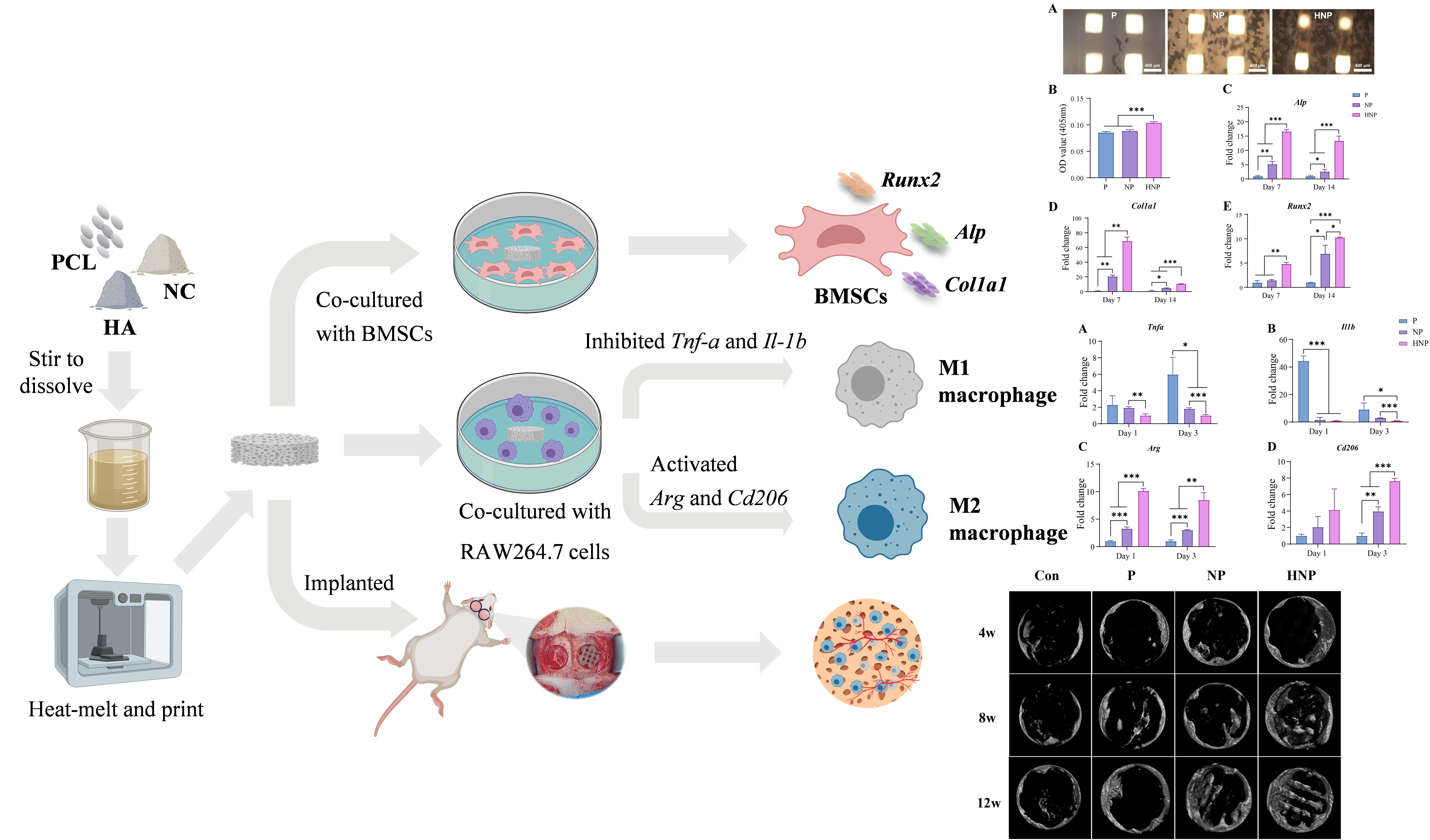
Excessive inflammation remains a major impediment to the clinical repair of critical-sized bone defects, with the immune micro-environment playing a pivotal role in osteogenesis. An appropriate local immune response following biomaterial implantation is essential for successful bone tissue regeneration. In this study, a hydroxyapatite/montmorillonite nanoclay/polycaprolactone (HNP) composite scaffold was designed and subsequently fabricated using three-dimensional (3D) printing, with the aim of modulating macrophage polarization and promoting bone regeneration. The resulting HNP scaffold exhibited favorable mechanical strength and significantly promoted bone marrow mesenchymal stem cell adhesion, proliferation, secretion of osteogenic cytokines, and osteogenic differentiation. Moreover, it modulated the bone immune micro-environment by suppressing M1 macrophage polarization and promoting a shift toward the M2 phenotype, thereby establishing a pro-osteogenic immune milieu. In vivo studies using a rat calvarial defect model demonstrated that, compared with other groups, the HNP scaffold markedly enhanced M2 macrophage polarization, promoted angiogenesis, and accelerated new bone formation. Overall, the 3D-printed HNP scaffold effectively regulated the immune micro-environment and facilitated both bone regeneration and neovascularization, highlighting its strong potential as a candidate for bone tissue engineering applications.
- Wubneh A, Tsekoura EK, Ayranci C, Uludağ H. Current state of fabrication technologies and materials for bone tissue engineering. Acta Biomater. 2018;80:1-30. doi: 10.1016/j.actbio.2018.09.031
- Zhang X, Yang Y, Yang Z, et al. Four-dimensional printing and shape memory materials in bone tissue engineering. Int J Mol Sci. 2023;24(1):814. doi: 10.3390/ijms24010814
- Wang W, Yeung KWK. Bone grafts and biomaterials substitutes for bone defect repair: a review. Bioact Mater. 2017;2(4):224-247. doi: 10.1016/j.bioactmat.2017.05.007
- Schmidt AH. Autologous bone graft: is it still the gold standard? Injury. 2021;52(Suppl 2):S18-S22. doi: 10.1016/j.injury.2021.01.043
- Feng Y, Zhu S, Mei D, et al. Application of 3D printing technology in bone tissue engineering: a review. Curr Drug Deliv. 2021;18(7):847-861. doi: 10.2174/1567201817999201113100322
- Sadowska JM, Ginebra M-P. Inflammation and biomaterials: role of the immune response in bone regeneration by inorganic scaffolds. J Mater Chem B. 2020;8(41):9404-9427. doi: 10.1039/d0tb01379j
- He J, Chen G, Liu M, et al. Scaffold strategies for modulating immune microenvironment during bone regeneration. Mater Sci Eng C Mater Biol Appl. 2020;108:110411. doi: 10.1016/j.msec.2019.110411
- Zheng Z-W, Chen Y-H, Wu D-Y, et al. Development of an accurate and proactive immunomodulatory strategy to improve bone substitute material-mediated osteogenesis and angiogenesis. Theranostics. 2018;8(19):5482-5500. doi: 10.7150/thno.28315
- Dutta SD, Ganguly K, Patil TV, Randhawa A, Lim KT. Unraveling the potential of 3D bioprinted immunomodulatory materials for regulating macrophage polarization: State-of-the-art in bone and associated tissue regeneration. Bioact Mater. 2023;28:284-310. doi: 10.1016/j.bioactmat.2023.05.014
- Yang N, Liu Y. The role of the immune microenvironment in bone regeneration. Int J Med Sci. 2021;18(16):3697-3707. doi: 10.7150/ijms.61080
- Lee J, Byun H, Madhurakkat Perikamana SK, Lee S, Shin H. Current advances in immunomodulatory biomaterials for bone regeneration. Adv Healthc Mater. 2019;8(4):e1801106. doi: 10.1002/adhm.201801106
- Xie Y, Hu C, Feng Y, et al. Osteoimmunomodulatory effects of biomaterial modification strategies on macrophage polarization and bone regeneration. Regen Biomater. 2020;7(3):233-245. doi: 10.1093/rb/rbaa006
- Locati M, Curtale G, Mantovani A. Diversity, mechanisms, and significance of macrophage plasticity. Annu Rev Pathol. 2020;15:123-147. doi: 10.1146/annurev-pathmechdis-012418-012718
- Tian Y, Li Y, Liu J, et al. Photothermal therapy with regulated Nrf2/NF-κB signaling pathway for treating bacteria-induced periodontitis. Bioact Mater. 2022;9:428-445. doi: 10.1016/j.bioactmat.2021.07.033
- Zheng K, Niu W, Lei B, Boccaccini AR. Immunomodulatory bioactive glasses for tissue regeneration. Acta Biomater. 2021;133:168-186. doi: 10.1016/j.actbio.2021.08.023
- Lin Z, Shen D, Zhou W, et al. Regulation of extracellular bioactive cations in bone tissue microenvironment induces favorable osteoimmune conditions to accelerate in situ bone regeneration. Bioact Mater. 2021;6(8):2315-2330. doi: 10.1016/j.bioactmat.2021.01.018
- Liu X, Chen M, Luo J, et al. Immunopolarization-regulated 3D printed-electrospun fibrous scaffolds for bone regeneration. Biomaterials. 2021;276:121037. doi: 10.1016/j.biomaterials.2021.121037
- Jin S, Yang R, Chu C, et al. Topological structure of electrospun membrane regulates immune response, angiogenesis and bone regeneration. Acta Biomater. 2021;129:148-158. doi: 10.1016/j.actbio.2021.05.042
- Arif ZU, Khalid MY, Noroozi R, Sadeghianmaryan A, Jalalvand M, Hossain M. Recent advances in 3D-printed polylactide and polycaprolactone-based biomaterials for tissue engineering applications. Int J Biol Macromol. 2022;218:930-968. doi: 10.1016/j.ijbiomac.2022.07.140
- Gómez-Lizárraga KK, Flores-Morales C, Del Prado-Audelo ML, Álvarez-Pérez MA, Piña-Barba MC, Escobedo C. Polycaprolactone- and polycaprolactone/ceramic-based 3D-bioplotted porous scaffolds for bone regeneration: a comparative study. Mater Sci Eng C. 2017;79:326-335. doi: 10.1016/j.msec.2017.05.003
- Nobles KP, Janorkar AV, Williamson RS. Surface modifications to enhance osseointegration–resulting material properties and biological responses. J Biomed Mater Res B Appl Biomater. 2021;109(11):1909-1923. doi: 10.1002/jbm.b.34835
- Druzian DM, Bonazza GKC, Sangoi GG, et al. Fabrication and properties of the montmorillonite/ nanobioglass hybrid reinforcement from agroindustrial waste for bone regeneration. ACS Appl Mater Interfaces. 2024;16(15):19391-19410. doi: 10.1021/acsami.4c02160
- Gaharwar AK, Cross LM, Peak CW, et al. 2D nanoclay for biomedical applications: regenerative medicine, therapeutic delivery, and additive manufacturing. Adv Mater. 2019;31(23):e1900332. doi: 10.1002/adma.201900332
- Katti KS, Jasuja H, Jaswandkar SV, Mohanty S, Katti DR. Nanoclays in medicine: a new frontier of an ancient medical practice. Mater Adv. 2022;3(20):7484-7500. doi: 10.1039/d2ma00528j
- Bee S-L, Abdullah MAA, Bee S-T, Sin LT, Rahmat AR. Polymer nanocomposites based on silylated-montmorillonite: a review. Prog Polym Sci. 2018;85:57-82. doi: 10.1016/j.progpolymsci.2018.07.003
- Li Y, Yang G, Wang Y, et al. Osteoimmunity-regulating nanosilicate-reinforced hydrogels for enhancing osseointegration. J Mater Chem B. 2023;11(41):9933-9949. doi: 10.1039/d3tb01509b
- Stodolak-Zych E, Kurpanik R, Dzierzkowska E, et al. Effects of montmorillonite and gentamicin addition on the properties of electrospun polycaprolactone fibers. Materials (Basel). 2021;14(22):6905. doi: 10.3390/ma14226905
- Sadeghianmaryan A, Yazdanpanah Z, Soltani YA, Sardroud HA, Nasirtabrizi MH, Chen X. Curcumin-loaded electrospun polycaprolactone/montmorillonite nanocomposite: wound dressing application with anti-bacterial and low cell toxicity properties. J Biomater Sci Polym Ed. 2020;31(2):169-187. doi: 10.1080/09205063.2019.1680928
- Szcześ A, Hołysz L, Chibowski E. Synthesis of hydroxyapatite for biomedical applications. Adv Colloid Interface Sci. 2017;249:321-330. doi: 10.1016/j.cis.2017.04.007
- Ribeiro N, Sousa A, Cunha-Reis C, et al. New prospects in skin regeneration and repair using nanophased hydroxyapatite embedded in collagen nanofibers. Nanomedicine. 2021;33:102353. doi: 10.1016/j.nano.2020.102353
- Helaehil JV, Lourenço CB, Huang B, et al. In vivo investigation of polymer-ceramic PCL/HA and PCL/β-TCP 3D composite scaffolds and electrical stimulation for bone regeneration. Polymers (Basel). 2021;14(1):65. doi: 10.3390/polym14010065
- Rezania N, Asadi-Eydivand M, Abolfathi N, Bonakdar S, Mehrjoo M, Solati-Hashjin M. Three-dimensional printing of polycaprolactone/hydroxyapatite bone tissue engineering scaffolds mechanical properties and biological behavior. J Mater Sci Mater Med. 2022;33(3):31. doi: 10.1007/s10856-022-06653-8
- Petretta M, Gambardella A, Desando G, et al. Multifunctional 3D-printed magnetic polycaprolactone/hydroxyapatite scaffolds for bone tissue engineering. Polymers (Basel). 2021;13(21):3825. doi: 10.3390/polym13213825
- Shang L, Shao J, Ge S. Immunomodulatory properties: the accelerant of hydroxyapatite-based materials for bone regeneration. Tissue Eng Part C Methods. 2022;28(8):377-392. doi: 10.1089/ten.TEC.2022.00111112
- Katti DR, Sharma A, Ambre AH, Katti KS. Molecular interactions in biomineralized hydroxyapatite amino acid modified nanoclay: in silico design of bone biomaterials. Mater Sci Eng C Mater Biol Appl. 2015;46:207-217. doi: 10.1016/j.msec.2014.07.057
- Bhowmick A, Banerjee SL, Pramanik N, et al. Organically modified clay supported chitosan/hydroxyapatite-zinc oxide nanocomposites with enhanced mechanical and biological properties for the application in bone tissue engineering. Int J Biol Macromol. 2018;106:11-19. doi: 10.1016/j.ijbiomac.2017.07.168
- Bhowmick A, Jana P, Pramanik N, et al. Multifunctional zirconium oxide doped chitosan based hybrid nanocomposites as bone tissue engineering materials. Carbohydr Polym. 2016;151:879-888. doi: 10.1016/j.carbpol.2016.06.034
- Cho SJ, Jung SM, Kang M, Shin HS, Youk JH. Preparation of hydrophilic PCL nanofiber scaffolds via electrospinning of PCL/PVP-b-PCL block copolymers for enhanced cell biocompatibility. Polymer. 2015/07/09/ 2015;69:95-102. doi: 10.1016/j.polymer.2015.05.037
- Neufurth M, Wang X, Wang S, et al. 3D printing of hybrid biomaterials for bone tissue engineering: calcium-polyphosphate microparticles encapsulated by polycaprolactone. Acta Biomater. 2017;64:377-388. doi: 10.1016/j.actbio.2017.09.031
- Liang H-Y, Lee W-K, Hsu J-T, et al. Polycaprolactone in bone tissue engineering: a comprehensive review of innovations in scaffold fabrication and surface modifications. J Funct Biomater. 2024;15(9):243. doi: 10.3390/jfb15090243
- Esmaeili J, Jalise SZ, Pisani S, et al. Development and characterization of Polycaprolactone/chitosan-based scaffolds for tissue engineering of various organs: a review. Int J Biol Macromol. 2024;272(Pt 2):132941. doi: 10.1016/j.ijbiomac.2024.132941
- Mousa M, Evans ND, Oreffo ROC, Dawson JI. Clay nanoparticles for regenerative medicine and biomaterial design: a review of clay bioactivity. Biomaterials. 2018;159:204-214. doi: 10.1016/j.biomaterials.2017.12.024
- Jansson M, Lenton S, Plivelic TS, Skepö M. Intercalation of cationic peptides within Laponite layered clay minerals in aqueous suspensions: the effect of stoichiometry and charge distance matching. J Colloid Interface Sci. 2019;557:767-776. doi: 10.1016/j.jcis.2019.09.055
- Baveloni FG, Riccio BVF, Di Filippo LD, Fernandes MA, Meneguin AB, Chorilli M. Nanotechnology-based drug delivery systems as potential for skin application: a review. Curr Med Chem. 2021;28(16):3216-3248. doi: 10.2174/0929867327666200831125656
- Sajjad W, Khan T, Ul-Islam M, et al. Development of modified montmorillonite-bacterial cellulose nanocomposites as a novel substitute for burn skin and tissue regeneration. Carbohydr Polym. 2019;206:548-556. doi: 10.1016/j.carbpol.2018.11.023
- Cui Z-K, Kim S, Baljon JJ, Wu BM, Aghaloo T, Lee M. Microporous methacrylated glycol chitosan-montmorillonite nanocomposite hydrogel for bone tissue engineering. Nat Commun. 2019;10(1):3523. doi: 10.1038/s41467-019-11511-3
- Sheng R, Chen J, Wang H, et al. Nanosilicate-reinforced silk fibroin hydrogel for endogenous regeneration of both cartilage and subchondral bone. Adv Healthc Mater. 2022;11(17):e2200602. doi: 10.1002/adhm.202200602
- Nitya G, Nair GT, Mony U, Chennazhi KP, Nair SV. In vitro evaluation of electrospun PCL/nanoclay composite scaffold for bone tissue engineering. J Mater Sci Mater Med. 2012;23(7):1749-1761. doi: 10.1007/s10856-012-4647-x
- Sukhanova A, Boyandin A, Ertiletskaya N, et al. Composite polymer granules based on poly-ε-caprolactone and montmorillonite prepared by solution-casting and melt extrusion. Polymers (Basel). 2023;15(20):4099. doi: 10.3390/polym15204099
- Zhu B, Bai T, Wang P, Wang Y, Liu C, Shen C. Selective dispersion of carbon nanotubes and nanoclay in biodegradable poly(ε-caprolactone)/poly(lactic acid) blends with improved toughness, strength and thermal stability. Int J Biol Macromol. 2020;153:1272-1280. doi: 10.1016/j.ijbiomac.2019.10.262
- Dhania S, Rani R, Kumar R, Thakur R. Fabricated polyhydroxyalkanoates blend scaffolds enhance cell viability and cell proliferation. J Biotechnol. 2023;361:30-40. doi: 10.1016/j.jbiotec.2022.11.014
- Boyan BD, Lotz EM, Schwartz Z. Roughness and hydrophilicity as osteogenic biomimetic surface properties. Tissue Eng Part A. 2017;23(23-24):1479-1489. doi: 10.1089/ten.TEA.2017.0048
- Ijaola AO, Akamo DO, Damiri F, et al. Polymeric biomaterials for wound healing applications: a comprehensive review. J Biomater Sci Polym Ed. 2022;33(15):1998-2050. doi: 10.1080/09205063.2022.2088528
- Yang H, Gao H, Wang Y. Hollow hydroxyapatite microsphere: a promising carrier for bone tissue engineering. J Microencapsul. 2016;33(5):421-426. doi: 10.1080/02652048.2016.1202347
- Huang H, Yang A, Li J, et al. Preparation of multigradient hydroxyapatite scaffolds and evaluation of their osteoinduction properties. Regen Biomater. 2022; 9:rbac001. doi: 10.1093/rb/rbac001
- Yan L, Wang J, Cai X, et al. Macrophage plasticity: signaling pathways, tissue repair, and regeneration. MedComm (2020). 2024;5(8):e658. doi: 10.1002/mco2.658
- Takayanagi H. Osteoimmunology: shared mechanisms and crosstalk between the immune and bone systems. Nat Rev Immunol. 2007;7(4):292-304. doi: 10.1038/nri2062.
- Li C, Xu MM, Wang K, Adler AJ, Vella AT, Zhou B. Macrophage polarization and meta-inflammation. Transl Res. 2018;191:29-44. doi: 10.1016/j.trsl.2017.10.004
- Shapouri-Moghaddam A, Mohammadian S, Vazini H, et al. Macrophage plasticity, polarization, and function in health and disease. J Cell Physiol. 2018;233(9): 6425-6440. doi: 10.1002/jcp.26429
- Wynn TA, Vannella KM. Macrophages in tissue repair, regeneration, and fibrosis. Immunity. 2016;44(3):450-462. doi: 10.1016/j.immuni.2016.02.015
- Rőszer T. Understanding the mysterious M2 macrophage through activation markers and effector mechanisms. Mediators Inflamm. 2015;2015:816460. doi: 10.1155/2015/816460
- Wynn TA, Chawla A, Pollard JW. Macrophage biology in development, homeostasis and disease. Nature. 2013;496(7446):445-455. doi: 10.1038/nature12034
- Mahon OR, Browe DC, Gonzalez-Fernandez T, et al. Nano-particle mediated M2 macrophage polarization enhances bone formation and MSC osteogenesis in an IL-10 dependent manner. Biomaterials. 2020;239:119833. doi: 10.1016/j.biomaterials.2020.119833
- Lee E-J, Jain M, Alimperti S. Bone microvasculature: stimulus for tissue function and regeneration. Tissue Eng Part B Rev. 2021;27(4):313-329. doi: 10.1089/ten.TEB.2020.0154
- Yu Y, Dai K, Gao Z, et al. Sulfated polysaccharide directs therapeutic angiogenesis via endogenous VEGF secretion of macrophages. Sci Adv. 2021;7(7):eabd8217. doi: 10.1126/sciadv.abd8217
- Poon B, Kha T, Tran S, Dass CR. Bone morphogenetic protein-2 and bone therapy: successes and pitfalls. J Pharm Pharmacol. 2016;68(2):139-147. doi: 10.1111/jphp.12506