

Characterization of radiation-induced esophagitis using explainable machine learning algorithms for lung cancer patients treated in the NRG/RTOG 0617clinical trial

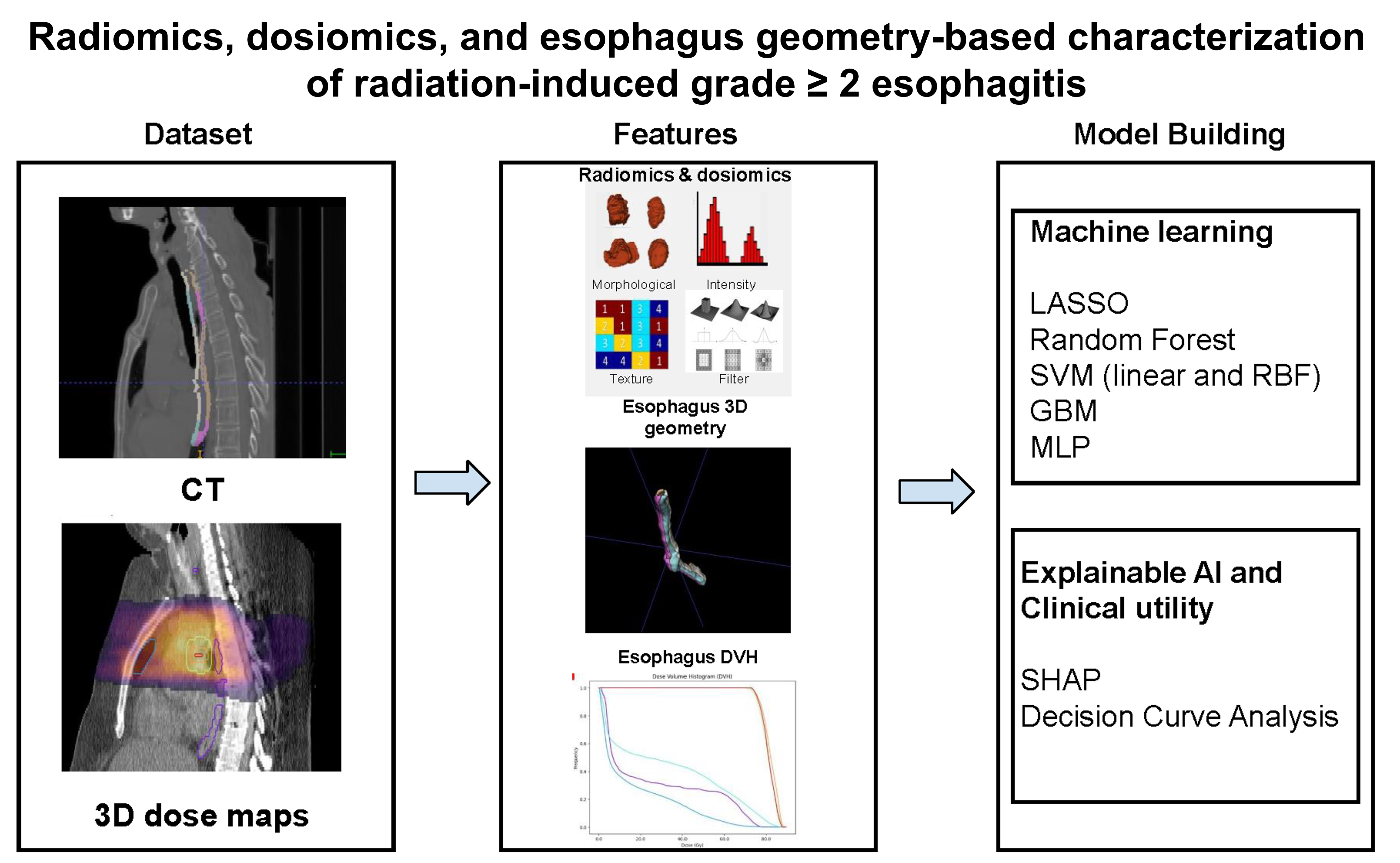
Limiting acute esophagitis remains a clinical challenge in the treatment of locally advanced non-small cell lung cancer using chemoradiotherapy. In this study, machine learning (ML) algorithms were used to predict acute esophagitis in patients (n = 451; training set 70% and independent test set 30%) treated in the Radiation Therapy Oncology Group 0617 clinical trial. Multiple ML models were trained to predict grade ≥ 2 esophagitis using clinical, radiomics, and dosimetric/dosiomics features. Model performance was evaluated using the area under the receiver operating characteristic curve (AUC). The best-performing model was explained using the Shapley additive explanation framework and decision curve analysis. The highest-accuracy model was based on radiomics–dosiomics features using the least absolute shrinkage and selection operator model (AUC = 0.70; 95% confidence interval (CI): 0.60–0.78). A novel model that accounts for esophageal geometry relative to tumor location achieved an AUC of 0.67 (95% CI: 0.58–0.76) using dose–volume histogram relationships. The ML models revealed important relationships that can guide radiation dose planning to minimize individual risk and facilitate personalized radiation therapy.
- Curran WJ, Paulus R, Langer CJ, et al. Sequential vs Concurrent Chemoradiation for Stage III Non–Small Cell Lung Cancer: Randomized Phase III Trial RTOG 9410. J Natl Cancer Inst. 2011;103(19):1452-1460. doi: 10.1093/jnci/djr325
- Rubin P, Constine LS, Marks LB, editors. ALERT—Adverse Late Effects of Cancer Treatment. Vol 1: General Concepts and Specific Precepts. Springer; 2014. doi: 10.1007/978-3-540-72314-1
- Bentzen SM, Constine LS, Deasy JO, et al. Quantitative analyses of normal tissue effects in the clinic (QUANTEC): an introduction to the scientific issues. Int J Radiat Oncol Biol Phys. 2010;76(3):S3-S9. doi: 10.1016/j.ijrobp.2009.09.040
- Chapet O, Kong FM, Lee JS, Hayman JA, Ten Haken RK. Normal tissue complication probability modeling for acute esophagitis in patients treated with conformal radiation therapy for non-small cell lung cancer. Radiother Oncol. 2005;77(2):176-181. doi: 10.1016/j.radonc.2005.10.001
- Bradley J, Deasy JO, Bentzen SM, El Naqa I. Dosimetric correlates for acute esophagitis in patients treated with radiotherapy for lung carcinoma. Int J Radiat Oncol Biol Phys. 2004;58(4):1106-1113. doi: 10.1016/j.ijrobp.2003.09.080
- El Naqa I, Bradley J, Blanco AI, et al. Multivariable modeling of radiotherapy outcomes, including dose-volume and clinical factors. Int J Radiat Oncol Biol Phys. 2006;64(4):1275-1286. doi: 10.1016/j.ijrobp.2005.11.022
- Wei X, Liu HH, Tucker SL, et al. Risk factors for acute esophagitis in non-small-cell lung cancer patients treated with concurrent chemotherapy and three-dimensional conformal radiotherapy. Int J Radiat Oncol Biol Phys. 2006;66(1):100-107. doi: 10.1016/j.ijrobp.2006.04.022
- Rodríguez N, Algara M, Foro P, et al. Predictors of acute esophagitis in lung cancer patients treated with concurrent three-dimensional conformal radiotherapy and chemotherapy. Int J Radiat Oncol Biol Phys. 2009;73(3):810-817. doi: 10.1016/j.ijrobp.2008.04.064
- Palma DA, Senan S, Oberije C, et al. Predicting esophagitis after chemoradiation therapy for non-small cell lung cancer: an individual patient data meta-analysis. Int J Radiat Oncol Biol Phys. 2013;87(4):690-696. doi: 10.1016/j.ijrobp.2013.07.029
- Kamran SC, Yeap BY, Ulysse CA, et al. Assessment of a contralateral esophagus-sparing technique in locally advanced lung cancer treated with high-dose chemoradiation: a phase 1 nonrandomized clinical trial. JAMA Oncol. 2021;7(6):910-914. doi: 10.1001/jamaoncol.2021.0281
- Ladbury C, Li R, Danesharasteh A, et al. Explainable artificial intelligence to identify dosimetric predictors of toxicity in patients with locally advanced non-small cell lung cancer: a secondary analysis of RTOG 0617. Int J Radiat Oncol Biol Phys. 2023;117(5):1287-1296. doi: 10.1016/j.ijrobp.2023.06.019
- Lambin P, Leijenaar RTH, Deist TM, et al. Radiomics: the bridge between medical imaging and personalized medicine. Nat Rev Clin Oncol. 2017;14(12):749-762. doi: 10.1038/nrclinonc.2017.141
- Leger S, Zwanenburg A, Pilz K, et al. A comparative study of machine learning methods for time-to-event survival data for radiomics risk modelling. Sci Rep. 2017;7(1):13206. doi: 10.1038/s41598-017-13448-3
- Giger ML. Computer-aided detection and diagnosis/radiomics/machine learning/deep learning in medical imaging. Med Phys. 2023;50(S1):50-53. doi: 10.1002/mp.16025
- Gillies RJ, Kinahan PE, Hricak H. Radiomics: images are more than pictures, they are data. Radiology. 2016;278(2):563-577. doi: 10.1148/radiol.2015151169
- Ma Z, Liang B, Wei R, et al. Enhanced prediction of postoperative radiotherapy-induced esophagitis in non-small cell lung cancer: dosiomic model development in a real-world cohort and validation in the PORT-C randomized controlled trial. Thorac Cancer. 2023;14(28):2839-2845. doi: 10.1111/1759-7714.15068
- Luna JM, Chao HH, Shinohara RT, et al. Machine learning highlights the deficiency of conventional dosimetric constraints for prevention of high-grade radiation esophagitis in non-small cell lung cancer treated with chemoradiation. Clin Transl Radiat Oncol. 2020;22:69-75. doi: 10.1016/j.ctro.2020.03.007
- Nair SS, Salazar RM, Xu T, et al. Multiomic random forest toxicity modeling of radiation esophagitis. Phys Imaging Radiat Oncol. 2025;36:100838. doi: 10.1016/j.phro.2025.100838
- Herr DJ, Yin H, Bergsma D, et al. Factors associated with acute esophagitis during radiation therapy for lung cancer. Radiother Oncol. 2024;197:110349. doi: 10.1016/j.radonc.2024.110349
- Bradley JD, Hu C, Komaki RR, et al. Long-term results of NRG Oncology RTOG 0617: standard- versus high-dose chemoradiotherapy with or without cetuximab for unresectable stage III non–small-cell lung cancer. J Clin Oncol. 2020;38(7):706-714. doi: 10.1200/JCO.19.01162
- Bradley JD, Paulus R, Komaki R, et al. Standard-dose versus high-dose conformal radiotherapy with concurrent and consolidation carboplatin plus paclitaxel with or without cetuximab for patients with stage IIIA or IIIB non-small-cell lung cancer (RTOG 0617): a randomised, two-by-two factorial phase 3 study. Lancet Oncol. 2015;16(2):187-199. doi: 10.1016/S1470-2045(14)71207-0
- Clark K, Vendt B, Smith K, et al. The Cancer Imaging Archive (TCIA): maintaining and operating a public information repository. J Digit Imaging. 2013;26(6):1045-1057. doi: 10.1007/s10278-013-9622-7
- Trotti A, Colevas AD, Setser A, et al. CTCAE v3.0: development of a comprehensive grading system for the adverse effects of cancer treatment. Semin Radiat Oncol. 2003;13(3):176-181. doi: 10.1016/S1053-4296(03)00031-6
- Monti S, Xu T, Mohan R, Liao Z, Palma G, Cella L. Radiation-induced esophagitis in non-small-cell lung cancer patients: voxel-based analysis and normal tissue complication probability modeling. Cancers (Basel). 2022;14(7):1833. doi: 10.3390/cancers14071833
- Drzymala RE, Mohan R, Brewster L, et al. Dose-volume histograms. Int J Radiat Oncol Biol Phys. 1991;21(1):71-78. doi: 10.1016/0360-3016(91)90168-4
- Zwanenburg A, Vallières M, Abdalah MA, et al. The Image Biomarker Standardization Initiative: standardized quantitative radiomics for high-throughput image-based phenotyping. Radiology. 2020;295(2):328-338. doi: 10.1148/radiol.2020191145
- Zwanenburg A, Leger S, Vallières M, Löck S. Image biomarker standardisation initiative. arXiv. Preprint posted online 2016. doi: 10.48550/arXiv.1612.07003
- Whybra P, Zwanenburg A, Andrearczyk V, et al. The Image Biomarker Standardization Initiative: standardized convolutional filters for reproducible radiomics and enhanced clinical insights. Radiology. 2024;310(2):e231319. doi: 10.1148/radiol.231319
- Dixon WJ, Massey FJ Jr. Introduction to Statistical Analysis. 2nd ed. McGraw-Hill; 1957.
- Chawla NV, Bowyer KW, Hall LO, Kegelmeyer WP. SMOTE: synthetic minority over-sampling technique. J Artif Intell Res. 2002;16:321-357. doi: 10.1613/jair.953
- Tibshirani R. Regression shrinkage and selection via the lasso. J R Stat Soc Series B Methodol. 1996;58(1):267-288. doi: 10.1111/j.2517-6161.1996.tb02080.x
- Breiman L. Random forests. Mach Learn. 2001;45(1):5-32. doi: 10.1023/A:1010933404324
- Wade C, Glynn K. Hands-On Gradient Boosting with XGBoost and scikit-learn: Perform Accessible Machine Learning and Extreme Gradient Boosting With Python. Packt Publishing; 2020.
- Aitkin M, Foxall R. Statistical modelling of artificial neural networks using the multi-layer perceptron. Stat Comput. 2003;13(3):227-239. doi: 10.1023/A:1024218716736
- Guyon I, Weston J, Barnhill S, Vapnik V. Gene selection for cancer classification using support vector machines. Mach Learn. 2002;46(1-3):389-422. doi: 10.1023/A:1012487302797
- Li Y, Luo Y. Performance-weighted-voting model: an ensemble machine learning method for cancer type classification using whole-exome sequencing mutation. Quant Biol. 2020;8(4):347-358. doi: 10.1007/s40484-020-0226-1
- Rainio O, Teuho J, Klén R. Evaluation metrics and statistical tests for machine learning. Sci Rep. 2024;14(1):6086. doi: 10.1038/s41598-024-56706-x
- Demler OV, Pencina MJ, D’Agostino RB Sr. Misuse of DeLong test to compare AUCs for nested models. Stat Med. 2012;31(23):2577-2587. doi: 10.1002/sim.5328
- Lundberg S, Lee SI. A unified approach to interpreting model predictions. arXiv. Preprint posted online 2017. doi: 10.48550/arXiv.1705.07874
- He H, Garcia EA. Learning from imbalanced data. IEEE Trans Knowl Data Eng. 2009;21(9):1263-1284. doi: 10.1109/TKDE.2008.239
- Pedregosa F, Varoquaux G, Gramfort A, et al. Scikit-learn: Machine learning in Python. arXiv. Preprint posted online 2012. doi: 10.48550/arXiv.1201.0490
- Vickers AJ, Elkin EB. Decision curve analysis: a novel method for evaluating prediction models. Med Decis Making. 2006;26(6):565-574. doi: 10.1177/0272989X06295361
- Moons KGM, Altman DG, Reitsma JB, et al. Transparent Reporting of a Multivariable Prediction Model for Individual Prognosis or Diagnosis (TRIPOD): explanation and elaboration. Ann Intern Med. 2015;162(1):W1-W73. doi: 10.7326/M14-0698
- Zheng X, Guo W, Wang Y, et al. Multi-omics to predict acute radiation esophagitis in patients with lung cancer treated with intensity-modulated radiation therapy. Eur J Med Res. 2023;28(1):126. doi: 10.1186/s40001-023-01041-6
- Wang K, Zhao J, Duan J, et al. Radiomic and dosimetric parameter-based nomogram predicts radiation esophagitis in patients with non-small cell lung cancer undergoing combined immunotherapy and radiotherapy. Front Oncol. 2024;14:1490348. doi: 10.3389/fonc.2024.1490348