

A multimodal image-based automated method for macular hole boundary detection

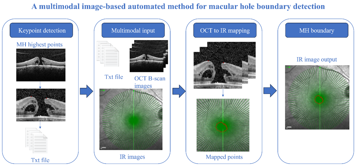
Accurate detection of the macular hole (MH) boundary is crucial for diagnosis and treatment planning. Manual annotation by ophthalmologists is time-consuming, poorly reproducible, and subject to substantial inter-observer variability. To address these challenges, this paper proposes an automated MH boundary detection method based on multimodal images. The method mainly consists of the following stages: (i) automatically localizing the scanning line center in the fundus infrared (IR) image using image processing techniques to provide a reference point for subsequent cross-modal mapping; (ii) introducing the YOLOv8-pose keypoint detection model into the task of MH highest point detection, achieving its automated localization in fundus optical coherence tomography (OCT) B-scan images; (iii) constructing a cross-modal mapping algorithm that, through a combination of position transformation and rotation, facilitates the mapping of the MH highest point from OCT B-scan images to IR images; (iv) designing a boundary fitting scheme that connects all mapped points with reference to adjacent scanning lines to form a closed polygon, thereby achieving MH boundary detection. Finally, a series of quantitative and qualitative experiments were conducted to evaluate the performance of the YOLOv8-pose-based keypoint detection model on public and clinical MH datasets for highest-point detection and to validate the effectiveness of the proposed method on a clinical dataset for MH boundary detection. This study presents a new approach to automated MH boundary detection and may facilitate the accurate diagnosis and treatment planning of MH.
- Tozzi L, Gius I, Greggio A, et al. Case report: spontaneous closure of idiopathic full-thickness macular hole and early development of lamellar hole. Optom Vis Sci. 2023;100(11):804–809. doi: 10.1097/OPX.0000000000002070
- Zhang Y, Li J, Yue H, et al. Clinical observation of anatomical and visual outcomes of macular hole after inverted internal limiting membrane flap in patients with idiopathic macular. J Healthc Eng. 2023;2023:5816473. doi: 10.1155/2023/5816473
- Wolf S, Wolf-Schnurrbusch U. Spectral-domain optical coherence tomography use in macular diseases: a review. Ophthalmologica. 2010;224(6):333–340. doi: 10.1159/000313814
- Singh VK, Kucukgoz B, Murphy DC, Xiong X, Steel DH, Obara B. Benchmarking automated detection of the retinal external limiting membrane in a 3D spectral domain optical coherence tomography image dataset of full thickness macular holes. Comput Biol Med. 2022;140:105070. doi: 10.1016/j.compbiomed.2021.105070
- Elhusseiny AM, Schwartz SG, Flynn HW Jr, Smiddy WE. Long-term outcomes after macular hole surgery. Ophthalmol Retina. 2020;4(4):369–376. doi: 10.1016/j.oret.2019.09.015
- Singh A, Kendal A, Trivedi D, Cazabon S. Patient expectation and satisfaction after macular hole surgery. Optom Vis Sci. 2011;88(2):312–316. doi: 10.1097/OPX.0b013e3182058fc0
- Kucukgoz B, Zou K, Murphy DC, Steel DH, Obara B, Fu H. Uncertainty-aware regression model to predict postoperative visual acuity in patients with macular holes. Comput Med Imaging Graph. 2025;119:102461. doi: 10.1016/j.compmedimag.2024.102461
- Lu J, Cheng Y, Hiya FE, et al. Deep-learning-based automated measurement of outer retinal layer thickness for use in the assessment of age-related macular degeneration, applicable to both swept-source and spectral-domain OCT imaging. Biomed Opt Express. 2024;15(1):413–427. doi: 10.1364/BOE.512359
- Herath HMSS, Yasakethu SLP, Madusanka N, Yi M, Lee BI. Comparative analysis of deep learning architectures for macular hole segmentation in OCT images: a performance evaluation of U-Net variants. J Imaging. 2025;11(2):53. doi: 10.3390/jimaging11020053
- Gribova V, Shalfeeva E. Explainable solutions from artificial intelligence for health-care support systems. Artif Intell Health. 2025;2(3):138–153. doi: 10.36922/aih.5736
- Gholami P, Sheikh Hassani M, Parthasarathy MK, Zelek JS, Lakshminarayanan V. Classification of optical coherence tomography images for diagnosing different ocular diseases. In: Multimodal Biomedical Imaging XIII. Vol 10487. SPIE; 2018:1048705. doi: 10.1117/12.2292520
- Asani B, Holmberg O, Schiefelbein JB, et al. Evaluation of OCT biomarker changes in treatment-naive neovascular AMD using a deep semantic segmentation algorithm. Eye (Lond). 2024;38(16):3180–3186. doi: 10.1038/s41433-024-03264-1
- Volkov E, Averkin A. Modification of U-Net architecture based on Gabor filters with trainable parameters to improve biomarker segmentation on OCT images. In: 2025 VI International Conference on Neural Networks and Neurotechnologies (NeuroNT). IEEE; 2025:66–68. doi: 10.1109/NeuroNT66873.2025.11049967
- Alekseev A, Bobe A. GaborNet: Gabor filters with learnable parameters in deep convolutional neural network. In: 2019 International Conference on Engineering and Telecommunication (EnT). IEEE; 2019:1–4. doi: 10.1109/EnT47717.2019.9030571
- Jebaseeli TJ, Haldorai A, Kim HJ. Diagnosis of Purtscher retinopathy from spectral-domain OCT images using panoptic segmentation technique. Traitement du Signal. 2025;42(3):1393–1407. doi: 10.18280/ts.420315
- Mukherjee S, De Silva T, Duic C, et al. Validation of deep learning–based automatic retinal layer segmentation algorithms for age-related macular degeneration with 2 spectral-domain OCT devices. Ophthalmol Sci. 2025;5(3):100670. doi: 10.1016/j.xops.2024.100670
- Cao H, Lian X, Chen L, Duan Z. A computationally efficient and lightweight model for high-accuracy OCT image classification. World Wide Web. 2025;28:35. doi: 10.1007/s11280-025-01348-w
- Sharma R, Gangrade J, Gangrade S, Mishra A, Kumar G, Gunjan VK. Modified EfficientNetB3 deep learning model to classify colour fundus images of eye diseases. In: 2023 IEEE 5th International Conference on Cybernetics, Cognition and Machine Learning Applications (ICCCMLA). IEEE; 2023:632–638. doi: 10.1109/ICCCMLA58983.2023.10346769
- Phu DN, Tran AQ, Tran NQ, Dang TH. The classification of optical coherence tomography images using machine learning for macular hole and diabetic macular edema diagnoses. In: 2023 12th International Conference on Control, Automation and Information Sciences (ICCAIS). IEEE; 2023:634–638. doi: 10.1109/ICCAIS59597.2023.10382327
- Mall PK, Singh PK, Yadav D. GLCM-based feature extraction and medical X-ray image classification using machine learning techniques. In: 2019 IEEE Conference on Information and Communication Technology (CICT). IEEE; 2019:1–6. doi: 10.1109/CICT48419.2019.9066263
- Shijin PS, Dharun VS. Extraction of texture features using GLCM and shape features using connected regions. Int J Eng Technol. 2016;8(6):2926–2930. doi: 10.21817/ijet/2016/v8i6/160806254
- Wang M, Lin T, Peng Y, et al. Self-guided optimization semi-supervised method for joint segmentation of macular hole and cystoid macular edema in retinal OCT images. IEEE Trans Biomed Eng. 2023;70(7):2013–2024. doi: 10.1109/TBME.2023.3234031
- Keller B, Cunefare D, Grewal DS, Mahmoud TH, Izatt JA, Farsiu S. Length-adaptive graph search for automatic segmentation of pathological features in optical coherence tomography images. J Biomed Opt. 2016;21(7):076015. doi: 10.1117/1.JBO.21.7.076015
- Wang Y, Zhen L, Tan T, et al. Geometric correspondence-based multimodal learning for ophthalmic image analysis. IEEE Trans Med Imaging. 2024;43(5):1945–1957. doi: 10.1109/TMI.2024.3352602
- Zhang H, Yang B, Li S, et al. Retinal OCT image segmentation with deep learning: a review of advances, datasets, and evaluation metrics. Comput Med Imaging Graph. 2025;123:102539. doi: 10.1016/j.compmedimag.2025.102539
- Zhang Y, Zhang Y, Li T, Shi M, Huang Y. Analysis of a multimodal ocular ultrasound image classification algorithm based on YOLO and VanillaNet. IEEE Access. 2025;13:148374–148383. doi: 10.1109/ACCESS.2025.3598747
- Wen PC, Guan Y, Li JQ, Mahmood T, Zhao YZ. A fundus image myopia diagnosis model based on homogeneous multimodal feature fusion. In: Hung JC, Yen NY, Chang JW, eds. Frontier Computing. Lecture Notes in Electrical Engineering. Vol 1031. Springer; 2023:39–51. doi: 10.1007/978-981-99-1428-9_5
- Huang SC, Pareek A, Seyyedi S, Banerjee I, Lungren MP. Fusion of medical imaging and electronic health records using deep learning: a systematic review and implementation guidelines. NPJ Digit Med. 2020;3:136. doi: 10.1038/s41746-020-00341-z
- Li Q, Li S, Wu Y, et al. Orientation-independent feature matching (OIFM) for multimodal retinal image registration. Biomed Signal Process Control. 2020;60:101957. doi: 10.1016/j.bspc.2020.101957
- Hossein-Nejad Z, Nasri M. A-RANSAC: adaptive random sample consensus method in multimodal retinal image registration. Biomed Signal Process Control. 2018;45:325–338. doi: 10.1016/j.bspc.2018.06.002
- Mur-Artal R, Tardós JD. ORB-SLAM2: an open-source SLAM system for monocular, stereo, and RGB-D cameras. IEEE Trans Robot. 2017;33(5):1255–1262. doi: 10.1109/TRO.2017.2705103
- de Vos BD, Berendsen FF, Viergever MA, Sokooti H, Staring M, Išgum I. A deep learning framework for unsupervised affine and deformable image registration. Med Image Anal. 2019;52:128–143. doi: 10.1016/j.media.2018.11.010
- Çatal O, Jansen W, Verbelen T, Dhoedt B, Steckel J. LatentSLAM: unsupervised multi-sensor representation learning for localization and mapping. In: 2021 IEEE International Conference on Robotics and Automation (ICRA). IEEE; 2021:6739–6745. doi: 10.1109/ICRA48506.2021.9560768
- Xie L, Wisse LEM, Pluta J, et al. Automated segmentation of medial temporal lobe subregions on in vivo T1-weighted MRI in early stages of Alzheimer’s disease. Hum Brain Mapp. 2019;40(12):3431–3451. doi: 10.1002/hbm.24607
- Park K, Sinha U, Barron JT, et al. Nerfies: deformable neural radiance fields. In: Proceedings of the IEEE/CVF International Conference on Computer Vision. IEEE/CVF; 2021:5865–5874.
- Gholami P, Roy P, Parthasarathy MK, Lakshminarayanan V. OCTID: optical coherence tomography image database. Comput Electr Eng. 2020;81:106532. doi: 10.1016/j.compeleceng.2019.106532
- Domingues NS. From engineering principles to healthcare practice: a hybrid reasoning framework for transparent clinical decision support. Artif Intell Health. 2026;3(2):025470102. doi: 10.36922/AIH025470102
- Abdusalomov A, Mirzakhalilov S, Umirzakova S, et al. Optimized lightweight architecture for coronary artery disease classification in medical imaging. Diagnostics. 2025;15(4):446. doi: 10.3390/diagnostics15040446
- Nambiar VB, Ramamurthy B, Veeresha P. Gender determination from periocular images using deep learning based EfficientNet architecture. Int J Math Comput Eng. 2024;2(1):59–70. doi: 10.2478/ijmce-2024-0005