

Seg-lite artificial intelligence: Scalable, explainable deep learning for ovarian cancer histopathology on low-resource systems

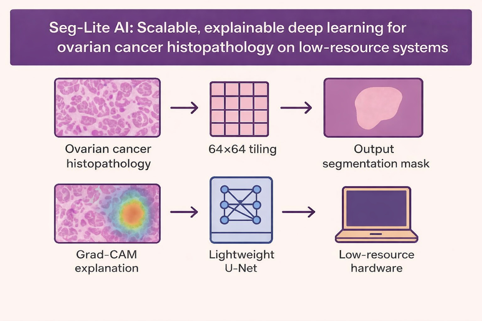
Histopathological examination remains essential for ovarian cancer diagnosis, yet routine assessment is time-consuming and depends on specialist interpretation, which may limit prompt clinical decision-making. This study proposes a compact deep-learning framework designed for automated ovarian cancer tissue segmentation that can function effectively on standard laptops and graphics processing units with limited VRAM. The model, a streamlined U-Net containing 237,457 parameters, was trained on 43,265 image patches at 64 × 64 resolution using a 70/15/15 train/validation/test split. Despite its small footprint, it achieved a Dice score, intersection-over-union, and pixel accuracy of 1.000 on the test set, converging in approximately 20 min over six epochs. Gradient-weighted class activation mapping visualization confirmed that the network consistently focused on morphologically relevant structures. These findings demonstrate that high-precision segmentation can be achieved without computationally expensive architectures, providing a practical and scalable solution for resource-limited clinical environments.
- Sung H, Ferlay J, Siegel RL, et al. Global cancer statistics 2020: GLOBOCAN estimates of incidence and mortality worldwide for 36 cancers in 185 countries. CA Cancer J Clin. 2021;71(3):209-249. doi: 10.3322/caac.21660
- Torre LA, Trabert B, DeSantis CE, et al. Ovarian cancer statistics, 2018. CA Cancer J Clin. 2018;68(4):284-296. doi: 10.3322/caac.21456
- Litjens G, Kooi T, Bejnordi BE, et al. A survey on deep learning in medical image analysis. Med Image Anal. 2017;42:60-88. doi: 10.1016/j.media.2017.07.005
- Elmore JG, Longton GM, Carney PA, et al. Diagnostic concordance among pathologists interpreting breast biopsy specimens. JAMA. 2015;313(11):1122-1132. doi: 10.1001/jama.2015.1405
- Esteva A, Kuprel B, Novoa RA, et al. Dermatologist-level classification of skin cancer with deep neural networks. Nature. 2017;542(7639):115-118. doi: 10.1038/nature21056
- Isensee F, Jaeger PF, Kohl SAA, Petersen J, Maier-Hein KH. nnU-Net: A self-configuring method for deep learning-based biomedical image segmentation. Nat Methods. 2021;18(2):203-211. doi: 10.1038/s41592-020-01008-z
- Ronneberger O, Fischer P, Brox T. U-Net: Convolutional networks for biomedical image segmentation. In: Medical Image Computing and Computer-Assisted Intervention (MICCAI). Berlin: Springer; 2015:234-241. doi: 10.1007/978-3-319-24574-4_28
- Cao H, Wang Y, Chen J, et al. Swin-Unet: Unet-like pure transformer for medical image segmentation. In: European Conference on Computer Vision Workshops. Springer; 2023:205-218. doi: 10.1007/978-3-031-25066-8_9
- Guo LY, Wu AH, Wang YX, Zhang LP, Chai H, Liang XF. Deep learning-based ovarian cancer subtypes identification using multi-omics data. BioData Min. 2020;13(1). doi: 10.1186/s13040-020-00222-x
- Wang S, Liu Z, Rong Y, et al. Deep learning provides a new computed tomography-based prognostic biomarker for recurrence prediction in high-grade serous ovarian cancer. Radiother Oncol. 2020;132:171-177. doi: 10.1016/j.radonc.2018.10.019
- Ho DJ, Chui MH, Vanderbilt CM, et al. Deep interactive learning-based ovarian cancer segmentation of H&E-stained whole slide images to study morphological patterns of BRCA mutation. J Pathol Inform. 2023;14:100160. doi: 10.1016/j.jpi.2022.100160
- Guetarni B, Windal F, Benhabiles H, et al. A Vision Transformer-Based Framework for Knowledge Transfer From Multi-Modal to Mono-Modal Lymphoma Subtyping Models. IEEE J Biomed Health Inform. 2024;28(9):5562- 5572. doi: 10.1109/jbhi.2024.3407878
- Kumar A, Singh SK, Saxena S, et al. Deep feature learning for histopathological image classification of canine mammary tumors and human breast cancer. Inf Sci. 2024;508:405-421. doi: 10.1016/j.ins.2019.08.072
- Wang CW, Chang CC, Lee YC et al. Weakly supervised deep learning for prediction of treatment effectiveness on ovarian cancer from histopathology images. Comput Med Imaging Graph. 2022;99:102093. doi: 10.1016/j.compmedimag.2022.102093
- Howard AG, Zhu M, Chen B, et al. MobileNets: Efficient Convolutional Neural Networks for Mobile Vision Applications. arXiv. Preprint posted online 2017. doi: 10.48550/arXiv.1704.04861
- Tan M, Le Q. EfficientNet: Rethinking model scaling for convolutional neural networks. In: International Conference on Machine Learning (ICML); 2019:6105-6114. Available from: https://proceedings.mlr.press/v97/tan19a.html
- Mehta S, Rastegari M. MobileViT: Light-weight, General-purpose, and Mobile-friendly Vision Transformer. arXiv. Preprint posted online 2021. doi: 10.48550/arXiv.2110.02178
- Vasu PKA, Gabriel J, Zhu J, Tuzel O, Ranjan A. FastViT: A fast hybrid vision transformer using structural reparameterization. In: Proceedings of the IEEE/CVF International Conference on Computer Vision; 2023:5785- 5795. doi: 10.1109/ICCV51070.2023.00532
- Hinton G, Vinyals O, Dean J. Distilling the knowledge in a neural network. arXiv. Preprint posted online 2015. Available from: https://arxiv.org/abs/1503.02531
- Banbury CR, Reddi VJ, Lam M, et al. Benchmarking TinyML Systems: Challenges and Direction. arXiv. Preprint posted online 2020. doi: 10.48550/ARXIV.2003.04821
- Selvaraju RR, Cogswell M, Das A, Vedantam R, Parikh D, Batra D. Grad-CAM: Visual Explanations from Deep Networks Via Gradient-Based Localization. In: Proceedings of the IEEE International Conference on Computer Vision; 2017. p. 618-626. doi: 10.1109/ICCV.2017.74
- Holzinger A, Biemann C, Pattichis CS, Kell DB. What do we need to build explainable AI systems for the medical domain? arXiv. Preprint posted online 2017. doi: 10.48550/arXiv.1712.09923
- Schlemper J, Oktay O, Schaap M, et al. Attention gated networks: Learning to leverage salient regions in medical images. Med Image Anal. 2019;53:197-207. doi: 10.1016/j.media.2019.01.012
- Ghorbani A, Wexler J, Zou JY, Kim B. Towards Automatic Concept-Based Explanations. In: Advances in Neural Information Processing Systems 32 (NeurIPS) Annual Conference on Neural Information Processing Systems; 2019.
- Wang X, Yang S, Zhang J, et al. TransPath: Transformer- Based Self-supervised Learning for Histopathological Image Classification. In: Medical Image Computing and Computer Assisted Intervention – MICCAI 2022 (Lecture Notes in Computer Science). Springer International Publishing; 2021:186-195. doi: 10.1007/978-3-030-87237-3_18
- Amann J, Blasimme A, Vayena E, Frey D, Madai VI. Explainability for artificial intelligence in healthcare: A multidisciplinary perspective. BMC Med Inform Decis Mak. 2020;20(1):310. doi: 10.1186/s12911-020-01332-6
- UBC Ovarian Cancer Subtype Classification and Outlier Detection (UBC-OCEAN). Kaggle. Available from: https:// www.kaggle.com/competitions/UBC-OCEAN
- Sørensen T. A method of establishing groups of equal amplitude in plant sociology based on similarity of species and its application to analyses of the vegetation on Danish commons. Biologiske Skrifter / Kongelige Danske Videnskabernes Selskab. 1948;5(4):1–34.
- Shashikala HK, Suresh MB. Performance Analysis of Segmentation Techniques for Knee Osteoarthritis Detection from X‐Ray Images. In: Integration of Federated Learning and Blockchain for Smart Cities. New Jersey, U.S: John Wiley and Sons; 2025:659-682. doi: 10.1002/9781394167760.ch23
- Chung NC, Miasojedow B, Startek M, Gambin A. Jaccard/ Tanimoto similarity test and estimation methods for biological presence-absence data. BMC Bioinformatics. 2019;20(Suppl 15):644. doi: 10.1186/s12859-019-3118-5
- Farahani H, Boschman J, Farnell D, et al. Deep learning-based histotype diagnosis of ovarian carcinoma whole-slide pathology images. Mod Pathol. 2022;35(12):1983–1990. doi: 10.1038/s41379-022-01146-z
- Yoo JJ, Namdar K, Khalvati F. Deep superpixel generation and clustering for weakly supervised segmentation of brain tumors in MR images. BMC Med Imaging. 2024;24(1):335. doi: 10.1186/s12880-024-01523-x
- Breen J, Allen K, Zucker K, et al. Artificial intelligence in ovarian cancer histopathology: A systematic review. NPJ Precis Oncol. 2023;7(1):83. doi: 10.1038/s41698-023-00432-6
- Saha AK, Rabbani M, Sum ASI, Mridha MF, Kabir MM. An enhanced deep learning model for accurate classification of ovarian cancer from histopathological images. Sci Rep. 2025;15(1):21860. doi: 10.1038/s41598-025-07903-9
- Asadi-Aghbolaghi M, Farahani H, Zhang A, et al. Machine Learning-driven Histotype Diagnosis of Ovarian Carcinoma: Insights from the OCEAN AI Challenge. medRxiv. Preprint posted online April 23, 2024. doi: 10.1101/2024.04.19.24306099
- Garcia-Atutxa I, Martínez-Más J, Bueno-Crespo A, Villanueva-Flores F. Early-fusion hybrid CNN-transformer models for multiclass ovarian tumor ultrasound classification. Front Artif Intell. 2025;8:1679310. doi: 10.3389/frai.2025.1679310
- Chong PL, Vaigeshwari V, Mohammed Reyasudin BK, et al. Integrating artificial intelligence in healthcare: Applications, challenges, and future directions. Future Sci OA. 2025;11(1):2527505. doi: 10.1080/20565623.2025.2527505
- Han F, Huang X, Wang X, et al. Artificial intelligence in orthopedic surgery: Current applications, challenges, and future directions. MedComm (2020). 2025;6(7):e70260. doi: 10.1002/mco2.70260
- Musthafa MM, Mahesh TR, Kumar VV, Guluwadi S. Enhancing brain tumor detection in MRI images through explainable AI using Grad-CAM with Resnet 50. BMC Med Imaging. 2024;24(1):107. doi: 10.1186/s12880-024-01292-7
- El-Khoury R, Zaatari G. The rise of AI-assisted diagnosis: Will pathologists be partners or bystanders? Diagnostics (Basel). 2025;15(18):2308. doi: 10.3390/diagnostics15182308
- Sajjad U, Rezapour M, Su Z, Tozbikian GH, Gurcan MN, Niazi MKK. NRK-ABMIL: Subtle metastatic deposits detection for predicting lymph node metastasis in breast cancer whole-slide images. Cancers (Basel). 2023;15(13):3428. doi: 10.3390/cancers15133428
- Neary-Zajiczek L, Beresna L, Razavi B, Pawar V, Shaw M, Stoyanov D. Minimum resolution requirements of digital pathology images for accurate classification. Med Image Anal. 2023;89:102891. doi: 10.1016/j.media.2023.102891
- Babaeipour R, Fox MS, Parraga G, Ouriadov A. Comparative analysis of foundational, advanced, and traditional deep learning models for hyperpolarized gas MRI lung segmentation: Robust performance in data-constrained scenarios. Bioengineering (Basel). 2025;12(10):1062. doi: 10.3390/bioengineering12101062
- Komura D, Ochi M, Ishikawa S. Machine learning methods for histopathological image analysis: Updates in 2024. Comput Struct Biotechnol J. 2025;27:383-400. doi: 10.1016/j.csbj.2024.12.033
- Wang CW, Firdi NP, Chu TC, et al. ATEC23 challenge: Automated prediction of treatment effectiveness in ovarian cancer using histopathological images. Med Image Anal. 2025;99:103342. doi: 10.1016/j.media.2024.103342
- Wang J, Zhang S, Li J, et al. Development and clinical validation of deep learning-based immunohistochemistry prediction models for subtyping and staging of gastrointestinal cancers. BMC Gastroenterol. 2025;25(1):494. doi: 10.1186/s12876-025-04045-0