
Loading the joint to protect it: Exercise effects on knee articular cartilage

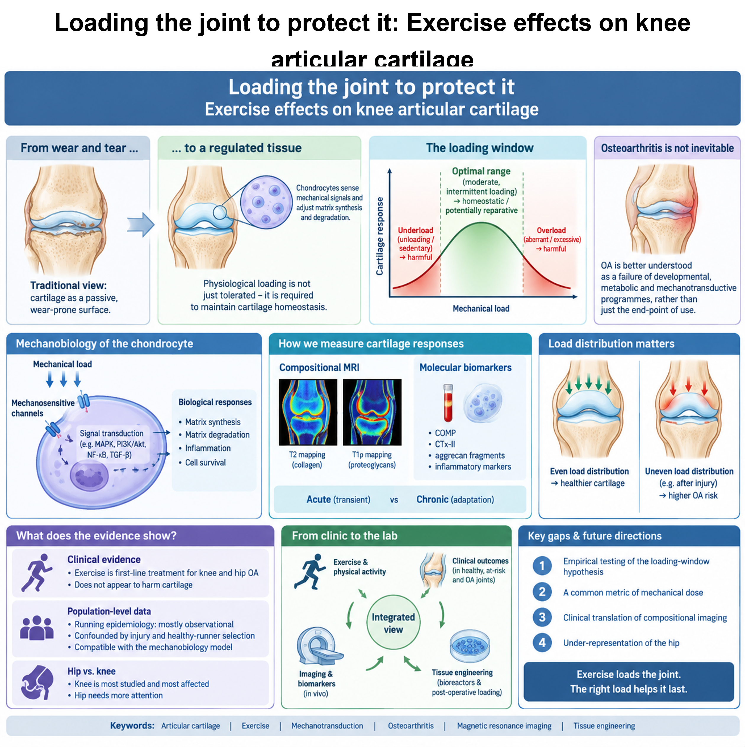
Articular cartilage was long viewed as an inert, wear-prone bearing surface whose lifespan is spent by mechanical use. Contemporary evidence characterises articular cartilage as a mechanically regulated tissue in which physiological loading contributes to matrix homeostasis and suggests that osteoarthritis is better understood as a multifactorial disorder involving developmental, metabolic, and mechanotransductive dysregulation rather than simply as a consequence of mechanical wear. This narrative review synthesises converging strands of evidence on how exercise, physical activity, and mechanical loading affect knee articular cartilage, using the hip as a comparator. We outline the mechanobiology of the chondrocyte, in which mechanosensitive channels and downstream pathways translate load magnitude, mode, and duration into context-dependent biological responses, and we propose, as an integrative hypothesis rather than an established quantitative relationship, a physiological loading window that is multidimensional rather than a single dose axis. We review how compositional magnetic resonance imaging and molecular biomarkers make cartilage responses measurable in vivo, and we distinguish transient acute responses from chronic adaptation. We argue that load distribution matters as much as load magnitude, drawing on post-traumatic osteoarthritis. We appraise the clinical evidence in people at risk of, or with, knee osteoarthritis, where exercise is a first-line treatment that does not appear to harm cartilage, and we examine whether population-level running epidemiology, which is observational and susceptible to injury-related confounding and healthy-runner selection, is compatible with this model. Finally, we connect exercise mechanobiology to cartilage tissue engineering, where bioreactor conditioning and post-operative loading pose the same dose problem. We identify priority gaps: empirical testing of the loading-window hypothesis, a common metric of mechanical dose, the clinical translation of compositional imaging, and the under-representation of the hip.
- Roos EM, Dahlberg L. Positive effects of moderate exercise on glycosaminoglycan content in knee cartilage: a four-month, randomized, controlled trial in patients at risk of osteoarthritis. Arthritis Rheum. 2005;52(11):3507-3514. doi: 10.1002/art.21415
- Stańczak M, Swinnen B, Surmacz J, et al. Knee joint response to mechanical loading: bounding mechanotransduction with rehabilitation. Cell Physiol Biochem. 2025;59(5):666-729. doi: 10.33594/000000818
- Scerif F, Eldridge SE. Osteoarthritis year in review 2025: biology. Osteoarthr Cartil. 2026;34(2):213-220. doi: 10.1016/j.joca.2025.12.013
- Bannuru RR, Osani MC, Vaysbrot EE, et al. OARSI guidelines for the non-surgical management of knee, hip, and polyarticular osteoarthritis. Osteoarthr Cartil. 2019;27(11):1578-1589. doi: 10.1016/j.joca.2019.06.011
- National Institute for Health and Care Excellence. Osteoarthritis in over 16s: diagnosis and management. NICE guideline [NG226]. London: NICE; 2022. Accessed September 21, 2026. https://www.nice.org.uk/guidance/ng226
- March L, Little C. Articular cartilage in health and disease. In: Sambrook P, Schrieber L, Taylor T, Ellis AM, eds. The Musculoskeletal System. 2nd ed. Churchill Livingstone; 2010:77-95. doi: 10.1016/B978-0-7020-3377-3.00006-8
- Han EH, Chen SS, Klisch SM, Sah RL. Contribution of proteoglycan osmotic swelling pressure to the compressive properties of articular cartilage. Biophys J. 2011;101(4):916-924. doi: 10.1016/j.bpj.2011.07.006
- Albano D, Viglino U, Esposito F, et al. Quantitative and compositional MRI of the articular cartilage: a narrative review. Tomography. 2024;10(7):949-969. doi: 10.3390/tomography10070072
- Zhang M, Meng N, Wang X, Chen W, Zhang Q. TRPV4 and PIEZO channels mediate the mechanosensing of chondrocytes to the biomechanical microenvironment. Membranes (Basel). 2022;12(2):237. doi: 10.3390/membranes12020237
- Lee W, Leddy HA, Chen Y, et al. Synergy between Piezo1 and Piezo2 channels confers high-strain mechanosensitivity to articular cartilage. Proc Natl Acad Sci USA. 2014;111(47):E5114-E5122. doi: 10.1073/pnas.1414298111
- O’Conor CJ, Leddy HA, Benefield HC, Liedtke WB, Guilak F. TRPV4-mediated mechanotransduction regulates the metabolic response of chondrocytes to dynamic loading. Proc Natl Acad Sci USA. 2014;111(4):1316-1321. doi: 10.1073/pnas.1319569111
- Lee W, Nims RJ, Savadipour A, et al. Inflammatory signaling sensitizes Piezo1 mechanotransduction in articular chondrocytes as a pathogenic feed-forward mechanism in osteoarthritis. Proc Natl Acad Sci USA. 2021;118(13):e2001611118. doi: 10.1073/pnas.2001611118
- Deng Y, Lu J, Li W, et al. Reciprocal inhibition of YAP/TAZ and NF-κB regulates osteoarthritic cartilage degradation. Nat Commun. 2018;9(1):4564. doi: 10.1038/s41467-018-07022-2
- Lories RJ, Corr M, Lane NE. To Wnt or not to Wnt: the bone and joint health dilemma. Nat Rev Rheumatol. 2013;9(6):328-339. doi: 10.1038/nrrheum.2013.25
- Vanwanseele B, Eckstein F, Knecht H, Stüssi E, Spaepen A. Knee cartilage of spinal cord-injured patients displays progressive thinning in the absence of normal joint loading and movement. Arthritis Rheum. 2002;46(8):2073-2078. doi: 10.1002/art.10462
- Hinterwimmer S, Krammer M, Krötz M, et al. Cartilage atrophy in the knees of patients after seven weeks of partial load bearing. Arthritis Rheum. 2004;50(8):2516-2520. doi: 10.1002/art.20378
- Rhee DK, Marcelino J, Baker M, et al. The secreted glycoprotein lubricin protects cartilage surfaces and inhibits synovial cell overgrowth. J Clin Invest. 2005;115(3):622-631. doi: 10.1172/jci200522263
- Li H, Yi G, Zhou D, et al. Lubricin in osteoarthritis: functions and therapeutic prospects. Front Immunol. 2026;17:1790804. doi: 10.3389/fimmu.2026.1790804
- Nugent GE, Aneloski NM, Schmidt TA, Schumacher BL, Voegtline MS, Sah RL. Dynamic shear stimulation of bovine cartilage biosynthesis of proteoglycan 4. Arthritis Rheum. 2006;54(6):1888-1896. doi: 10.1002/art.21831
- Nugent-Derfus GE, Takara T, O’Neill JK, et al. Continuous passive motion applied to whole joints stimulates chondrocyte biosynthesis of PRG4. Osteoarthr Cartil. 2007;15(5):566-574. doi: 10.1016/j.joca.2006.10.015
- Sah RL, Kim YJ, Doong JY, Grodzinsky AJ, Plaas AH, Sandy JD. Biosynthetic response of cartilage explants to dynamic compression. J Orthop Res. 1989;7(5):619-636. doi: 10.1002/jor.1100070502
- Coburn SL, Crossley KM, Kemp JL, et al. Immediate and delayed effects of joint loading activities on knee and hip cartilage: a systematic review and meta-analysis. Sports Med Open. 2023;9(1):56. doi: 10.1186/s40798-023-00602-7
- Chen M, Qiu L, Shen S, et al. The influences of walking, running and stair activity on knee articular cartilage: quantitative MRI using T1 rho and T2 mapping. PLoS ONE. 2017;12(11):e0187008. doi: 10.1371/journal.pone.0187008
- Dreiner M, Munk T, Zaucke F, Liphardt AM, Niehoff A. Relationship between different serum cartilage biomarkers in the acute response to running and jumping in healthy male individuals. Sci Rep. 2022;12(1):6434. doi: 10.1038/s41598-022-10310-z
- Hao HQ, Zhang JF, He QQ, Wang Z. Cartilage oligomeric matrix protein, C-terminal cross-linking telopeptide of type II collagen, and matrix metalloproteinase-3 as biomarkers for knee and hip osteoarthritis (OA) diagnosis: a systematic review and meta-analysis. Osteoarthr Cartil. 2019;27(5):726-736. doi: 10.1016/j.joca.2018.10.009
- Mündermann A, Dyrby CO, Andriacchi TP, King KB. Serum concentration of cartilage oligomeric matrix protein (COMP) is sensitive to physiological cyclic loading in healthy adults. Osteoarthr Cartil. 2005;13(1):34-38. doi: 10.1016/j.joca.2004.09.007
- Kong SY, Stabler TV, Criscione LG, Elliott AL, Jordan JM, Kraus VB. Diurnal variation of serum and urine biomarkers in patients with radiographic knee osteoarthritis. Arthritis Rheum. 2006;54(8):2496-2504. doi: 10.1002/art.21977
- Zhang Y, Huang Y, Zhang L, et al. Effects of long-term running on the structure and biochemical composition of knee cartilage in males: a cross-sectional study. Quant Imaging Med Surg. 2024;14(8):6036-6047. doi: 10.21037/qims-23-1563
- Sophia Fox AJ, Bedi A, Rodeo SA. The basic science of articular cartilage: structure, composition, and function. Sports Health. 2009;1(6):461-468. doi: 10.1177/1941738109350438
- Li G, Li JS, Torriani M, Hosseini A. Short-term contact kinematic changes and longer-term biochemical changes in the cartilage after ACL reconstruction: a pilot study. Ann Biomed Eng. 2018;46(11):1797-1805. doi: 10.1007/s10439-018-2079-6
- Poulsen E, Goncalves GH, Bricca A, Roos EM, Thorlund JB, Juhl CB. Knee osteoarthritis risk is increased 4-6 fold after knee injury: a systematic review and meta-analysis. Br J Sports Med. 2019;53(23):1454-1463. doi: 10.1136/bjsports-2018-100022
- Alfayyadh A. Molecular cartilage pathology following anterior cruciate ligament reconstruction: a systematic review of MRI studies. Front Surg. 2026;13:1714750. doi: 10.3389/fsurg.2026.1714750
- Kuenze C, Collins K, Pfeiffer KA, Lisee C. Assessing physical activity after ACL injury: moving beyond return to sport. Sports Health. 2022;14(2):197-204. doi: 10.1177/19417381211025307
- Whittaker JL, Culvenor AG, Juhl CB, et al. OPTIKNEE 2022: consensus recommendations to optimise knee health after traumatic knee injury to prevent osteoarthritis. Br J Sports Med. 2022;56(24):1393-1405. doi: 10.1136/bjsports-2022-106299
- Pfeiffer SJ, Spang J, Nissman D, et al. Gait mechanics and T1ρ MRI of tibiofemoral cartilage 6 months after ACL reconstruction. Med Sci Sports Exerc. 2019;51(4):630-639. doi: 10.1249/MSS.0000000000001834
- Teng HL, Wu D, Su F, et al. Gait characteristics associated with a greater increase in medial knee cartilage T1ρ and T2 relaxation times in patients undergoing anterior cruciate ligament reconstruction. Am J Sports Med. 2017;45(14):3262-3271. doi: 10.1177/0363546517723007
- Andriacchi TP, Mündermann A, Smith RL, Alexander EJ, Dyrby CO, Koo S. A framework for the in vivo pathomechanics of osteoarthritis at the knee. Ann Biomed Eng. 2004;32(3):447-457. doi: 10.1023/B:ABME.0000017541.82498.37
- Kurt C, Gök A. Effect of knee joint meniscus tears on joint cartilage contact and pressure with finite element analysis. Biomedicines. 2026;14(4):869. doi: 10.3390/biomedicines14040869
- Ferreira V, Machado L, Vilaça A, Xará-Leite F, Roriz P. Can slight variations to lateral wedge insoles induce significant biomechanical changes in patients with knee osteoarthritis? Biomechanics. 2022;2(3):342-351. doi: 10.3390/biomechanics2030027
- Bricca A, Juhl CB, Steultjens M, Wirth W, Roos EM. Impact of exercise on articular cartilage in people at risk of, or with established, knee osteoarthritis: a systematic review of randomised controlled trials. Br J Sports Med. 2019;53(15):940-947. doi: 10.1136/bjsports-2017-098661
- Kraus VB, Sprow K, Powell KE, et al. Effects of physical activity in knee and hip osteoarthritis: a systematic umbrella review. Med Sci Sports Exerc. 2019;51(6):1324-1339. doi: 10.1249/MSS.0000000000001944
- Messier SP, Mihalko SL, Beavers DP, et al. Effect of high-intensity strength training on knee pain and knee joint compressive forces among adults with knee osteoarthritis: the START randomized clinical trial. JAMA. 2021;325(7):646-657. doi: 10.1001/jama.2021.0411
- Petrigna L, Roggio F, Trovato B, Zanghì M, Guglielmino C, Musumeci G. How Physical Activity Affects Knee Cartilage and a Standard Intervention Procedure for an Exercise Program: A Systematic Review. Healthcare (Basel). 2022;10(10):1821. doi: 10.3390/healthcare10101821
- Esculier JF, Krowchuk NM, Li LC, Taunton JE, Hunt MA. What are the perceptions about running and knee joint health among the public and healthcare practitioners in Canada? PLoS ONE. 2018;13(10):e0204872. doi: 10.1371/journal.pone.0204872
- Alentorn-Geli E, Samuelsson K, Musahl V, Green CL, Bhandari M, Karlsson J. The association of recreational and competitive running with hip and knee osteoarthritis: a systematic review and meta-analysis. J Orthop Sports Phys Ther. 2017;47(6):373-390. doi: 10.2519/jospt.2017.7137
- Burfield M, Sayers M, Buhmann R. The association between running volume and knee osteoarthritis prevalence: a systematic review and meta-analysis. Phys Ther Sport. 2023;61:1-10. doi: 10.1016/j.ptsp.2023.02.003
- Hartwell MJ, Tanenbaum JE, Chiampas G, Terry MA, Tjong VK. Does running increase the risk of hip and knee arthritis? A survey of 3804 marathon runners. Sports Health. 2024;16(4):622-629. doi: 10.1177/19417381231190876
- Gates LS, Perry TA, Golightly YM, et al. Recreational physical activity and risk of incident knee osteoarthritis: an international meta-analysis of individual participant-level data. Arthritis Rheumatol. 2022;74(4):612-622. doi: 10.1002/art.42001
- Coburn SL, Crossley KM, Kemp JL, et al. Is running good or bad for your knees? A systematic review and meta-analysis of cartilage morphology and composition changes in the tibiofemoral and patellofemoral joints. Osteoarthr Cartil. 2023;31(2):144-157. doi: 10.1016/j.joca.2022.09.013
- Khan MCM, O’Donovan J, Charlton JM, Roy JS, Hunt MA, Esculier JF. The influence of running on lower limb cartilage: a systematic review and meta-analysis. Sports Med. 2022;52(1):55-74. doi: 10.1007/s40279-021-01533-7
- Dong X, Li C, Liu J, et al. The effect of running on knee joint cartilage: a systematic review and meta-analysis. Phys Ther Sport. 2021;47:147-155. doi: 10.1016/j.ptsp.2020.11.030
- Lo GH, Musa SM, Driban JB, et al. Running does not increase symptoms or structural progression in people with knee osteoarthritis: data from the osteoarthritis initiative. Clin Rheumatol. 2018;37(9):2497-2504. doi: 10.1007/s10067-018-4121-3
- Anderson DE, Johnstone B. Dynamic mechanical compression of chondrocytes for tissue engineering: a critical review. Front Bioeng Biotechnol. 2017;5:76. doi: 10.3389/fbioe.2017.00076
- Fransen M, McConnell S, Hernandez-Molina G, Reichenbach S. Exercise for osteoarthritis of the hip. Cochrane Database Syst Rev. 2014;(4):CD007912. doi: 10.1002/14651858.CD007912.pub2
- Griffin DR, Dickenson EJ, O’Donnell J, et al. The Warwick Agreement on femoroacetabular impingement syndrome (FAI syndrome): an international consensus statement. Br J Sports Med. 2016;50(19):1169-1176. doi: 10.1136/bjsports-2016-096743
- Dhillon J, Fasulo SM, Kraeutler MJ, Belk JW, McCulloch PC, Scillia AJ. The most common rehabilitation protocol after matrix-assisted autologous chondrocyte implantation is immediate partial weight-bearing and continuous passive motion. Arthrosc Sports Med Rehabil. 2022;4(6):e2115-e2123. doi: 10.1016/j.asmr.2022.09.007
- Hashemi-Afzal F, Fallahi H, Bagheri F, Collins MN, Baghaban Eslaminejad M, Seitz H. Advancements in hydrogel design for articular cartilage regeneration: a comprehensive review. Bioact Mater. 2025;43:1-31. doi: 10.1016/j.bioactmat.2024.09.005
- Ebert JR, Robertson WB, Lloyd DG, Zheng MH, Wood DJ, Ackland T. Traditional vs accelerated approaches to post-operative rehabilitation following matrix-induced autologous chondrocyte implantation (MACI): comparison of clinical, biomechanical and radiographic outcomes. Osteoarthr Cartil. 2008;16(10):1131-1140. doi: 10.1016/j.joca.2008.03.010
- Steinecker-Frohnwieser B, Lohberger B, Toegel S, et al. Activation of the mechanosensitive ion channels Piezo1 and TRPV4 in primary human healthy and osteoarthritic chondrocytes exhibits ion channel crosstalk and modulates gene expression. Int J Mol Sci. 2023;24(9):7868. doi: 10.3390/ijms24097868
- Glatt V, Evans CH, Stoddart MJ. Regenerative rehabilitation: the role of mechanotransduction in orthopaedic regenerative medicine. J Orthop Res. 2019;37(6):1263-1269. doi: 10.1002/jor.24205