

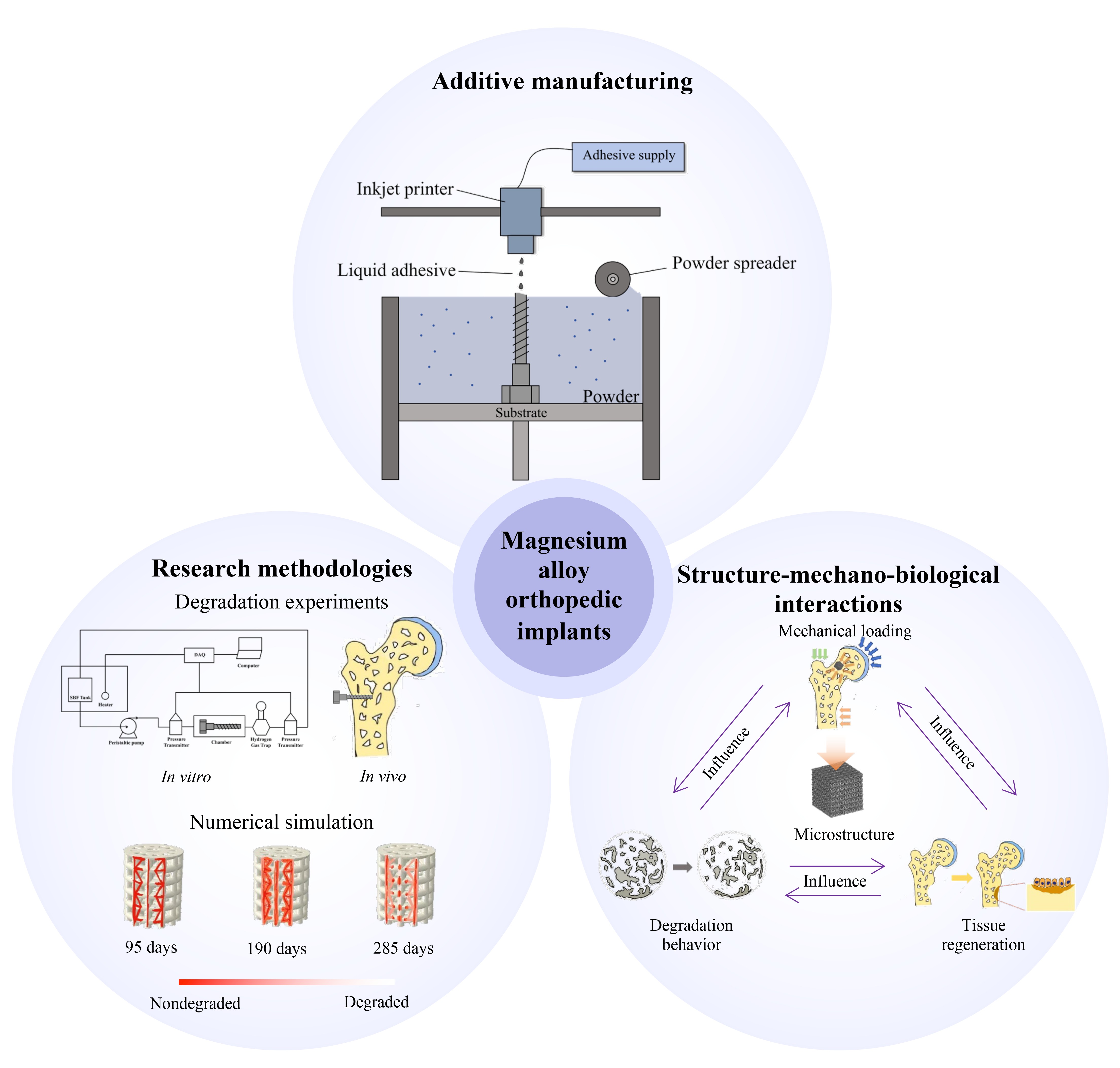
Influence of porous structures on the degradation behavior of additively manufactured magnesium and magnesium alloy orthopedic implants

The escalating incidence of bone defects has prompted a substantial demand for orthopedic implants, and additively manufactured biodegradable porous magnesium and magnesium alloy orthopedic implants have demonstrated significant potential for clinical applications. However, the mismatch between degradation-induced changes in mechanical properties and tissue regeneration remains a major challenge hindering their applications. As porous structure is a critical factor influencing the degradation behavior of magnesium/magnesium alloy orthopedic implants, this study aims to comprehensively review the current state of research in this area. The degradation behavior of magnesium/magnesium alloy orthopedic implants has been investigated using both experimental and numerical simulation methods. Degradation experiments have enabled direct observations of the influences of structures on degradation behavior and underlying mechanisms. Numerical simulations have been employed to analyze the stress and strain distributions within the structure during degradation and surrounding tissue regeneration, facilitating the investigation of the “structure-stress-tissue regeneration” regulation on degradation. Porous structures play critical roles in regulating mechanical properties, bearing physiological loads, and establishing a localized mechanical microenvironment of magnesium/magnesium alloy orthopedic implants. Design variables, including porosity, specific surface area, pore size, shape, and interconnectivity, influence the macroscopic mechanical properties, structural deformation, stress distribution, and contact with surrounding tissues, thereby regulating degradation behavior and tissue regeneration of implants. However, models that quantitatively describe the “porous structural variables-degradation-tissue regeneration” interaction remain to be developed. This study systematically summarizes the influences of porous structures on the degradation behavior of additively manufactured magnesium/ magnesium alloy orthopedic implants and the “structure-mechanics-degradation-biology” interaction mechanisms. This review provides a systematic understanding of the state-of-the-art research and future directions to guide the development and applications of orthopedic implants.
- Hu X, Lin Z, He J, et al. Recent progress in 3D printing degradable polylactic acid‐based bone repair scaffold for the application of cancellous bone defect. MedComm. 2022;1(1):e14. doi: 10.1002/mba2.14
- Habibovic P. Strategic directions in osteoinduction and biomimetics. Tissue Eng. 2017;23(23-24):1295-1296. doi: 10.1089/ten.tea.2017.0430
- Yang Y, He C, Yang W, et al. Mg bone implant: Features, developments and perspectives. Mater Des. 2020;185:108259. doi: 10.1016/j.matdes.2019.108259
- Wang W, Yeung KW. Bone grafts and biomaterials substitutes for bone defect repair: A review. Bioact Mater. 2017;2(4):224-247. doi: 10.1016/j.bioactmat.2017.05.007
- Block MS, Kent JN. Sinus augmentation for dental implants: The use of autogenous bone. J Oral Maxillofac Surg. 1997;55(11):1281-1286. doi: 10.1016/S0278-2391(97)90185-3
- Sakkas A, Wilde F, Heufelder M, Winter K, Schramm A. Autogenous bone grafts in oral implantology-is it still a “gold standard”? A consecutive review of 279 patients with 456 clinical procedures. Int J Implant Dent. 2017;3:1-17. doi: 10.1186/s40729-017-0084-4
- Younger EM, Chapman MW. Morbidity at bone graft donor sites. J Orthop Trauma. 1989;3(3):192-195. doi: 10.1097/00005131-198909000-00002
- Robinson PG, Abrams GD, Sherman SL, Safran MR, Murray IR. Autologous bone grafting. Oper Techn Sport Med. 2020;28(4):150780. doi: 10.1016/j.otsm.2020.150780
- Sumner DR, Galante JO. Determinants of stress shielding: Design versus materials versus interface.Clin Orthop Relat Res. 1992;274:202-212.
- Huiskes R, Weinans H, Van Rietbergen B. The relationship between stress shielding and bone resorption around total hip stems and the effects of flexible materials. Clin Orthop Relat Res. 1992;274:124-134.
- Sundfeldt M, Carlsson LV, Johansson CB, Thomsen P, Gretzer C. Aseptic loosening, not only a question of wear: A review of different theories. Acta Orthop. 2006;77(2):177- 197. doi: 10.1080/17453670610045902
- Yaszemski MJ, Payne RG, Hayes WC, Langer R, Mikos AG. Evolution of bone transplantation: Molecular, cellular and tissue strategies to engineer human bone. Biomaterials. 1996;17(2):175-185. doi: 10.1016/0142-9612(96)85762-0
- Roshan-Ghias A, Lambers FM, Gholam-Rezaee M, Müller R, Pioletti DP. In vivo loading increases mechanical properties of scaffold by affecting bone formation and bone resorption rates. Bone. 2011;49(6):1357-1364. doi: 10.1016/j.bone.2011.09.040
- Kroeze RJ, Helder MN, Govaert LE, Smit TH. Biodegradable polymers in bone tissue engineering. Materials. 2009;2(3):833-856. doi: 10.3390/ma2030833
- Giarmatzis G, Jonkers I, Wesseling M, Van Rossom S, Verschueren S. Loading of hip measured by hip contact forces at different speeds of walking and running. J Bone Miner Res. 2015;30(8):1431-1440. doi: 10.1002/jbmr.2483
- Hart NH, Newton RU, Tan J, et al. Biological basis of bone strength: Anatomy, physiology and measurement. J Musculoskelet Neuronal Interact. 2020;20(3):347-371.
- Bakhtiari H, Nouri A, Khakbiz M, Tolouei-Rad M. Fatigue behaviour of load-bearing polymeric bone scaffolds: A review. Acta Biomater. 2023;172:16-37. doi: 10.1016/j.actbio.2023.09.048
- Bandyopadhyay A, Mitra I, Goodman SB, Kumar M, Bose S. Improving biocompatibility for next generation of metallic implants. Prog Mater Sci. 2023;133:101053. doi: 10.1016/j.pmatsci.2022.101053
- Lee BH, Lee C, Kim DG, Choi K, Lee KH, Do Kim Y. Effect of surface structure on biomechanical properties and osseointegration. Mater Sci Eng C. 2008;28(8):1448-1461. doi: 10.1016/j.msec.2008.03.015
- Shah FA, Thomsen P, Palmquist A. Osseointegration and current interpretations of the bone-implant interface. Acta Biomater. 2019;84:1-15. doi: 10.1016/j.actbio.2018.11.018
- Heary RF, Parvathreddy N, Sampath S, Agarwal N. Elastic modulus in the selection of interbody implants. J Spine Surg. 2017;3(2):163. doi: 10.21037/jss.2017.05.01
- Yang W, Chen H, Bai H, et al. Additive manufactured osseointegrated screws with hierarchical design. Bio-Des Manuf. 2024;7(2):206-235. doi: 10.1007/s42242-024-00269-3
- Agarwal R, Gupta V, Singh J. Additive manufacturing-based design approaches and challenges for orthopaedic bone screws: A state-of-the-art review. J Braz Soc Mech Sci Eng. 2022;44(1):37. doi: 10.1007/s40430-021-03331-8
- Wang X, Xu S, Zhou S, et al. Topological design and additive manufacturing of porous metals for bone scaffolds and orthopaedic implants: A review. Biomaterials. 2016;83:127- 141. doi: 10.1016/j.biomaterials.2016.01.012
- Zhang J, Shen Y, Sun Y, et al. Design and mechanical testing of porous lattice structure with independent adjustment of pore size and porosity for bone implant. J Mater Res Technol. 2022;18:3240-3255. doi: 10.1016/j.jmrt.2022.04.002
- Mandal S, Das V, Debata M, et al. Study of pore morphology, microstructure, and cell adhesion behaviour in porous Ti-6Al-4V scaffolds. Emergent Mater. 2019;2:453-462. doi: 10.1007/s42247-019-00055-3
- Zhang Z, Jones D, Yue S, et al. Hierarchical tailoring of strut architecture to control permeability of additive manufactured titanium implants. Mater Sci Eng C. 2013;33(7):4055-4062. doi: 10.1016/j.msec.2013.05.050
- Chua C. The design of scaffolds for use in tissue engineering. Part I. Traditional factors. Tissue Eng. 2001;7:679-689. doi: 10.1089/107632701753337645
- Zhang Q, Zhou J, Zhi P, et al. 3D printing method for bone tissue engineering scaffold. Med Novel Technol Devices. 2023;17:100205. doi: 10.1016/j.medntd.2022.100205
- Ansari N, Alabtah FG, Albakri MI, Khraisheh M. Post processing of additive manufactured Mg alloys: Current status, challenges, and opportunities. J Magnesium Alloys. 2024;12(4):1283-1310. doi: 10.1016/j.jma.2024.04.017
- Guddati S, Kiran ASK, Leavy M, Ramakrishna S. Recent advancements in additive manufacturing technologies for porous material applications. Int J Adv Manuf Tech. 2019;105(1):193-215. doi: 10.1007/s00170-019-04116-z
- Prakash KS, Nancharaih T, Rao VS. Additive manufacturing techniques in manufacturing-an overview. Mater Today Proc. 2018;5(2):3873-3882. doi: 10.1016/j.matpr.2017.11.642
- Nahlieli O. Complications of sialendoscopy: Personal experience, literature analysis, and suggestions. J Oral Maxillofac Surg. 2015;73(1):75-80. doi: 10.1016/j.joms.2014.07.028
- Chen Y, Li W, Zhang C, Wu Z, Liu J. Recent developments of biomaterials for additive manufacturing of bone scaffolds. Adv Healthcare Mater. 2020;9(23): e2000724. doi: 10.1002/adhm.202000724
- Germaini MM, Belhabib S, Guessasma S, Deterre R, Corre P, Weiss P. Additive manufacturing of biomaterials for bone tissue engineering-A critical review of the state of the art and new concepts. Prog Mater Sci. 2022;130:100963. doi: 10.1016/j.pmatsci.2022.100963
- Cockerill I, Su Y, Sinha S, et al. Porous zinc scaffolds for bone tissue engineering applications: A novel additive manufacturing and casting approach. Mater Sci Eng C. 2020;110:110738. doi: 10.1016/j.msec.2020.110738
- Gartzke AK, Julmi S, Klose C, et al. A simulation model for the degradation of magnesium-based bone implants. J Mech Behav Biomed Mater. 2020;101:103411. doi: 10.1016/j.jmbbm.2019.103411
- Nasr Azadani M, Zahedi A, Bowoto OK, Oladapo BI. A review of current challenges and prospects of magnesium and its alloy for bone implant applications. Prog Biomater. 2022;11(1):1-26. doi: 10.1007/s40204-022-00182-x
- Cheng A, Schwartz Z, Kahn A, et al. Advances in porous scaffold design for bone and cartilage tissue engineering and regeneration. Tissue Eng Part B Rev. 2019;25(1):14-29. doi: 10.1089/ten.teb.2018.0119
- Garimella A, Ghosh SB, Bandyopadhyay-Ghosh S. A comprehensive study of the impact on the microstructure and corrosion behavior of magnesium alloy-based porous bone implants. Mater Today Proc. 2023;9(3):1249-1282. doi: 10.1016/j.matpr.2023.04.027
- Zhao D, Yu K, Sun T, et al. Material-structure-function integrated additive manufacturing of degradable metallic bone implants for load‐bearing applications. Adv Funct Mater. 2023;33(16):2213128. doi: 10.1002/adfm.202213128
- Parai R, Bandyopadhyay-Ghosh S. Engineered bio-nanocomposite magnesium scaffold for bone tissue regeneration. J Mech Behav Biomed Mater. 2019;96:45-52. doi: 10.1016/j.jmbbm.2019.04.019
- Sarian MN, Iqbal N, Sotoudehbagha P, et al. Potential bioactive coating system for high-performance absorbable magnesium bone implants. Bioact Mater. 2022;12:42-63. doi: 10.1016/j.bioactmat.2021.10.034
- Krämer M, Schilling M, Eifler R, et al. Corrosion behavior, biocompatibility and biomechanical stability of a prototype magnesium-based biodegradable intramedullary nailing system. Mater Sci Eng C Mater Biol Appl. 2016;59:129-135. doi: 10.1016/j.msec.2015.10.006
- Ghazizadeh E, Jabbari A, Sedighi M. In vitro corrosion-fatigue behavior of biodegradable Mg/HA composite in simulated body fluid. J Magnesium Alloys. 2021;9(6):2169-2184. doi: 10.1016/j.jma.2021.03.027
- Jing X, Ding Q, Wu Q, et al. Magnesium-based materials in orthopaedics: Material properties and animal models. Biomater Transl. 2021;2(3):197-213. doi: 10.12336/biomatertransl.2021.03.004
- Saad APM, Syahrom A. Study of dynamic degradation behaviour of porous magnesium under physiological environment of human cancellous bone. Corros Sci. 2018;131:45-56. doi: 10.1016/j.corsci.2017.10.026
- Peng B, Xu H, Song F, Wen P, Tian Y, Zheng Y. Additive manufacturing of porous magnesium alloys for biodegradable orthopedic implants: Process, design, and modification. J Mater Sci Technol. 2024;182:79-110. doi: 10.1016/j.jmst.2023.08.072
- Bielik M, Neubauer E, Kitzmantel M, Neubauer I, Kozeschnik E. Numerical simulation and experimental characterization of a single-seam plasma wire arc additive manufacturing process for Ti-6Al-4V. Mater Sci Addit Manuf. 2025;4(3):025140021. doi: 10.36922/MSAM025140021
- Shahed KS, Groeneveld-Meijer W, Lear M, Schreiber J, Manogharan G. Powder spreading behavior of bimodal ceramics in the binder jetting process. Mater Sci Addit Manuf. 2025;4(2):025110016. doi: 10.36922/MSAM025110016
- Chen J, Chen B. Progress in additive manufacturing of magnesium alloys: A review. Materials. 2024;17(15):3851. doi: 10.3390/ma17153851
- Zaitceva M, Borisov A, Popovich A, Sufiiarov V. Selective laser melting of ferritic/martensitic oxide dispersion-strengthened steel: Processing, microstructure, and mechanical properties. Mater Sci Addit Manuf. 2025;4(1):025060004. doi: 10.36922/MSAM025060004
- Li X, Fang X, Zhang M, Wang B, Huang K. Enhanced strength-ductility synergy of magnesium alloy fabricated by ultrasound assisted directed energy deposition. J Mater Sci Technol. 2024;178:247-261. doi: 10.1016/j.jmst.2023.09.021
- Cheng S, Liu F, Xu Y, et al. Effects of arc oscillation on microstructure and mechanical properties of AZ31 magnesium alloy prepared by CMT wire-arc directed energy deposition. Mater Sci Eng A. 2023;864:144539. doi: 10.1016/j.msea.2022.144539
- Gieseke M, Noelke C, Kaierle S, Wesling V, Haferkamp H. Selective laser melting of magnesium and magnesium alloys. Magnes Technol. 2013:65-68. doi: 10.1007/978-3-319-48150-0_11
- Prashanth KG, Scudino S, Klauss HJ, et al. Microstructure and mechanical properties of Al-12Si produced by selective laser melting: Effect of heat treatment. Mater Sci Eng A. 2014;590:153-160. doi: 10.1016/j.msea.2013.10.023
- Allavikutty R, Gupta P, Santra TS, Rengaswamy J. Additive manufacturing of Mg alloys for biomedical applications: Current status and challenges. Curr Opin Biomed Eng. 2021;18:100276. doi: 10.1016/j.cobme.2021.100276
- Gao C, Wang C, Jin H, et al. Additive manufacturing technique-designed metallic porous implants for clinical application in orthopedics. RSC Adv. 2018;8(44):25210-25227. doi: 10.1039/c8ra04815k
- Dong J, Li Y, Lin P, et al. Solvent-cast 3D printing of magnesium scaffolds. Acta Biomater. 2020;114:497-514. doi: 10.1016/j.actbio.2020.08.002
- Takagi H, Sasahara H, Abe T, et al. Material-property evaluation of magnesium alloys fabricated using wire-and-arc-based additive manufacturing. Addit Manuf. 2018;24:498-507. doi: 10.1016/j.addma.2018.10.026
- Karunakaran R, Ortgies S, Tamayol A, Bobaru F, Sealy MP. Additive manufacturing of magnesium alloys. Bioact Mater. 2020;5(1):44-54. doi: 10.1016/j.bioactmat.2019.12.004
- Manjhi SK, Sekar P, Bontha S, Balan A. Additive manufacturing of magnesium alloys: Characterization and post-processing. Int J Lightweight Mater Manuf. 2024;7(1):184-213. doi: 10.1016/j.ijlmm.2023.06.004
- Kleger N, Cihova M, Masania K, Studart AR, Löffler JF. 3D printing of salt as a template for magnesium with structured porosity. Adv Mater. 2019;31(37):1903783. doi: 10.1002/adma.201903783
- Wolff M, Mesterknecht T, Bals A, Ebel T, Willumeit- Römer R. FFF of Mg-alloys for biomedical application. Magnes Technol. 2019:43-49. doi: 10.1007/978-3-030-05789-3_8
- Farag M, Yun HS. Effect of gelatin addition on fabrication of magnesium phosphate-based scaffolds prepared by additive manufacturing system. Mater Lett. 2014;132:111-115. doi: 10.1016/j.matlet.2014.06.055
- Wei D, Anniyaer A, Koizumi Y, et al. On microstructural homogenization and mechanical properties optimization of biomedical Co-Cr-Mo alloy additively manufactured by using electron beam melting. Addit Manuf. 2019;28:215-227. doi: 10.1016/j.addma.2019.05.010
- Badkoobeh F, Mostaan H, Rafiei M, Bakhsheshi-Rad HR, RamaKrishna S, Chen X. Additive manufacturing of biodegradable magnesium-based materials: Design strategies, properties, and biomedical applications. J Magnesium Alloys. 2023;11(3):801-839. doi: 10.1016/j.jma.2022.12.001
- Zhang WN, Wang LZ, Feng ZX, Chen YM. Research progress on selective laser melting (SLM) of magnesium alloys: A review. Optik. 2020;207:163842. doi: 10.1016/j.ijleo.2019.163842
- Zumdick NA, Jauer L, Kersting LC, Kutz TN, Schleifenbaum JH, Zander D. Additive manufactured WE43 magnesium: A comparative study of the microstructure and mechanical properties with those of powder extruded and as-cast WE43. Mater Charact. 2019;147:384-397. doi: 10.1016/j.matchar.2018.11.011
- Guo Y, Quan G, Jiang Y, Ren L, Fan L, Pan H. Formability, microstructure evolution and mechanical properties of wire arc additively manufactured AZ80M magnesium alloy using gas tungsten arc welding. J Magnesium Alloys. 2021;9(1):192-201. doi: 10.1016/j.jma.2020.01.003
- Juan C. Microstructure and mechanical properties of high strength Mg-15Gd-1Zn-0.4 Zr alloy additive-manufactured by selective laser melting process. T Nonferr Metal Soc. 2021;31(7):1969-1978. doi: 10.1016/S1003-6326(21)65630-3
- Zhang XL, Zhang ZT. Influence of sub-rapid solidification on microstructure and mechanical properties of AZ61A magnesium alloy. T Nonferr Metal Soc. 2008;18:S86-S90. doi: 10.1016/s1003-6326(10)60180-x
- Kokubo T, Ito S, Shigematsu M, Sanka S, Yamamuro T. Fatigue and life-time of bioactive glass-ceramic AW containing apatite and wollastonite. J Mater Sci. 1987;22:4067-4070. doi: 10.1007/BF01133359
- Dutta S, Devi KB, Roy M. Processing and degradation behavior of porous magnesium scaffold for biomedical applications. Adv Powder Technol. 2017;28(12):3204-3212. doi: 10.1016/j.apt.2017.09.024
- Zhuang H, Han Y, Feng A. Preparation, mechanical properties and in vitro biodegradation of porous magnesium scaffolds. Mater Sci Eng C. 2008;28(8):1462-1466. doi: 10.1016/j.msec.2008.04.001
- Wang C, Liu J, Min S, et al. The effect of pore size on the mechanical properties, biodegradation and osteogenic effects of additively manufactured magnesium scaffolds after high temperature oxidation: An in vitro and in vivo study. Bioact Mater. 2023;28:537-548. doi: 10.1016/j.bioactmat.2023.06.009
- Kopp A, Derra T, Müther M, et al. Influence of design and postprocessing parameters on the degradation behavior and mechanical properties of additively manufactured magnesium scaffolds. Acta Biomater. 2019;98:23-35. doi: 10.1016/j.actbio.2019.04.012
- Zhen Z, Xi TF, Zheng YF. A review on in vitro corrosion performance test of biodegradable metallic materials. T Nonferr Metal Soc. 2013;23(8):2283-2293. doi: 10.1016/s1003-6326(13)62730-2
- Abidin NIZ, Rolfe B, Owen H, et al. The in vivo and in vitro corrosion of high-purity magnesium and magnesium alloys WZ21 and AZ91. Corros Sci. 2013;75:354-366. doi: 10.1016/j.corsci.2013.06.019
- Hou R, Feyerabend F, Helmholz H, Garamus VM, Willumeit-Römer R. Effects of proteins on magnesium degradation-static vs. dynamic conditions. J Magnesium Alloys. 2023;11(4):1332-1342. doi: 10.1016/j.jma.2021.07.021
- Ng W, Chiu K, Cheng F. Effect of pH on the in vitro corrosion rate of magnesium degradable implant material. Mater Sci Eng C. 2010;30(6):898-903. doi: 10.1016/j.msec.2010.04.003
- Marco I, Feyerabend F, Willumeit-Römer R, Van der Biest O. Degradation testing of Mg alloys in Dulbecco’s modified eagle medium: Influence of medium sterilization. Mater Sci Eng C Mater Biol Appl. 2016;62:68-78. doi: 10.1016/j.msec.2016.01.039
- Jia G, Chen C, Zhang J, et al. In vitro degradation behavior of Mg scaffolds with three-dimensional interconnected porous structures for bone tissue engineering. Corros Sci. 2018;144:301-312. doi: 10.1016/j.corsci.2018.09.001
- Saad APM, Jasmawati N, Harun MN, et al. Dynamic degradation of porous magnesium under a simulated environment of human cancellous bone. Corros Sci. 2016;112:495-506. doi: 10.1016/j.corsci.2016.08.017
- Wang Y, Huang H, Jia G, Zeng H, Yuan G. Fatigue and dynamic biodegradation behavior of additively manufactured Mg scaffolds. Acta Biomater. 2021;135:705-722. doi: 10.1016/j.actbio.2021.08.040
- Zhao F, Vaughan TJ, Mcnamara LM. Multiscale fluid-structure interaction modelling to determine the mechanical stimulation of bone cells in a tissue engineered scaffold. Biomech Model Mechanobiol. 2015;14:231-243. doi: 10.1007/s10237-014-0599-z
- Witte F, Fischer J, Nellesen J, et al. In vitro and in vivo corrosion measurements of magnesium alloys. Biomaterials. 2006;27(7):1013-1018. doi: 10.1016/j.biomaterials.2005.07.037
- Kleer-Reiter N, Julmi S, Feichtner F, et al. Biocompatibility and degradation of the open-pored magnesium scaffolds LAE442 and La2. Materials. 2021;16(3):035037. doi: 10.1088/1748-605x/abf5c5
- Li Y, Zhou J, Pavanram P, et al. Additively manufactured biodegradable porous magnesium. Acta Biomater. 2018;67:378-392. doi: 10.1016/j.actbio.2017.12.008
- Yin Yee Chin P, Cheok Q, Glowacz A, Caesarendra W. A review of in-vivo and in-vitro real-time corrosion monitoring systems of biodegradable metal implants. Appl Sci. 2020;10(9):3141. doi: 10.3390/app10093141
- Bobe K, Willbold E, Morgenthal I, et al. In vitro and in vivo evaluation of biodegradable, open-porous scaffolds made of sintered magnesium W4 short fibres. Acta Biomater. 2013;9(10):8611-8623. doi: 10.1016/j.actbio.2013.03.035
- Kim JA, Lim J, Naren R, Yun HS, Park EK. Effect of the biodegradation rate controlled by pore structures in magnesium phosphate ceramic scaffolds on bone tissue regeneration in vivo. Acta Biomater. 2016;44:155-167. doi: 10.1016/j.actbio.2016.08.039
- Feyerabend F. In vitro analysis of magnesium corrosion in orthopaedic biomaterials. Biomater Bone Regener. 2014: 225-269. doi: 10.1533/9780857098104.2.225
- Kirkland N, Birbilis N, Staiger M. Assessing the corrosion of biodegradable magnesium implants: A critical review of current methodologies and their limitations. Acta Biomater. 2012;8(3):925-936. doi: 10.1016/j.actbio.2011.11.014
- Saad AP, Prakoso AT, Sulong M, Basri H, Wahjuningrum DA, Syahrom A. Impacts of dynamic degradation on the morphological and mechanical characterisation of porous magnesium scaffold. Biomech Model Mechanobiol. 2019;18:797-811. doi: 10.1007/s10237-018-01115-z
- Liu Y, Yang Z, Tan L, Li H, Zhang Y. An animal experimental study of porous magnesium scaffold degradation and osteogenesis. Braz J Med Biol Res. 2014;47(8):715-720. doi: 10.1590/1414-431x20144009
- Zhao D, Wang T, Nahan K, et al. In vivo characterization of magnesium alloy biodegradation using electrochemical H2 monitoring, ICP-MS, and XPS. Acta Biomater. 2017;50:556-565. doi: 10.1016/j.actbio.2017.01.024
- Zhao D, Wang T, Hoagland W, et al. Visual H2 sensor for monitoring biodegradation of magnesium implants in vivo. Acta Biomater. 2016;45:399-409. doi: 10.1016/j.actbio.2016.08.049
- Abdalla M, Joplin A, Elahinia M, Ibrahim H. Corrosion modeling of magnesium and its alloys for biomedical applications. Corros Mater Degrad. 2020;1(2):11. doi: 10.3390/cmd1020011
- Barzegari M, Mei D, Lamaka SV, Geris L. Computational modeling of degradation process of biodegradable magnesium biomaterials. Corros Sci. 2021;190:109674. doi: 10.1016/j.corsci.2021.109674
- Grogan JA, Leen SB, McHugh PE. A physical corrosion model for bioabsorbable metal stents. Acta Biomater. 2014;10(5):2313-2322. doi: 10.1016/j.actbio.2013.12.059
- Bajger P, Ashbourn J, Manhas V, Guyot Y, Lietaert K, Geris L. Mathematical modelling of the degradation behaviour of biodegradable metals. Biomech Model Mechanobiol. 2017;16:227-238. doi: 10.1007/s10237-016-0812-3
- Grogan J, O’Brien B, Leen S, McHugh P. A corrosion model for bioabsorbable metallic stents. Acta Biomater. 2011;7(9):3523-3533. doi: 10.1016/j.actbio.2011.05.032
- Grogan JA, Leen SB, McHugh PE. Optimizing the design of a bioabsorbable metal stent using computer simulation methods. Biomaterials. 2013;34(33):8049-8060. doi: 10.1016/j.biomaterials.2013.07.010
- Gastaldi D, Sassi V, Petrini L, Vedani M, Trasatti S, Migliavacca F. Continuum damage model for bioresorbable magnesium alloy devices-Application to coronary stents. J Mech Behav Biomed Mater. 2011;4(3):352-365. doi: 10.1016/j.jmbbm.2010.11.003
- Wilder JW, Clemons C, Golovaty D, Kreider KL, Young GW, Lillard RS. An adaptive level set approach for modeling damage due to galvanic corrosion. J Eng Math. 2015;91:121-142.doi: 10.1007/s10665-014-9732-3
- Gao Y, Wang L, Gu X, Chu Z, Guo M, Fan Y. A quantitative study on magnesium alloy stent biodegradation. J Biomech. 2018;74:98-105. doi: 10.1016/j.jbiomech.2018.04.027
- Duddu R. Numerical modeling of corrosion pit propagation using the combined extended finite element and level set method. Comput Mech. 2014;54(3):613-627. doi: 10.1007/s00466-014-1010-8
- Vijayaraghavan V, Garg A, Gao L, Vijayaraghavan R. Finite element based physical chemical modeling of corrosion in magnesium alloys. Metals. 2017;7(3):83. doi: 10.3390/met7030083
- Quinn C, Van Gaalen K, McHugh PE, Kopp A, Vaughan TJ. An enhanced phenomenological model to predict surface-based localised corrosion of magnesium alloys for medical use. J Mech Behav Biomed Mater. 2023;138:105637. doi: 10.1016/j.jmbbm.2022.105637
- Boland EL, Shirazi RN, Grogan JA, McHugh PE. Mechanical and corrosion testing of magnesium WE43 specimens for pitting corrosion model calibration. Adv Eng Mater. 2018;20(10):1800656. doi: 10.1002/adem.201800656
- Sanz-Herrera JA, Reina-Romo E, Boccaccini AR. In silico design of magnesium implants: Macroscopic modeling. J Mech Behav Biomed Mater. 2018;79:181-188. doi: 10.1016/j.jmbbm.2017.12.016
- Amerinatanzi A, Mehrabi R, Ibrahim H, Dehghan A, Shayesteh Moghaddam N, Elahinia M. Predicting the biodegradation of magnesium alloy implants: Modeling, parameter identification, and validation. Bioengineering. 2018;5(4):105. doi: 10.3390/bioengineering5040105
- Shen Z, Zhao M, Bian D, et al. Predicting the degradation behavior of magnesium alloys with a diffusion-based theoretical model and in vitro corrosion testing. J Mater Sci Technol. 2019;35(7):1393-1402. doi: 10.1016/j.jmst.2019.02.004
- Putra RU, Basri H, Prakoso AT, et al. Level of activity changes increases the fatigue life of the porous magnesium scaffold, as observed in dynamic immersion tests, over time. Sustainability. 2023;15(1):823. doi: 10.3390/su15010823
- Sanz-Herrera JA, Reina-Romo E. Continuum modeling and simulation in bone tissue engineering. Appl Sci. 2019;9(18):3674. doi: 10.3390/app9183674
- Ahmed H, Bedding-Tyrrell M, Deganello D, Xia Z, Xiong Y, Zhao F. Efficient calculation of fluid-induced wall shear stress within tissue engineering scaffolds by an empirical model. Med Novel Technol Devices. 2023;18:100223. doi: 10.1016/j.medntd.2023.100223
- Chalisgaonkar R. Insight in applications, manufacturing and corrosion behaviour of magnesium and its alloys-A review. Mater Today Proc. 2020;26:1060-1071. doi: 10.1016/j.matpr.2020.02.211
- Dong J, Lin T, Shao H, et al. Advances in degradation behavior of biomedical magnesium alloys: A review. J Alloys Compd. 2022;908:164600. doi: 10.1016/j.jallcom.2022.164600
- Chen J, Tan L, Yu X, Etim IP, Ibrahim M, Yang K. Mechanical properties of magnesium alloys for medical application: A review. J Mech Behav Biomed Mater. 2018;87:68-79. doi: 10.1016/j.jmbbm.2018.07.022
- Erdmann N, Angrisani N, Reifenrath J, et al. Biomechanical testing and degradation analysis of MgCa0. 8 alloy screws: A comparative in vivo study in rabbits. Acta Biomater. 2011;7(3):1421-1428. doi: 10.1016/j.actbio.2010.10.031
- Singh S, Manoj Kumar R, Kuntal KK, et al. Sol-gel derived hydroxyapatite coating on Mg-3Zn alloy for orthopedic application. JOM. 2015;67(4):702-712. doi: 10.1007/s11837-015-1364-1
- Gu X, Xie X, Li N, Zheng Y, Qin L. In vitro and in vivo studies on a Mg-Sr binary alloy system developed as a new kind of biodegradable metal. Acta Biomater. 2012;8(6):2360-2374. doi: 10.1016/j.actbio.2012.02.018
- Niu J, Xiong M, Guan X, et al. The in vivo degradation and bone-implant interface of Mg-Nd-Zn-Zr alloy screws: 18 months post-operation results. Corros Sci. 2016;113:183-187. doi: 10.1016/j.corsci.2016.10.009
- Gawlik MM, Wiese B, Desharnais V, Ebel T, Willumeit- Römer R. The effect of surface treatments on the degradation of biomedical Mg alloys-a review paper. Materials. 2018;11(12):2561. doi: 10.3390/ma11122561
- Saket M, Amini R, Kardar P, Ganjaee M. The chemical treatment of the AZ31-Magnesium alloy surface by a high-performance corrosion protective praseodymium (III)- based film. Mater Chem Phys. 2021;260:124113. doi: 10.1016/j.matchemphys.2020.124113
- Riaz U, Shabib I, Haider W. The current trends of Mg alloys in biomedical applications-A review. J Biomed Mater Res B Appl Biomater. 2019;107(6):1970-1996. doi: 10.1002/jbm.b.34290
- Esmaily M, Svensson J, Fajardo S, et al. Fundamentals and advances in magnesium alloy corrosion. Prog Mater Sci. 2017;89:92-193. doi: 10.1016/j.pmatsci.2017.04.011
- Bergmann G, Graichen F, Rohlmann A, et al. Realistic loads for testing hip implants. Biomed Mater Eng. 2010;20(2):65- 75. doi: 10.3233/bme-2010-0616
- Li X, Chu C, Chu PK. Effects of external stress on biodegradable orthopedic materials: A review. Bioact Mater. 2016;1(1):77-84. doi: 10.1016/j.bioactmat.2016.09.002
- Wu H, Wang X, Wang G, et al. Advancing scaffold-assisted modality for in situ osteochondral regeneration: A shift from biodegradable to bioadaptable. Adv Mater. 2024;36(47):2407040. doi: 10.1002/adma.202407040
- Callens SJ, Fan D, van Hengel IA, et al. Emergent collective organization of bone cells in complex curvature fields. Nat Commun. 2023;14(1):855. doi: 10.1038/s41467-023-36436-w
- Otsuki B, Takemoto M, Fujibayashi S, Neo M, Kokubo T, Nakamura T. Pore throat size and connectivity determine bone and tissue ingrowth into porous implants: Three-dimensional micro-CT based structural analyses of porous bioactive titanium implants. Biomaterials. 2006;27(35):5892- 5900. doi: 10.1016/j.biomaterials.2006.08.013
- Egan PF, Gonella VC, Engensperger M, Ferguson SJ, Shea K. Computationally designed lattices with tuned properties for tissue engineering using 3D printing. PLoS One. 2017;12(8):e0182902. doi: 10.1371/journal.pone.0182902
- Aghion E, Perez Y. Effects of porosity on corrosion resistance of Mg alloy foam produced by powder metallurgy technology. Mater Charact. 2014;96:78-83. doi: 10.1016/j.matchar.2014.07.012
- Jia G, Huang H, Niu J, et al. Exploring the interconnectivity of biomimetic hierarchical porous Mg scaffolds for bone tissue engineering: Effects of pore size distribution on mechanical properties, degradation behavior and cell migration ability. J Magn Alloys. 2021;9(6):1954-1966. doi: 10.1016/j.jma.2021.02.001
- Li Z, Chen Z, Chen X, Zhao R. Multi-objective optimization for designing porous scaffolds with controllable mechanics and permeability: A case study on triply periodic minimal surface scaffolds. Compos Struct. 2024;333:117923. doi: 10.1016/j.compstruct.2024.117923
- Chen Y, Zhou S, Li Q. Microstructure design of biodegradable scaffold and its effect on tissue regeneration. Biomaterials. 2011;32(22):5003-5014. doi: 10.1016/j.biomaterials.2011.03.064
- Truscello S, Kerckhofs G, Van Bael S, Pyka G, Schrooten J, Van Oosterwyck H. Prediction of permeability of regular scaffolds for skeletal tissue engineering: A combined computational and experimental study. Acta Biomater. 2012;8(4):1648-1658. doi: 10.1016/j.actbio.2011.12.021
- Jeong CG, Hollister SJ. Mechanical, permeability, and degradation properties of 3D designed poly (1, 8 octanediol‐co‐citrate) scaffolds for soft tissue engineering. J Biomed Mater Res B Appl Biomater. 2010;93(1):141-149. doi: 10.1002/jbm.b.31568
- Guo X, Zheng X, Yang Y, Yang X, Yi Y. Mechanical behavior of TPMS-based scaffolds: A comparison between minimal surfaces and their lattice structures. SN Appl Sci. 2019;1(10):1145. doi: 10.1007/s42452-019-1167-z
- Cheng MQ, Wahafu T, Jiang GF, et al. A novel open-porous magnesium scaffold with controllable microstructures and properties for bone regeneration. Sci Rep. 2016;6(1):24134. doi: 10.1038/srep24134
- Yan Y, Kang Y, Li D, et al. Microstructure, mechanical properties and corrosion behavior of porous Mg-6 wt.% Zn scaffolds for bone tissue engineering. J Mater Eng Perform. 2018;27:970-984. doi: 10.1007/s11665-018-3189-x
- Wang C, Min S, Liu J, et al. Effect of pore geometry on properties of high-temperature oxidized additively manufactured magnesium scaffolds. J Magn Alloys. 2023;12(11):4509-4520. doi: 10.1016/j.jma.2023.08.016
- Augustin J, Feichtner F, Waselau AC, et al. Effect of pore size on tissue ingrowth and osteoconductivity in biodegradable Mg alloy scaffolds. J Appl Biomater Funct Mater. 2022;20:22808000221078168. doi: 10.1177/22808000221078168
- Shi Y, Xu W, Che H, et al. The effect of topological design on the degradation behavior of additively manufactured porous zinc alloy. NPJ Mater Degrad. 2024;8(1):42. doi: 10.21203/rs.3.rs-3460164/v1
- Motaharinia A, Drelich JW, Sharif S, et al. Overview of porous magnesium-based scaffolds: development, properties and biomedical applications. Mater Futures. 2025;4(1):012401. doi: 10.1088/2752-5724/ad9493
- Antoniac I, Manescu V, Paltanea G, et al. Additive manufactured magnesium-based scaffolds for tissue engineering. Materials. 2022;15(23):8693. doi: 10.3390/ma15238693
- Bonithon R, Lupton C, Roldo M, et al. Open-porous magnesium-based scaffolds withstand in vitro corrosion under cyclic loading: A mechanistic study. Bioact Mater. 2023;19:406-417. doi: 10.1016/j.bioactmat.2022.04.012
- Guo L, Zhang X, Zhang Z, Hao Z. Degradation characteristics of high-purity magnesium implants under single static and cyclic compressive loads in vivo and in vitro. J Magn Alloys. 2025;13:1480-1494. doi: 10.1016/j.jma.2024.12.014
- Schmidt M, Waselau AC, Feichtner F, et al. In vivo investigation of open-pored magnesium scaffolds LAE442 with different coatings in an open wedge defect. J Appl Biomater Funct Mater. 2022;20:22808000221142679. doi: 10.1177/22808000221142679
- Ye J, Miao B, Xiong Y, et al. 3D printed porous magnesium metal scaffolds with bioactive coating for bone defect repair: Enhancing angiogenesis and osteogenesis. J Nanobiotechnol. 2025;23(1):160. doi: 10.1186/s12951-025-03222-3
- Zhang Y, Ding Y, Wang J, et al. Study on degradation behavior of porous magnesium alloy scaffold loaded with rhBMP-2 and repair of bone defects. J Mater Res Technol. 2024;30:6498-6507. doi: 10.1016/j.jmrt.2024.04.227
- Lv Z, Peng B, Ye Y, et al. Bolstered bone regeneration by multiscale customized magnesium scaffolds with hierarchical structures and tempered degradation. Bioact Mater. 2025;46:457-475. doi: 10.1016/j.bioactmat.2024.12.002
- Gong T, Lu Y, Cheng L. Comparison of the biomechanical performance of three spinal implants for treating the wedge-shaped burst fractures. Med Novel Technol Devices. 2022;13:100109. doi: 10.1016/j.medntd.2021.100109
- Lei B, Gao X, Zhang R, Yi X, Zhou Q. In situ magnesium phosphate/polycaprolactone 3D-printed scaffold induce bone regeneration in rabbit maxillofacial bone defect model. Mater Des. 2022;215:110477. doi: 10.1016/j.matdes.2022.110477
- Huang X, Lou Y, Duan Y, et al. Biomaterial scaffolds in maxillofacial bone tissue engineering: A review of recent advances. Bioact Mater. 2024;33:129-156. doi: 10.1016/j.bioactmat.2023.10.031
- Koo Y, Lee HB, Dong Z, et al. The effects of static and dynamic loading on biodegradable magnesium pins in vitro and in vivo. Sci Rep. 2017;7(1):14710. doi: 10.1038/s41598-017-14836-5
- Foroughi AH, Valeri C, Razavi MJ. A review of computational optimization of bone scaffold architecture: Methods, challenges, and perspectives. Prog Biomed Eng. 2024;12:212. doi: 10.1088/2516-1091/ad879a
- Bonfanti S, Hiemer S, Zulkarnain R, Guerra R, Zaiser M, Zapperi S. Computational design of mechanical metamaterials. Nat Comput Sci. 2024;4(8):574-583. doi: 10.1038/s43588-024-00672-x
- Qian C, Kaminer I, Chen H. A guidance to intelligent metamaterials and metamaterials intelligence. Nat Commun. 2025;16(1):1154. doi: 10.1038/s41467-025-56122-3