

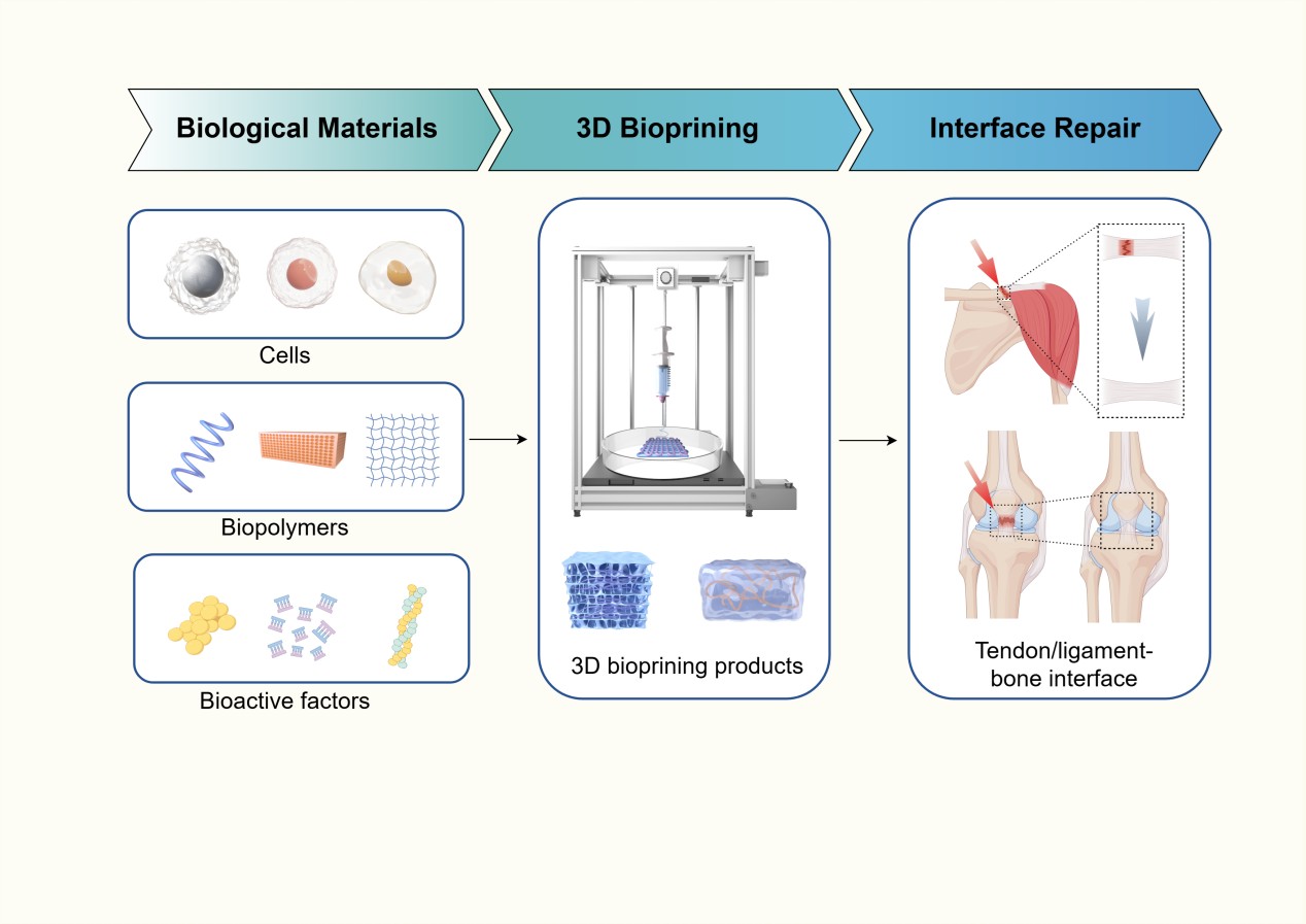
Three-dimensional bioprinting in tendon/ ligament–bone interface regeneration: From design innovations to performance enhancement

The tendon/ligament–bone (T/L–B) interface represents a critical junction where tendons and ligaments anchor to bone and is characterized by a complex, graded structure. Pathological conditions caused by aging, lifestyle factors, or trauma can severely impair this interface, leading to functional deficits and a significant decline in quality of life. However, replicating the intricate structural and biological features of the native T/L–B interface remains a major challenge with conventional fabrication methods. In this context, three-dimensional (3D) bioprinting has emerged as a promising approach for tissue repair and regeneration. This review aims to summarize the application of 3D bioprinting technologies in the reconstruction of the T/L–B interface. The review first provides a brief overview of the biology of the T/L–B interface. It then examines recent innovations in 3D printing technologies, biomaterials, and gradient structure design applied to interface regeneration. The review also explores strategies for optimizing the mechanical performance and bioactivity of 3D-bioprinted scaffolds for T/L–B interface regeneration. Finally, it highlights current challenges and future directions for advancing 3D bioprinting in this field. This review provides new insights into the clinical translation of 3D-bioprinted T/L–B interface constructs and may inform the future development of next-generation orthopedic implants.
- Wang D, Zhang X, Huang S, et al. Engineering multi-tissue units for regenerative medicine: Bone-tendon-muscle units of the rotator cuff. Biomaterials. 2021;272:120789. doi: 10.1016/j.biomaterials.2021.120789
- Rossetti L, Kuntz LA, Kunold E, et al. The microstructure and micromechanics of the tendon-bone insertion. Nat Mater. 2017;16(6):664–670. doi: 10.1038/nmat4863
- Moffat KL, Sun WH, Pena PE, et al. Characterization of the structure-function relationship at the ligament-to-bone interface. Proc. Natl Acad Sci USA. 2008;105(23):7947–7952. doi: 10.1073/pnas.0712150105
- Zhu C, Qiu J, Thomopoulos S, Xia Y. Augmenting tendon-to-bone repair with functionally graded scaffolds. Adv. Healthcare Mater. 2021;10(9):e2002269. doi: 10.1002/adhm.202002269
- Wang H, He K, Cheng CK. The structure, biology, and mechanical function of tendon/ligament-bone interfaces. Tissue Eng Part B Rev. 2024;30(5):545–558. doi: 10.1089/ten.TEB.2023.0295
- Zhong S, Lan Y, Liu J, et al. Advances focusing on the application of decellularization methods in tendon-bone healing. J Adv Res. 2025;67:361–372. doi: 10.1016/j.jare.2024.01.020
- Lu HH, Thomopoulos S. Functional attachment of soft tissues to bone: Development, healing, and tissue engineering. Annu Rev Biomed Eng. 2013;15:201–226. doi: 10.1146/annurev-bioeng-071910-124656
- Khalak FA, Decuyper JM, Khalak KA, Alonso SR, Saenz- Del-Burgo L, Pedraz Muñoz JL. 3D bioprinting approaches for musculoskeletal interfaces in tissue engineering. Int J Pharm. 2025;682:125939. doi: 10.1016/j.ijpharm.2025.125939
- Maffulli N, Longo UG, Gougoulias N, Caine D, Denaro V. Sport injuries: A review of outcomes. Br Med Bull. 2011;97:47–80. doi: 10.1093/bmb/ldq026
- Bedi A, Bishop J, Keener J, et al. Rotator cuff tears. Nat Rev Dis Primers. 2024;10(1):8. doi: 10.1038/s41572-024-00492-3
- Maniar N, Cole MH, Bryant AL, Opar DA. Muscle force contributions to anterior cruciate ligament loading. Sports Med (Auckland, NZ). 2022;52(8):1737–1750. doi: 10.1007/s40279-022-01674-3
- Bram JT, Magee LC, Mehta NN, Patel NM, Ganley TJ. Anterior cruciate ligament injury incidence in adolescent athletes: A systematic review and meta-analysis. Am J Sports Med. 2021;49(7):1962–1972. doi: 10.1177/0363546520959619
- Font Tellado S, Balmayor ER, Van Griensven M. Strategies to engineer tendon/ligament-to-bone interface: Biomaterials, cells and growth factors. Adv Drug Deliv Rev. 2015;94:126–40. doi: 10.1016/j.addr.2015.03.004
- Lei T, Zhang T, Ju W, et al. Biomimetic strategies for tendon/ ligament-to-bone interface regeneration. Bioact Mater. 2021;6(8):2491–2510. doi: 10.1016/j.bioactmat.2021.01.022
- Chen S, McCarthy A, John JV, Su Y, Xie J. Converting 2D nanofiber membranes to 3D hierarchical assemblies with structural and compositional gradients regulates cell behavior. Adv Mater (Deerfield Beach, Fla). 2020;32(43):e2003754. doi: 10.1002/adma.202003754
- Yang R, Zheng Y, Zhang Y, et al. Bipolar metal flexible electrospun fibrous membrane based on metal-organic framework for gradient healing of tendon-to-bone interface regeneration. Adv Healthc Mater. 2022;11(12):e2200072. doi: 10.1002/adhm.202200072
- Chen Y, Li Y, Zhu W, Liu Q. Biomimetic gradient scaffolds for the tissue engineering and regeneration of rotator cuff enthesis. Biofabrication. 2024;16(3). doi: 10.1088/1758-5090/ad467d
- Chen K, Liu Z, Zhou X, et al. Hierarchy reproduction: Multiphasic strategies for tendon/ligament-bone junction repair. Biomat Res. 2025;29:0132. doi: 10.34133/bmr.0132
- Fang L, Lin X, Xu R, et al. Advances in the development of gradient scaffolds made of nano-micromaterials for musculoskeletal tissue regeneration. Nano-Micro Lett. 2024;17(1):75. doi: 10.1007/s40820-024-01581-4
- Ostrovidov S, Salehi S, Costantini M, et al. 3D bioprinting in skeletal muscle tissue engineering. Small (Weinheim an der Bergstrasse, Germany). 2019;15(24):e1805530. doi: 10.1002/smll.201805530
- Mandrycky C, Wang Z, Kim K, Kim DH. 3D bioprinting for engineering complex tissues. Biotechnol Adv. 2016;34(4):422–434. doi: 10.1016/j.biotechadv.2015.12.011
- Mei Q, Rao J, Bei HP, Liu Y, Zhao X. 3D bioprinting photo-crosslinkable hydrogels for bone and cartilage repair. Int J Bioprint. 2021;7(3):367. doi: 10.18063/ijb.v7i3.367
- Fang W, Yang M, Wang L, et al. Hydrogels for 3D bioprinting in tissue engineering and regenerative medicine: Current progress and challenges. Int J Bioprint. 2023;9(5):759. doi: 10.18063/ijb.759
- Park W, Gao G, Cho DW. Tissue-specific decellularized extracellular matrix bioinks for musculoskeletal tissue regeneration and modeling using 3d bioprinting technology. Int J Mol Sci. 2021;22(15). doi: 10.3390/ijms22157837
- Altunbek M, Afghah F, Caliskan OS, Yoo JJ, Koc B. Design and bioprinting for tissue interfaces. Biofabrication. 2023;15(2). doi: 10.1088/1758-5090/acb73d
- Bai X, Yang Y, Chu J, Deng Y, Li M, Yang H. 3D bioprinting patient-specific grafts for tendon/ligament repair in motion: Emerging trends and challenges. Front Bioeng Biotechnol. 2025;13:1643430. doi: 10.3389/fbioe.2025.1643430
- Xu T, Rao J, Mo Y, et al. 3D printing in musculoskeletal interface engineering: Current progress and future directions. Adv Drug Deliv Rev. 2025;219:115552. doi: 10.1016/j.addr.2025.115552
- Chae S, Cho DW. Biomaterial-based 3D bioprinting strategy for orthopedic tissue engineering. Acta Biomater. 2023;156:4–20. doi: 10.1016/j.actbio.2022.08.004
- Chen P, Cui L, Fu SC, et al. The 3D-printed PLGA scaffolds loaded with bone marrow-derived mesenchymal stem cells augment the healing of rotator cuff repair in the rabbits. Cell Transplant. 2020;29:963689720973647. doi: 10.1177/0963689720973647
- Micalizzi S, Russo L, Giacomelli C, et al. Multimaterial and multiscale scaffold for engineering enthesis organ. Int J Bioprint. 2023;9(5):763. doi: 10.18063/ijb.763
- Murphy SV, Atala A. 3D bioprinting of tissues and organs. Nat Biotechnol. 2014;32(8):773–785. doi: 10.1038/nbt.2958
- Schwab A, Levato R, D’Este M, Piluso S, Eglin D, Malda J. Printability and shape fidelity of bioinks in 3d bioprinting. Chem Rev. 2020;120(19):11028–11055. doi: 10.1021/acs.chemrev.0c00084
- Lee SJ, Jeong W, Atala A. 3D bioprinting for engineered tissue constructs and patient-specific models: Current Progress and prospects in clinical applications. Adv Mater (Deerfield Beach, Fla). 2024;36(49):e2408032. doi: 10.1002/adma.202408032
- Malda J, Visser J, Melchels FP, et al. 25th anniversary article: Engineering hydrogels for biofabrication. Adv Mater (Deerfield Beach, Fla). 2013;25(36):5011–5028. doi: 10.1002/adma.201302042
- Derby B. Printing and prototyping of tissues and scaffolds. Science (New York, NY). 2012;338(6109):921–926. doi: 10.1126/science.1226340
- Li L, Wang P, Liang H, et al. Design of a Haversian system-like gradient porous scaffold based on triply periodic minimal surfaces for promoting bone regeneration. J Adv Res. 2023;54:89–104. doi: 10.1016/j.jare.2023.01.004
- Liu X, Hao M, Chen Z, et al. 3D bioprinted neural tissue constructs for spinal cord injury repair. Biomaterials. 2021;272:120771. doi: 10.1016/j.biomaterials.2021.120771
- Shapira A, Dvir T. 3D tissue and organ printing-hope and reality. Adv Sci (Weinheim, Baden-Wurttemberg, Germany). 2021;8(10):2003751. doi: 10.1002/advs.202003751
- Matai I, Kaur G, Seyedsalehi A, McClinton A, Laurencin CT. Progress in 3D bioprinting technology for tissue/ organ regenerative engineering. Biomaterials. 2020; 226:119536. doi: 10.1016/j.biomaterials.2019.119536
- Wang Z, Kapadia W, Li C, et al. Tissue-specific engineering: 3D bioprinting in regenerative medicine. J Control Rel: Offic J Control Rel Soc. 10 2021;329:237–256. doi: 10.1016/j.jconrel.2020.11.044
- Akbari M, Khademhosseini A. Tissue bioprinting for biology and medicine. Cell. 2022;185(15):2644–2648. doi: 10.1016/j.cell.2022.06.015
- Liu W, Zhang YS, Heinrich MA, et al. Rapid continuous multimaterial extrusion bioprinting. Adv Mater (Deerfield Beach, Fla). 2017;29(3). doi: 10.1002/adma.201604630
- Jiang X, Kong Y, Kuss M, et al. 3D bioprinting of multilayered scaffolds with spatially differentiated ADMSCs for rotator cuff tendon-to-bone interface regeneration. Appl Mater Today. 2022;27:101510. doi: 10.1016/j.apmt.2022.101510
- Ker ED, Nain AS, Weiss LE, et al. Bioprinting of growth factors onto aligned sub-micron fibrous scaffolds for simultaneous control of cell differentiation and alignment. Biomaterials. 2011;32(32):8097–9107. doi: 10.1016/j.biomaterials.2011.07.025
- Miri AK, Nieto D, Iglesias L, et al. Microfluidics-enabled multimaterial maskless stereolithographic bioprinting. Adv Mater (Deerfield Beach, Fla). 2018;30(27):e1800242. doi: 10.1002/adma.201800242
- Xie X, Wang Y, Li Z, et al. Recent advances in gradient biomimetic scaffolds for tendon-bone interface regeneration. Front Bioeng Biotechnol. 2025;13:1629816. doi: 10.3389/fbioe.2025.1629816
- Du L, Qin C, Zhang H, et al. Multicellular Bioprinting of Biomimetic Inks for Tendon-to-Bone Regeneration. Adv Sci (Weinheim, Baden-Wurttemberg, Germany). 2023;10(21):e2301309. doi: 10.1002/advs.202301309
- Chae S, Sun Y, Choi YJ, Ha DH, Jeon I, Cho DW. 3D cell-printing of tendon-bone interface using tissue-derived extracellular matrix bioinks for chronic rotator cuff repair. Biofabrication. 2021;13(3). doi: 10.1088/1758-5090/abd159
- Zhu Y, Yu X, Liu H, et al. Strategies of functionalized GelMA-based bioinks for bone regeneration: Recent advances and future perspectives. Bioact Mater. 2024;38:346–373. doi: 10.1016/j.bioactmat.2024.04.032
- Abbasi N, Ivanovski S, Gulati K, Love RM, Hamlet S. Role of offset and gradient architectures of 3-D melt electrowritten scaffold on differentiation and mineralization of osteoblasts. Biomater Res. 2020;24:2. doi: 10.1186/s40824-019-0180-z
- Zhang X, Song W, Han K, et al. Three-dimensional bioprinting of a structure-, composition-, and mechanics-graded biomimetic scaffold coated with specific decellularized extracellular matrix to improve the tendon-to-bone healing. ACS Appl Mater Interfaces. 2023;15(24):28964–28980. doi: 10.1021/acsami.3c03793
- Chae S, Yong U, Park W, et al. 3D cell-printing of gradient multi-tissue interfaces for rotator cuff regeneration. Bioact. Mater. 2023;19:611–625. doi: 10.1016/j.bioactmat.2022.05.004
- Rak Kwon D, Jung S, Jang J, Park GY, Suk Moon Y, Lee SC. A 3-Dimensional bioprinted scaffold with human umbilical cord blood-mesenchymal stem cells improves regeneration of chronic full-thickness rotator cuff tear in a rabbit model. Am J Sports Med. 2020;48(4):947–958. doi: 10.1177/0363546520904022
- Du L, Wu J, Han Y, Wu C. Immunomodulatory multicellular scaffolds for tendon-to-bone regeneration. Sci. Adv. 2024;10(10):eadk6610. doi: 10.1126/sciadv.adk6610
- Anjum S, Li T, Saeed M, Ao Q. Exploring polysaccharide and protein-enriched decellularized matrix scaffolds for tendon and ligament repair: A review. Int J Biol Macromol. 2024;254(Pt 2):127891. doi: 10.1016/j.ijbiomac.2023.127891
- Cao Y, Yang S, Zhao D, et al. Three-dimensional printed multiphasic scaffolds with stratified cell-laden gelatin methacrylate hydrogels for biomimetic tendon-to-bone interface engineering. J Orthop Transl. 2020;23:89–100. doi: 10.1016/j.jot.2020.01.004
- Han J, Han SC, Kim YK, et al. Bioactive scaffold with spatially embedded growth factors promotes bone-to-tendon interface healing of chronic rotator cuff tear in rabbit model. Am J Sports Med. 2023;51(9):2431–2442. doi: 10.1177/03635465231180289
- Bai L, Han Q, Meng Z, et al. Bioprinted living tissue constructs with layer-specific, growth factor-loaded microspheres for improved enthesis healing of a rotator cuff. Acta Biomater. 2022;154:275–289. doi: 10.1016/j.actbio.2022.10.058
- Tarafder S, Brito JA, Minhas S, Effiong L, Thomopoulos S, Lee CH. In situ tissue engineering of the tendon-to-bone interface by endogenous stem/progenitor cells. Biofabrication. 2019;12(1):015008. doi: 10.1088/1758-5090/ab48ca
- Lui H, Bindra R, Baldwin J, Ivanovski S, Vaquette C. Additively manufactured multiphasic bone-ligament-bone scaffold for scapholunate interosseous ligament reconstruction. Adv Healthcare Mater. 2019;8(14):e1900133. doi: 10.1002/adhm.201900133
- Feng F, He J, Li J, Mao M, Li D. Multicomponent bioprinting of heterogeneous hydrogel constructs based on microfluidic printheads. Int J Bioprint. 2019;5(2):202. doi: 10.18063/ijb.v5i2.202
- Beldjilali-Labro M, Garcia Garcia A, Farhat F, et al. Biomaterials in tendon and skeletal muscle tissue engineering: Current trends and challenges. Materials (Basel, Switzerland). 2018;11(7). doi: 10.3390/ma11071116
- Ma D, Wang J, Zheng M, et al. Degradation behavior of ZE21C magnesium alloy suture anchors and their effect on ligament-bone junction repair. Bioact. Mater. 2023;26:128–141. doi: 10.1016/j.bioactmat.2023.02.021
- Dong Y, Li J, Jiang Q, et al. Structure, ingredient, and function-based biomimetic scaffolds for accelerated healing of tendon-bone interface. J Orthop Transl. 2024;48:70–88. doi: 10.1016/j.jot.2024.07.007
- Zhang X, Li K, Wang C, et al. Facile and rapid fabrication of a novel 3D-printable, visible light-crosslinkable and bioactive polythiourethane for large-to-massive rotator cuff tendon repair. Bioact Mater. 2024;37:439–458. doi: 10.1016/j.bioactmat.2024.03.036
- Liu F, Luo S, Li J, et al. Laser-induced 3D graphene enabled polymer composites with improved mechanical and electrical properties toward multifunctional performance. Adv Sci (Weinheim, Baden-Wurttemberg, Germany). 2025:e09039. doi: 10.1002/advs.202509039
- Huang W, Zhang J, Singh V, et al. Digital light 3D printing of a polymer composite featuring robustness, self-healing, recyclability and tailorable mechanical properties. Addit Manuf. 2023;61:None. doi: 10.1016/j.addma.2022.103343
- Wang J, Wu Y, Li G, et al. Engineering large-scale self-mineralizing bone organoids with bone matrix-inspired hydroxyapatite hybrid bioinks. Adv Mater (Deerfield Beach, Fla). 2024;36(30):e2309875. doi: 10.1002/adma.202309875
- Barrera Bernal JL, Gaytán Salvatella Í, Del Campo BIM, Alvarez Perez MA, Masuoka-Ito D. Synthesis of hydroxyapatite-gelatin composite hydrogel for bone tissue application. Gels (Basel, Switzerland). 2025;11(8). doi: 10.3390/gels11080630
- Silva M, Gomes S, Correia C, et al. Biocompatible 3D-printed tendon/ligament scaffolds based on polylactic acid/graphite nanoplatelet composites. Nanomaterials (Basel, Switzerland). 2023;13(18). doi: 10.3390/nano13182518
- Balestri W, Hickman GJ, Morris RH, Hunt JA, Reinwald Y. Triphasic 3D in vitro model of bone-tendon-muscle interfaces to study their regeneration. Cells. 2023;12(2). doi: 10.3390/cells12020313
- Gu J, Zhang Q, Geng M, et al. Construction of nanofibrous scaffolds with interconnected perfusable microchannel networks for engineering of vascularized bone tissue. Bioact. Mater. 2021;6(10):3254–3268. doi: 10.1016/j.bioactmat.2021.02.033
- Hann SY, Cui H, Esworthy T, et al. Dual 3D printing for vascularized bone tissue regeneration. Acta Biomater. 2021;123:263–274. doi: 10.1016/j.actbio.2021.01.012
- Luo Y, Zhang T, Lin X. 3D printed hydrogel scaffolds with macro pores and interconnected microchannel networks for tissue engineering vascularization. Chem Eng J. 2022;430:132926. doi: 10.1016/j.cej.2021.132926
- Kim W, Kwon DR, Lee H, et al. 3D bioprinted multi-layered cell constructs with gradient core-shell interface for tendon-to-bone tissue regeneration. Bioact Mater. 2025; 43:471–490. doi: 10.1016/j.bioactmat.2024.10.002
- Zhou B, Jiang X, Zhou X, et al. GelMA-based bioactive hydrogel scaffolds with multiple bone defect repair functions: Therapeutic strategies and recent advances. Biomater Res. 2023;27(1):86. doi: 10.1186/s40824-023-00422-6
- Shanto PC, Park S, Fahad MAA, Park M, Lee BT. 3D bio-printed proteinaceous bioactive scaffold loaded with dual growth factor enhanced chondrogenesis and in situ cartilage regeneration. Bioact Mater. 2025;46:365–385. doi: 10.1016/j.bioactmat.2024.12.021
- Choe G, Lee M, Oh S, et al. Three-dimensional bioprinting of mesenchymal stem cells using an osteoinductive bioink containing alginate and BMP-2-loaded PLGA nanoparticles for bone tissue engineering. Biomater Adv. 2022; 136:212789. doi: 10.1016/j.bioadv.2022.212789
- Lu J, Gao Y, Cao C, et al. 3D bioprinted scaffolds for osteochondral regeneration: advancements and applications. Mater Today Bio. 2025;32:101834. doi: 10.1016/j.mtbio.2025.101834
- Yu K, Huangfu H, Qin Q, et al. Application of bone marrow-derived macrophages combined with bone mesenchymal stem cells in dual-channel three-dimensional bioprinting scaffolds for early immune regulation and osteogenic induction in rat calvarial defects. ACS Appl Mater Interf. 2022;14(41):47052–47065. doi: 10.1021/acsami.2c13557
- Liang W, Ao R, Xu M, et al. Bifunctional adECM bioscaffold with STIM1-ASCs and IGF-2 promotes functional masseter VML repair via myogenesis and fibrosis suppression. Bioact Mater. 2025;54:466–491. doi: 10.1016/j.bioactmat.2025.08.019
- Thattaruparambil Raveendran N, Vaquette C, Meinert C, Samuel Ipe D, Ivanovski S. Optimization of 3D bioprinting of periodontal ligament cells. Dent Mater: Offic Publ Acad Dent Mater. 2019;35(12):1683–1694. doi: 10.1016/j.dental.2019.08.114
- Jiang X, Wu S, Kuss M, et al. 3D printing of multilayered scaffolds for rotator cuff tendon regeneration. Bioact Mater. 2020;5(3):636–643. doi: 10.1016/j.bioactmat.2020.04.017
- Park SH, Choi YJ, Moon SW, et al. Three-dimensional bio-printed scaffold sleeves with mesenchymal stem cells for enhancement of tendon-to-bone healing in anterior cruciate ligament reconstruction using soft-tissue tendon graft. Arthroscopy. 2018;34(1):166–179. doi: 10.1016/j.arthro.2017.04.016
- Ni Y, Tian B, Lv J, et al. 3D-printed PCL scaffolds loaded with bFGF and BMSCs enhance tendon-bone healing in rat rotator cuff tears by immunomodulation and osteogenesis promotion. ACS Biomater Sci Eng. 2025;11(2): 1123–1139. doi: 10.1021/acsbiomaterials.4c02340
- Wang L, Wang C, Wu S, Fan Y, Li X. Influence of the mechanical properties of biomaterials on degradability, cell behaviors and signaling pathways: Current progress and challenges. Biomater Sci. 2020;8(10):2714–2733. doi: 10.1039/d0bm00269k
- Jia B, Huang H, Dong Z, et al. Degradable biomedical elastomers: Paving the future of tissue repair and regenerative medicine. Chem Soc Rev. 2024;53(8):4086–4153. doi: 10.1039/d3cs00923h
- Li C, Guo C, Fitzpatrick V, et al. Design of biodegradable, implantable devices towards clinical translation. Nat Rev Mater. 2020;5(1):61–81. doi: 10.1038/s41578-019-0150-z
- L GE, Farias NC, J SC, et al. Designing sustainable polymer blends: Tailoring mechanical properties and degradation behaviour in PHB/PLA/PCL blends in a seawater environment. Polymers. 2023;15(13). doi: 10.3390/polym15132874
- Zhang X, Wu Y, Han K, et al. 3-Dimensional bioprinting of a tendon stem cell-derived exosomes loaded scaffold to bridge the unrepairable massive rotator cuff tear. Am J Sports Med. 2024;52(9):2358–2371. doi: 10.1177/03635465241255918
- Chen P, Cui L, Chen G, et al. The application of BMP-12- overexpressing mesenchymal stem cells loaded 3D-printed PLGA scaffolds in rabbit rotator cuff repair. Int J Biol Macromol. 2019;138:79–88. doi: 10.1016/j.ijbiomac.2019.07.041
- Golafshan N, Castilho M, Daghrery A, et al. Composite graded melt electrowritten scaffolds for regeneration of the periodontal ligament-to-bone interface. ACS Appl Mater Interf. 2023;15(10):12735–12749. doi: 10.1021/acsami.2c21256
- Yu C, Chen R, Chen J, et al. Enhancing tendon-bone integration and healing with advanced multi-layer nanofiber-reinforced 3D scaffolds for acellular tendon complexes. Mater Today Bio. 2024;26:101099. doi: 10.1016/j.mtbio.2024.101099
- Luo W, Wang Y, Han Q, et al. Advanced strategies for constructing interfacial tissues of bone and tendon/ligament. J Tissue Eng. 2022;13:20417314221144714. doi: 10.1177/20417314221144714
- Tang Y, Wang Z, Xiang L, Zhao Z, Cui W. Functional biomaterials for tendon/ligament repair and regeneration. Regener Biomater. 2022;9:rbac062. doi: 10.1093/rb/rbac062
- Steltzer SS, Abraham AC, Killian ML. Interfacial tissue regeneration with bone. Curr Osteop Rep. 2024;22(2):290–298. doi: 10.1007/s11914-024-00859-1
- Golman M, Abraham AC, Kurtaliaj I, et al. Toughening mechanisms for the attachment of architectured materials: The mechanics of the tendon enthesis. Sci Adv. 2021;7(48):eabi5584. doi: 10.1126/sciadv.abi5584
- Li Y, Zhou M, Zheng W, Yang J, Jiang N. Scaffold-based tissue engineering strategies for soft-hard interface regeneration. Regener Biomater. 2023;10:rbac091. doi: 10.1093/rb/rbac091
- Chen C, Shi Q, Li M, et al. Engineering an enthesis-like graft for rotator cuff repair: An approach to fabricate highly biomimetic scaffold capable of zone-specifically releasing stem cell differentiation inducers. Bioact Mater. 2022;16:451–471. doi: 10.1016/j.bioactmat.2021.12.021
- Sun Han Chang RA, Shanley JF, Kersh ME, Harley BAC. Tough and tunable scaffold-hydrogel composite biomaterial for soft-to-hard musculoskeletal tissue interfaces. Sci Adv. 2020;6(34):eabb6763. doi: 10.1126/sciadv.abb6763
- Rose JC, De Laporte L. Hierarchical design of tissue regenerative constructs. Adv Healthc Mater. 2018;7(6):e1701067. doi: 10.1002/adhm.201701067
- Kang HW, Lee SJ, Ko IK, Kengla C, Yoo JJ, Atala A. A 3D bioprinting system to produce human-scale tissue constructs with structural integrity. Nat Biotechnol. 2016;34(3):312–319. doi: 10.1038/nbt.3413
- Potyondy T, Uquillas JA, Tebon PJ, et al. Recent advances in 3D bioprinting of musculoskeletal tissues. Biofabrication. 2021;13(2). doi: 10.1088/1758-5090/abc8de
- Xie X, Cai J, Li D, et al. Multiphasic bone-ligament-bone integrated scaffold enhances ligamentization and graft-bone integration after anterior cruciate ligament reconstruction. Bioact Mater. 2024;31:178–191. doi: 10.1016/j.bioactmat.2023.08.004
- Pitta Kruize C, Panahkhahi S, Putra NE, et al. Biomimetic approaches for the design and fabrication of bone-to-soft tissue interfaces. ACS Biomater Sci Eng. 2023;9(7): 3810–3831. doi: 10.1021/acsbiomaterials.1c00620
- Aykora D, Taşçı B, Şahin MZ, et al. Tendon regeneration deserves better: Focused review on In vivo models, artificial intelligence and 3D bioprinting approaches. Front Bioeng Biotechnol. 2025;13:1580490. doi: 10.3389/fbioe.2025.1580490
- Gracey E, Burssens A, Cambré I, et al. Tendon and ligament mechanical loading in the pathogenesis of inflammatory arthritis. Nat Rev Rheumatol. 2020;16(4):193–207. doi: 10.1038/s41584-019-0364-x
- Bertsch P, Diba M, Mooney DJ, Leeuwenburgh SCG. Self-healing injectable hydrogels for tissue regeneration. Chem Rev. 2023;123(2):834–873. doi: 10.1021/acs.chemrev.2c00179
- Li Y, Chen C, Jiang J, et al. Bioactive film-guided soft-hard interface design technology for multi-tissue integrative regeneration. Adv Sci (Weinheim, Baden-Wurttemberg, Germany). 2022;9(15):e2105945. doi: 10.1002/advs.202105945
- Xiao L, Sun Y, Liao L, Su X. Response of mesenchymal stem cells to surface topography of scaffolds and the underlying mechanisms. J Mater Chem B. 2023;11(12):2550–2567. doi: 10.1039/d2tb01875f
- Agarwal T, Onesto V, Banerjee D, et al. 3D bioprinting in tissue engineering: Current state-of-the-art and challenges towards system standardization and clinical translation. Biofabrication. 2025;17(4). doi: 10.1088/1758-5090/ade47a
- Raees S, Ullah F, Javed F, et al. Classification, processing, and applications of bioink and 3D bioprinting: A detailed review. Int J Biol Macromol. 2023;232:123476. doi: 10.1016/j.ijbiomac.2023.123476
- Wang Z, Xiang L, Lin F, Tang Y, Cui W. 3D bioprinting of emulating homeostasis regulation for regenerative medicine applications. J Control Rel: Offic J Control Rel Soc. 2023;353:147–165. doi: 10.1016/j.jconrel.2022.11.035
- Yuan Z, Bai X, Li S, et al. Multimaterial and multidimensional bioprinting in regenerative medicine: Advances, limitations, and future directions. Adv Healthc Mater. 2025;14(18):e2500475. doi: 10.1002/adhm.202500475
- Mozammal HMD, Lee H. A comprehensive review in the advancements of bioprinting for tissue engineering using polysaccharide biomaterials and a future strategies. Int J Biol Macromol. 2025;322(Pt 3):146667. doi: 10.1016/j.ijbiomac.2025.146667
- Li Y, Wu J, He C, et al. 3D prestress bioprinting of directed tissues. Adv Healthc Mater. 2023;12(28):e2301487. doi: 10.1002/adhm.202301487