

Evaluation of biomechanical properties and early osseointegration of biomimetic bone trabecular Ti6Al4V scaffolds based on Voronoi design

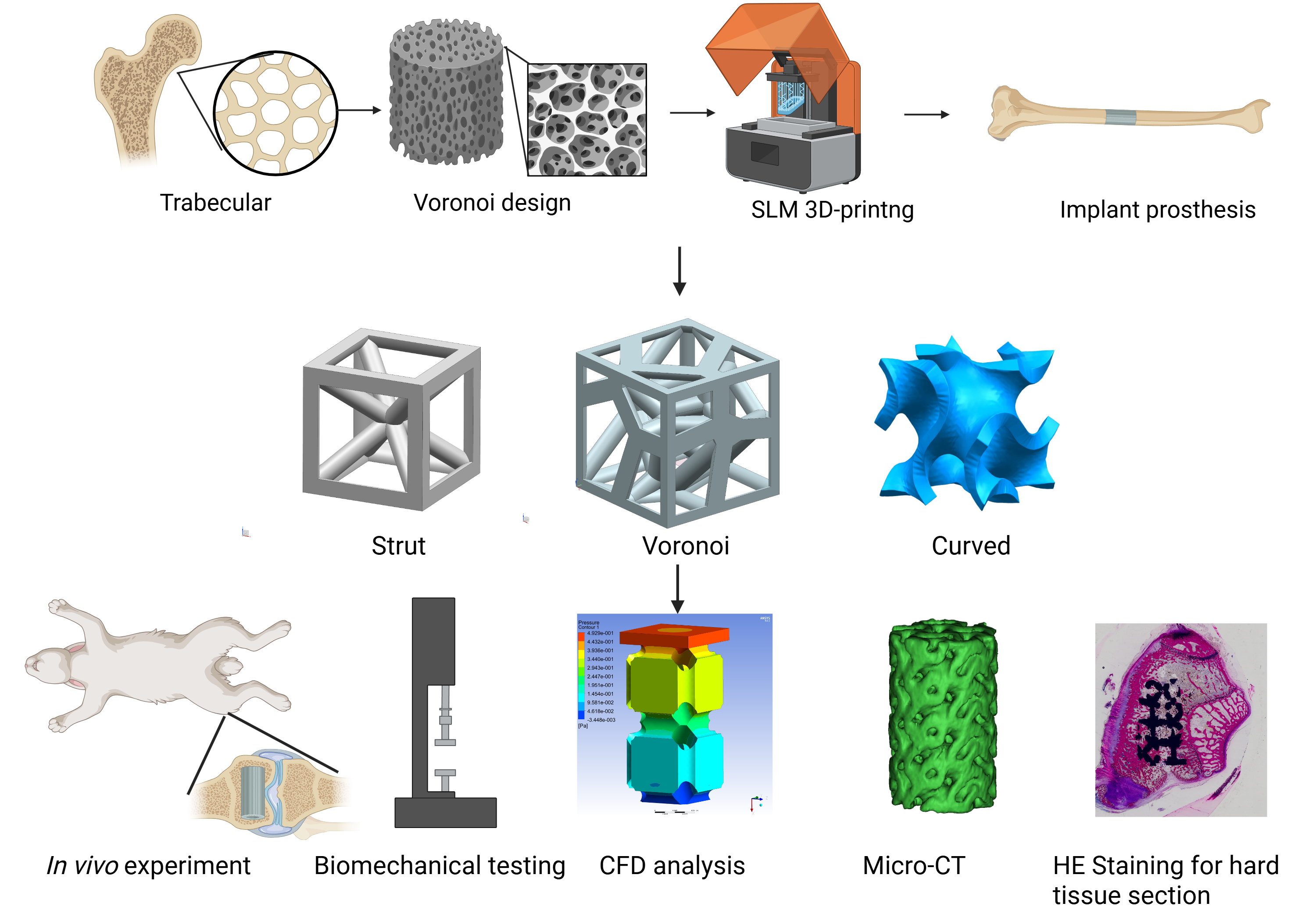
Ti6Al4V scaffolds demonstrate significant translational potential for bone defect reconstruction by virtue of their exceptional biocompatibility and corrosion resistance. However, achieving concurrent osseointegration enhancement and mechanical compatibility with native cancellous bone remains a critical design constraint. A trabecular bone-mimetic porous Ti6Al4V scaffold was fabricated via Voronoi-tessellated computer-aided design and selective laser melting. Precise modulation of pore architecture enabled controlled porosity. The mechanical properties of the scaffold were characterized through compression testing. Early-stage in vivo osseointegration was evaluated at weeks 4 and 12 in a rabbit femoral condyle defect model using histomorphometry and micro-computed tomography, with comparisons made against conventional strut-based and G-curved lattice structures. The Voronoi scaffold demonstrated an elastic modulus and yield strength comparable to cancellous bone, thereby mitigating stress-shielding effects. Additionally, according to the results from the biomechanics, computational fluid dynamics, and in vivo analyses, the scaffold demonstrated significantly enhanced osteogenic potential and superior bone-implant interface integration compared to the strut and triply periodic minimal surface (TPMS) designs. In conclusion, the Voronoi design provides an effective biomimetic strategy for fabricating porous titanium alloy bone scaffolds with enhanced osteogenic properties, which embody higher potential than conventional struts and TPMS structures in facilitating bone defect repair.
- Jing Z, Zhang T, Xiu P, et al. Functionalization of 3D-printed titanium alloy orthopedic implants: a literature review. Biomed Mater. 2020;15(5):052003. doi: 10.1088/1748-605X/ab9078
- Li L, Shi J, Zhang K, et al. Early osteointegration evaluation of porous Ti6Al4V scaffolds designed based on triply periodic minimal surface models. J Orthop Translat. 2019;19:94-105. doi: 10.1016/j.jot.2019.03.003
- Revilla-León M, Meyer MJ, Özcan M. Metal additive manufacturing technologies: literature review of current status and prosthodontic applications. Int J Comput Dent. 2019;22(1):55-67.
- Alammar A, Kois JC, Revilla-León M, Att W. Additive Manufacturing Technologies: Current Status and Future Perspectives. J Prosthodont. 2022;31(S1):4-12. doi: 10.1111/jopr.13477
- Liu F, Ran Q, Zhao M, Zhang T, Zhang DZ, Su Z. Additively manufactured continuous cell-size gradient porous scaffolds: pore characteristics, mechanical properties and biological responses in vitro. Materials (Basel). 2020;13(11):2589. doi: 10.3390/ma13112589
- Ran Q, Yang W, Hu Y, et al. Osteogenesis of 3D printed porous Ti6Al4V implants with different pore sizes. J Mech Behav Biomed Mater. 2018;84:1-11. doi: 10.1016/j.jmbbm.2018.04.010
- Arjunan A, Demetriou M, Baroutaji A, Wang C. Mechanical performance of highly permeable laser melted Ti6Al4V bone scaffolds. J Mech Behav Biomed Mater. 2020;102:103517. doi: 10.1016/j.jmbbm.2019.103517
- Wang H, Su K, Su L, Liang P, Ji P, Wang C. The effect of 3D-printed Ti(6)Al(4)V scaffolds with various macropore structures on osteointegration and osteogenesis: a biomechanical evaluation. J Mech Behav Biomed Mater. 2018;88:488-496. doi: 10.1016/j.jmbbm.2018.08.049
- Gryko A, Prochor P, Sajewicz E. Finite element analysis of the influence of porosity and pore geometry on mechanical properties of orthopaedic scaffolds. J Mech Behav Biomed Mater. 2022;132:105275. doi: 10.1016/j.jmbbm.2022.105275
- Van Bael S, Chai YC, Truscello S, et al. The effect of pore geometry on the in vitro biological behavior of human periosteum-derived cells seeded on selective laser-melted Ti6Al4V bone scaffolds. Acta Biomater. 2012;8(7):2824-34. doi: 10.1016/j.actbio.2012.04.001
- Chen Z, Yan X, Yin S, et al. Influence of the pore size and porosity of selective laser melted Ti6Al4V ELI porous scaffold on cell proliferation, osteogenesis and bone ingrowth. Mater Sci Eng C Mater Biol Appl. 2020;106:110289. doi: 10.1016/j.msec.2019.110289
- Coburn B, Salary RR. Mechanical characterization of porous bone-like scaffolds with complex microstructures for bone regeneration. Bioengineering (Basel). 2025;12(4):416. doi: 10.3390/bioengineering12040416
- Kumar PV, Pal S, Birru AK, Jaganathan BG, Muthu N. 3D-printed TPMS-structured hybrid PLA/MgTiO(3) scaffolds: Synergizing bioactivity and antibacterial performance for bone regeneration. Biomater Adv. 2025;177:214370. doi: 10.1016/j.bioadv.2025.214370
- Pazhamannil RV, Alkhedher M. Advances in additive manufacturing for bone tissue engineering: materials, design strategies, and applications. Biomed Mater. 2024;20(1). doi: 10.1088/1748-605X/ad9dce
- Ma J, Li Y, Mi Y, et al. Novel 3D printed TPMS scaffolds: microstructure, characteristics, and applications in bone regeneration. J Tissue Eng. 2024;15:20417314241263689. doi: 10.1177/20417314241263689
- Ye J, Miao B, Xiong Y, et al. 3D printed porous magnesium metal scaffolds with bioactive coating for bone defect repair: enhancing angiogenesis and osteogenesis. J Nanobiotechnology. 2025;23(1):160. doi: 10.1186/s12951-025-03222-3
- Hao W, Yongtao L, Jian J, Hanxing Z. Data-driven inverse design of novel spinodoid bone scaffolds with highly matched mechanical properties in three orthogonal directions. Mater Des. 2025;251:113697. doi: 10.1016/j.matdes.2025.113697
- Amini AR, Laurencin CT, Nukavarapu SP. Bone tissue engineering: recent advances and challenges. Crit Rev Biomed Eng. 2012;40(5):363-408. doi: 10.1615/critrevbiomedeng.v40.i5.10
- Deng F, Liu L, Li Z, Liu J. 3D printed Ti6Al4V bone scaffolds with different pore structure effects on bone ingrowth. J Biol Eng. 2021;15(1):4. doi: 10.1186/s13036-021-00255-8
- Vossenberg P, Higuera GA, van Straten G, van Blitterswijk CA, van Boxtel AJ. Darcian permeability constant as indicator for shear stresses in regular scaffold systems for tissue engineering. Biomech Model Mechanobiol. 2009;8(6):499-507. doi: 10.1007/s10237-009-0153-6
- Sinha R, Le Gac S, Verdonschot N, van den Berg A, Koopman B, Rouwkema J. Endothelial cell alignment as a result of anisotropic strain and flow induced shear stress combinations. Sci Rep. 2016;6:29510. doi: 10.1038/srep29510
- Ali D. Effect of scaffold architecture on cell seeding efficiency: a discrete phase model CFD analysis. Comput Biol Med. 2019;109:62-69. doi: 10.1016/j.compbiomed.2019.04.025
- Melchels FP, Tonnarelli B, Olivares AL, et al. The influence of the scaffold design on the distribution of adhering cells after perfusion cell seeding. Biomaterials. 2011;32(11): 2878-2884. doi: 10.1016/j.biomaterials.2011.01.023
- Beaudoin AJ, Mihalko WM, Krause WR. Finite element modelling of polymethylmethacrylate flow through cancellous bone. J Biomech. 1991;24(2):127-136. doi: 10.1016/0021-9290(91)90357-s
- Sun J, Chen C, Zhang B, Yao C, Zhang Y. Advances in 3D-printed scaffold technologies for bone defect repair: materials, biomechanics, and clinical prospects. Biomed Eng Online. 2025;24(1):51. doi: 10.1186/s12938-025-01381-w
- Zhang L, Yang G, Johnson BN, Jia X. Three-dimensional (3D) printed scaffold and material selection for bone repair. Acta Biomater. 2019;84:16-33. doi: 10.1016/j.actbio.2018.11.039
- Attarilar S, Ebrahimi M, Djavanroodi F, Fu Y, Wang L, Yang J. 3D printing technologies in metallic implants: a thematic review on the techniques and procedures. Int J Bioprint. 2021;7(1):306. doi: 10.18063/ijb.v7i1.306
- Distefano F, Pasta S, Epasto G. Titanium lattice structures produced via additive manufacturing for a bone scaffold: a review. J Funct Biomater. 2023;14(3):125. doi: 10.3390/jfb14030125
- Elhattab K, Hefzy MS, Hanf Z, et al. Biomechanics of additively manufactured metallic scaffolds-a review. Materials (Basel). 2021;14(22):6833. doi: 10.3390/ma14226833
- Wang C, Xu D, Lin L, et al. Large-pore-size Ti6Al4V scaffolds with different pore structures for vascularized bone regeneration. Mater Sci Eng C Mater Biol Appl. 2021;131:112499. doi: 10.1016/j.msec.2021.112499
- Zhao F, Xiong Y, Ito K, van Rietbergen B, Hofmann S. Porous geometry guided micro-mechanical environment within scaffolds for cell mechanobiology study in bone tissue engineering. Front Bioeng Biotechnol. 2021;9:736489. doi: 10.3389/fbioe.2021.736489
- Fonseca H, Moreira-Gonçalves D, Coriolano HJ, Duarte JA. Bone quality: the determinants of bone strength and fragility. Sports Med. 2014;44(1):37-53. doi: 10.1007/s40279-013-0100-7
- Müller R. Hierarchical microimaging of bone structure and function. Nat Rev Rheumatol. 2009;5(7):373-381. doi: 10.1038/nrrheum.2009.107
- Gómez S, Vlad MD, López J, Fernández E. Design and properties of 3D scaffolds for bone tissue engineering. Acta Biomater. 2016;42:341-350. doi: 10.1016/j.actbio.2016.06.032
- Zhao Z, Li J, Yao D, Wei Y. Mechanical and permeability properties of porous scaffolds developed by a Voronoi tessellation for bone tissue engineering. J Mater Chem B. 2022;10(46):9699-9712. doi: 10.1039/d2tb01478e
- Zou S, Gong H, Gao J. Additively manufactured multilevel voronoi-lattice scaffolds with bonelike mechanical properties. ACS Biomater Sci Eng. 2022;8(7):3022-3037. doi: 10.1021/acsbiomaterials.1c01482
- Mamuti M, Chao L, Tian Z. Analysis of mechanical characteristics and permeability of TPMS and Voronoi porous structure for bone scaffold. Comput Methods Biomech Biomed Engin. 2024:1-14. doi: 10.1080/10255842.2024.2358378
- Zhu L, Liang H, Lv F, et al. Design and compressive fatigue properties of irregular porous scaffolds for orthopedics fabricated using selective laser melting. ACS Biomater Sci Eng. 2021;7(4):1663-1672. doi: 10.1021/acsbiomaterials.0c01392
- Morgan EF, Unnikrisnan GU, Hussein AI. Bone mechanical properties in healthy and diseased states. Annu Rev Biomed Eng. 2018;20:119-143. doi: 10.1146/annurev-bioeng-062117-121139
- Channasanon S, Kaewkong P, Chantaweroad S, et al. Scaffold geometry and computational fluid dynamics simulation supporting osteogenic differentiation in dynamic culture. Comput Methods Biomech Biomed Engin. 2024;27(5):587-598. doi: 10.1080/10255842.2023.2195961
- Prakoso AT, Basri H, Adanta D, et al. The effect of tortuosity on permeability of porous scaffold. Biomedicines. 2023;11(2):427. doi: 10.3390/biomedicines11020427
- Lai R, Jiang J, Huo Y, et al. Design of novel graded bone scaffolds based on triply periodic minimal surfaces with multi-functional pores. Front Bioeng Biotechnol. 2025;13:1503582. doi: 10.3389/fbioe.2025.1503582
- Pei T, Su G, Yang J, et al. Fluid shear stress regulates osteogenic differentiation via annexina6-mediated autophagy in MC3T3-E1 cells. Int J Mol Sci. 2022;23(24):15702. doi: 10.3390/ijms232415702
- Zhao Y, Richardson K, Yang R, et al. Notch signaling and fluid shear stress in regulating osteogenic differentiation. Front Bioeng Biotechnol. 2022;10:1007430. doi: 10.3389/fbioe.2022.1007430
- Ferguson BM, Clark JR, Li Q. Scaffold geometries designed to promote bone ingrowth by enhancing mechanobiological stimulation and biotransportation - a multiobjective optimisation approach. J Mech Behav Biomed Mater. 2025;164:106883. doi: 10.1016/j.jmbbm.2024.106883
- Liu B, Feng J, Chen J, He Y, Fu J. A topology optimisation-based design method for 3D Voronoi porous structures and its application for medical pillows. Virtual Phys Prototyp. 2023;18(1):e2285392. doi: 10.1080/17452759.2023.2285392