

AI-driven integration of multimodal CT/MRI imaging and 3D printing in medicine: Advances, clinical applications, and future directions

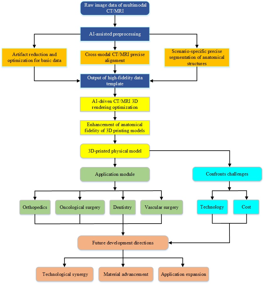
The integration of artificial intelligence (AI)-enabled multimodal computed tomography (CT)/magnetic resonance imaging (MRI) medical imaging and three-dimensional (3D) printing technology represents a pivotal direction in medical engineering for advancing precision diagnosis and therapy. Multimodal data fusion serves as the primary strategy to enhance the accuracy of 3D-printed models; however, cross-modal data fusion is hindered by inherent technical challenges, including failures in feature alignment and discrepancies in the physical properties of imaging datasets. In recent years, the advancement and seamless integration of AI technology have emerged as the core link bridging the entire workflow, from multimodal CT/MRI imaging acquisition to 3D printing, offering novel paradigms for the development of high-precision 3D printing technology in clinical settings. This review systematically elaborates on AI’s core technical underpinnings for multimodal imaging and 3D printing: AI effectively mitigates integration and adaptation hurdles arising from intrinsic discrepancies in data source characteristics through three key pathways—artifact reduction and optimization of raw imaging data, precise cross-modal registration, and fine-grained segmentation of anatomical structures. Furthermore, AI-driven optimization of 3D rendering effects, combined with four-view projection, significantly enhances the fidelity of anatomical detail reproduction, thereby minimizing the matching error between 3D-printed models and in vivo physical entities. Subsequently, the review details the clinical application value of multimodal 3D printing technology across key medical specialties, including orthopedics, oncological surgery, dentistry, and vascular surgery, while concomitantly highlighting prevailing challenges in technical translation and clinical adoption. Finally, it outlines future development directions from three critical dimensions: technological synergy (among AI, imaging, and 3D printing), material advancement (targeting durability and functional adaptability), and application expansion (to underserved clinical scenarios such as rehabilitation). This work aims to provide a comprehensive reference for accelerating the clinical translation of this interdisciplinary technology.
- Farid AR, Comtesse S, Sagi HC, et al. Enabling technology in fracture surgery: state of the art. J Bone Joint Surg. 2025;107(14):1636-1647. doi:10.2106/JBJS.24.00938
- Streckenbach A, Schubert N, Streckenbach F, et al. Current state and outlook in medical 3D printing and the role of radiology. RofoFortschrRontg. 2025;197(7):770-780. doi:10.1055/a2436-7185
- Santiago FR, Ramos-Bossini AJL, Wáng YXJ, Barbero JPM, Espinosa JG, Martínez AM. The value of magnetic resonance imaging and computed tomography in the study of spinal disorders. Quant Imaging Med Surg. 2022;12(7):3947. doi:10.21037/qims-2022-04
- Cejvanovic S, Sheikh Z, Hamann S, Subramanian PS. Imaging the brain: diagnosis aided by structural features on neuroimaging studies. Eye. 2024;38(12):2380-2391. doi:10.1038/s41433-024-03142-w
- Martucci M, Russo R, Schimperna F, et al. Magnetic resonance imaging of primary adult brain tumors: state of the art and future perspectives. Biomedicines. 2023;11(2):364. doi:10.3390/biomedicines11020364
- Schultz CH, Fairley R, Murphy LSL, Doss M. The risk of cancer from CT scans and other sources of low-dose radiation: a critical appraisal of methodologic quality. Prehosp Disaster Med. 2020;35(1):3-16. doi:10.1017/S1049023X1900520X
- Lyu P, Li Z, Chen Y, et al. Deep learning reconstruction CT for liver metastases: low-dose dual-energy vs standard-dose single-energy. Eur Radiol. 2024;34(1):28-38. doi:10.1007/s00330-023-10033-3
- Usman M, Latif S, Asim M, Lee BD, Qadir J. Retrospective motion correction in multishot MRI using generative adversarial network. Sci Rep. 2020;10(1):4786. doi:10.1038/s41598-020-61705-9
- Wu L, Gao C, Wu T, et al. Magnetic resonance imaging in the clinical evaluation of lung disorders: current status and future prospects. J Magn Reson Imaging. 2025;62(5): 1260-1279. doi:10.1002/jmri.29802
- Kuiper RJA, Colaris JW, Stockmans F, et al. Impact of bone and cartilage segmentation from CT and MRI on both bone forearm osteotomy planning. Int J Comput Assist Radiol Surgery. 2023;18(12):2307-2318. doi:10.1007/s11548-023-02929-8
- Jacobson N, Carerra E, Smith L, et al. Defining soft tissue: bitmap printing of soft tissue for surgical planning. 3D Print Addit Manuf. 2022;9(6):461-472. doi:10.1089/3dp.2021.0141
- Marro A, Bandukwala T, Mak W, Three-dimensional printing and medical imaging: a review of the methods and applications. Curr Probl Diagn Radiol. 2016;45(1):2-9. doi: 10.1067/j.cpradiol.2015.07.009
- Ng WL, An J, Chua CK. Process, material, and regulatory considerations for 3D printed medical devices and tissue constructs. Engineering. 2024;36(1):146-166. doi:10.1016/j.eng.2024.01.028
- Illi J, Bernhard B, Nguyen C, et al. Translating imaging into 3D printed cardiovascular phantoms: a systematic review of applications, technologies, and validation. JACC Basic Transl Sci. 2022;7(10):1050-1062. doi:10.1016/j.jacbts.2022.01.002
- Qin Y, Xu Z, Wang X, Skare M. Artificial intelligence and economic development: an evolutionary investigation and systematic review. J Knowl Econ. 2024;15:1736-1770. doi:10.1007/s13132-023-01183-2
- Mendoza-Cerezo L, Jesús MRR, Macías-García A, Marcos- Romero AC, Díaz-Parralejo A. Evolution of bioprinting and current applications. Int J Bioprint. 2023;9(4):367-382. doi:10.18063/ijb.742
- Tappa K, Bird JE, Arribas EM, Santiago L. Multimodality imaging for 3D printing and surgical rehearsal in complex spine surgery. RadioGraphics. 2024;44(3):e230116. doi:10.1148/rg.230116
- Albertini JN, Derycke L, Millon A, Soler R. Digital twin and artificial intelligence technologies for predictive planning of endovascular procedures. Semin Vasc Surg. 2024;37(3):306-313. doi:10.1053/j.semvascsurg.2024.07.002
- Dai Y, Wang P, Mishra A, et al. 3D bioprinting and artificial intelligence‐assisted biofabrication of personalized oral soft tissue constructs. Adv Healthc Mater. 2025;14(13):2402727. doi:10.1002/adhm.202402727
- Jeon K, Park WY, Kahn CE Jr, Nagy P, You SC, Yoon SH. Advancing medical imaging research through standardization: The path to rapid development, rigorous validation, and robust reproducibility. Invest Radiol. 2025;60(1):1-10. doi:10.1097/RLI.0000000000001106
- Huang H, Liu B, Xu Y, Zhou W. Synthetic-to-real domain adaptation with deep learning for fitting the intravoxel incoherent motion model of diffusion‐weighted imaging. Med Phys. 2023;50(3):1614-1622. doi:10.1002/mp.16031
- Chau RCW, Hsung RTC, McGrath C, Pow EHN, Lam WYH. Accuracy of artificial intelligence-designed single-molar dental prostheses: a feasibility study. J Prosthet Dent. 2024;131(6):1111-1117. doi:10.1016/j.prosdent.2022.12.004
- Chen Z, Pawar K, Ekanayake M, Pain C, Zhong S, Egan GF. Deep learning for image enhancement and correction in magnetic resonance imaging—state-of-the-art and challenges. J Digit Imaging. 2023;36(1):204-230. doi:10.1007/s10278-022-00721-9
- Yu H, Dong X, Li H, et al. Application of artificial intelligence deep learning-based multimodal CT-MRI images automatic fusion and segmentation in preoperative planning for anterior cruciate ligament reconstruction. Chin J Bone Joint Surg. 2025;18(1):27-35. doi:10.3969/j.issn.2095-9958.2025.01.05
- Wan Z, Li H, Shi N, Liu Y, Liu F. Intelligent evaluation of tumor calcification areas based on whole slide images. Laser Optoelectron Prog. 2024;61(22):2217001. doi:10.3788/LOP240787
- Agarwal M, Saba L, Gupta SK, et al. Wilson disease tissue classification and characterization using seven artificial intelligence models embedded with 3D optimization paradigm on a weak training brain magnetic resonance imaging datasets: a supercomputer application. Med Biol Eng Comput. 2021;59(3):511-533. doi:10.1007/s11517-021-02322-0
- Jia G, Huang X, Tao S, et al. Artificial intelligence-based medical image segmentation for 3D printing and naked eye 3D visualization. Intell Med. 2022;2(1):48-53. doi:10.1016/j.imed.2021.04.001
- Liang Y, Wang Q, Zhang YW, et al. A pilot study on clinical application of three-dimensional morphological completion of lesioned mandibles assisted by generative adversarial networks. Chin J Stomatol. 2024;59(12):1213-1220. doi:10.3760/cma.j.cn112144-20240930-00367.
- Wenran HU, Rong FU. Trans-YOLOv5: a YOLOv5-based prior transformer network model for automated detection of abnormal cells or clumps in cervical cytology images. J South Med Univ. 2024;44(7):1217-1226. doi:10.12122/j.issn.1673-4254.2024.07.01
- Xiaoyun Z, Shuxiong B, Shuai H. CT multiple-level reconstruction for preoperational estimation of facet joint violation in lumbar percutaneous pedicle screw placement. Chin J Tissue Eng Res. 2020;24(21):3347-3352. doi:10.3969/j.issn.2095-4344.2683
- Bhardwaj N, Sood M, Gill SS. 3D-bioprinting and AI-empowered anatomical structure designing: a review. Curr Med Imaging. 2024;20(1):e15734056259274. doi:10.2174/0115734056259274231019061329
- Luo M, Zhou N, Wang T, et al. Bi-constraints diffusion: a conditional diffusion model with degradation guidance for metal artifact reduction. IEEE Trans Med Imaging. 2025;44(9):3552-3562. doi:10.1109/TMI.2024.3442950
- Liu Z, Zhu Z, Zheng S, Liu Y, Zhou J, Zhao Y. Margin preserving self-paced contrastive learning towards domain adaptation for medical image segmentation. IEEE J Biomed Health Inform. 2022;26(2):638-647. doi:10.1109/JBHI.2022.3140853
- Zhou Y, Yan Z, Hubert P. An artifactual fibre overlap removal algorithm for micro-computed tomography image post-processing and 3D microstructure generation with graphics processing unit acceleration. Mater Des. 2024;247: 113376. doi:10.1016/j.matdes.2024.113376
- Ogbuanya CE, Obayi A, Larabi-Marie-Sainte S, Saad AO, Berriche L. A hybrid optimization approach for accelerated multimodal medical image fusion. PLoS One. 2025;20(7):e0324973. doi:10.1371/journal.pone.0324973
- Huang H, Zheng H, Lin L, et al. Medical image segmentation with deep atlas prior. IEEE Trans Med Imaging. 2021;40(12):3519-3530. doi:10.1109/TMI.2021.3089661
- Zhang J, Zhang N, Li Y, Zhou J, Cao D, He K. Research progress of 3D printing-based construction of experimental physical models for medical imaging devices. Chin Med Equip J. 2022;43(8):1-6. doi:10.19745/j.1003-8868.2022173
- Yang H, Yang R, Zhu H, et al. Design of multi-modal vertebral phantom based on tissue equivalence and 3D printing technology. Chin J Med Phys. 2016;33(9):6. doi:10.3969/j.issn.1005-202X.2016.09.011
- Yusuff H, Zorn PE, Giraudeau C, et al. Development of a cost-effective 3D-printed MRI phantom to enhance teaching of system performance and image quality concepts. Magn Reson Mater Phys Biol Med. 2025;38(3):561-574. doi:10.1007/s10334-024-01217-z
- Hindi OA, Pinarbasi B, Bakici M, et al. In situ bioprinting enhances bone regeneration in a live animal model of craniofacial defects. ACS Biomater Sci & Eng. 2025;11(12):5027-5037. doi:10.1021/acsbiomaterials.5c00780
- Lee D, Ruf M, Karadimitriou N, et al. Development of stochastically reconstructed 3D porous media micromodels using additive manufacturing: numerical and experimental validation. Sci Rep. 2024;14(1):9375. doi:10.1038/s41598-024-60075-w
- Zhang Z, Chen L, Sheng W, et al. Application of artificial intelligence in the diagnosis and treatment of lumbar disc herniation: evolution towards standardization, efficiency, and precision of diagnosis and treatment methods. Chin J Tissue Eng Res. 2025;29(29):6269-6276. doi:10.12307/2025.759
- Wang L, Wang H, Jiang Y, et al. The efficacy and dosimetry analysis of CT-guided 125I seed implantation assisted with 3D-printing non-co-planar template in locally recurrent rectal cancer. Radiat Oncol. 2020;15(1):179-183. doi:10.1186/s13014-020-01607-2
- Gomez-Feria J, Narros J L, Go´ mez Ciriza G, et al. 3D printing of diffuse low-grade gliomas involving eloquent cortical areas and subcortical functional pathways: technical note. World Neurosurg. 2021;147:164-171. doi:10.1016/j.wneu.2020.12.082
- Mangano FG, Yang KR, Lerner H, Admakin O, Mangano C. Artificial intelligence and mixed reality for dental implant planning: a technical note. Clin Implant Dent Relat Res. 2024;26(5):942-953. doi:10.1111/cid.13357
- Winkel DJ, Mujagic E, Staub D, Harder D, Bremerich J, Obmann MM. Multimodal imaging and 3D printing of a thoracoabdominal aortic aneurysm eroding the spine. Radiol Case Rep. 2023;18(2):657-660. doi:10.1016/j.radcr.2022.11.020.
- Minkenberg JG, Bender L, Franz C, et al. Accurate full-scale patient-specific Circle of Willis models including aneurysms: a novel manufacturing approach. PLoS One. 2025;20(7):e0328300. doi:10.1371/journal.pone.0328300
- Holmes T W, Yin Z, Bujila R, et al. Ultrahigh-resolution K-edge imaging of coronary arteries with prototype deep-silicon photon-counting CT: initial results in phantoms. Radiology. 2024;311(3):e231598. doi:10.1148/radiol.231598.
- Sato K, Yamashiro A, Koyama T. Material investigation for the development of non-rigid phantoms for CT-MRI image registration. Nihon Hoshasen Gijutsu Gakkai Zasshi. 2022;78(6):615-624. doi:10.6009/jjrt.2022-1241
- Wang X, Zhao F, Zhen P. Physicochemical properties of 3D printed bioactive glass/hydroxyapatite bone repair materials. J Ningxia Med Univ. 2022;44(5):511-516. doi:10.16050/j.cnki.issn1674-6309.2022.05.013
- Lichtenstein J, Heinzel S, Kurz B, et al. Are we ready for virtual planning and 3D-printed titanium plates in zygomatic fractures? A feasibility study based on 16 post-mortem fractures. J Craniomaxillofac Surg. 2025;53(3):325-331. doi:10.1016/j.jcms.2024.11.004
- Jin Z, Zhang Z, Shao X, Gu GX. Monitoring anomalies in 3D bioprinting with deep neural networks. ACS Biomater Sci Eng. 2023;9(7):3945-3952. doi:10.1021/acsbiomaterials.0c01761
- Zang XL, Sun J, Li YL, et al. 3D-bioprinting manufacturing polylactic-co-glycolic acid/nano-hydroxyapatite scaffold/ bone morphogenetic protein-2 sustained release composite. Chin J Tissue Eng Res. 2016;20(16):2405-2411. doi:10.3969/j.issn.2095-4344.2016.16.017
- Wu BY, Ye K, Chen JH, et al. Biocompatibility of 3D printed polyetheretherketone/hydroxyapatite composites. Chin J Tissue Eng Res. 2023;27(12):1932-1937. doi:10.12307/2023.079
- Li P, Cheng W, Wang JQ, et al. Osteogenesis effect of bone marrow mesenchymal stem cells combined with bionic bone scaffold. Henan Med Res. 2022;31(7):1153-1158. doi:10.3969/j.issn.1004-437X.2022.07.001
- Hu CR, Qiu B, Zhou ZX, Yang Y, Li J. In vitro biocompatibility of 3D printed polycaprolactone/nano-hydroxyapatite composite scaffold with bone marrow mesenchymal stem cells. Chin J Tissue Eng Res. 2020;24(4):589-595. doi:10.3969/j.issn.2095-4344.1880
- Liu D, Qin H, Wang YX, et al. 3D-printed hydroxyapatite/ polylactic acid network composites for skull defects. Chin J Tissue Eng Res. 2019;23(6):833-837. doi:10.3969/j.issn.2095-4344.0676
- Silva G, Ashford R. Using artificial intelligence to predict outcomes of operatively managed neck of femur fractures. Br J Hosp Med. 2024;85(6):1-12. doi:10.12968/hmed.2024.0034
- Fogarasi M, Coburn J C, Ripley B. Algorithms used in medical image segmentation for 3D printing and how to understand and quantify their performance. 3D Print Med. 2022;8(1):18. doi:10.1186/s41205-022-00145-9
- Yang B, Gong K, Liu H, Li Q, Zhu W. Anatomically guided pet image reconstruction using conditional weakly-supervised multi-task learning integrating self-attention. IEEE Trans Med Imaging. 2024;43(6):2098-2112. doi:10.1109/TMI.2024.3356189