

Dual-nozzle 3D-printed calcium sulfate/ polylactic acid scaffold incorporating linezolid microspheres for effective repair of femoral condyle defects in rats

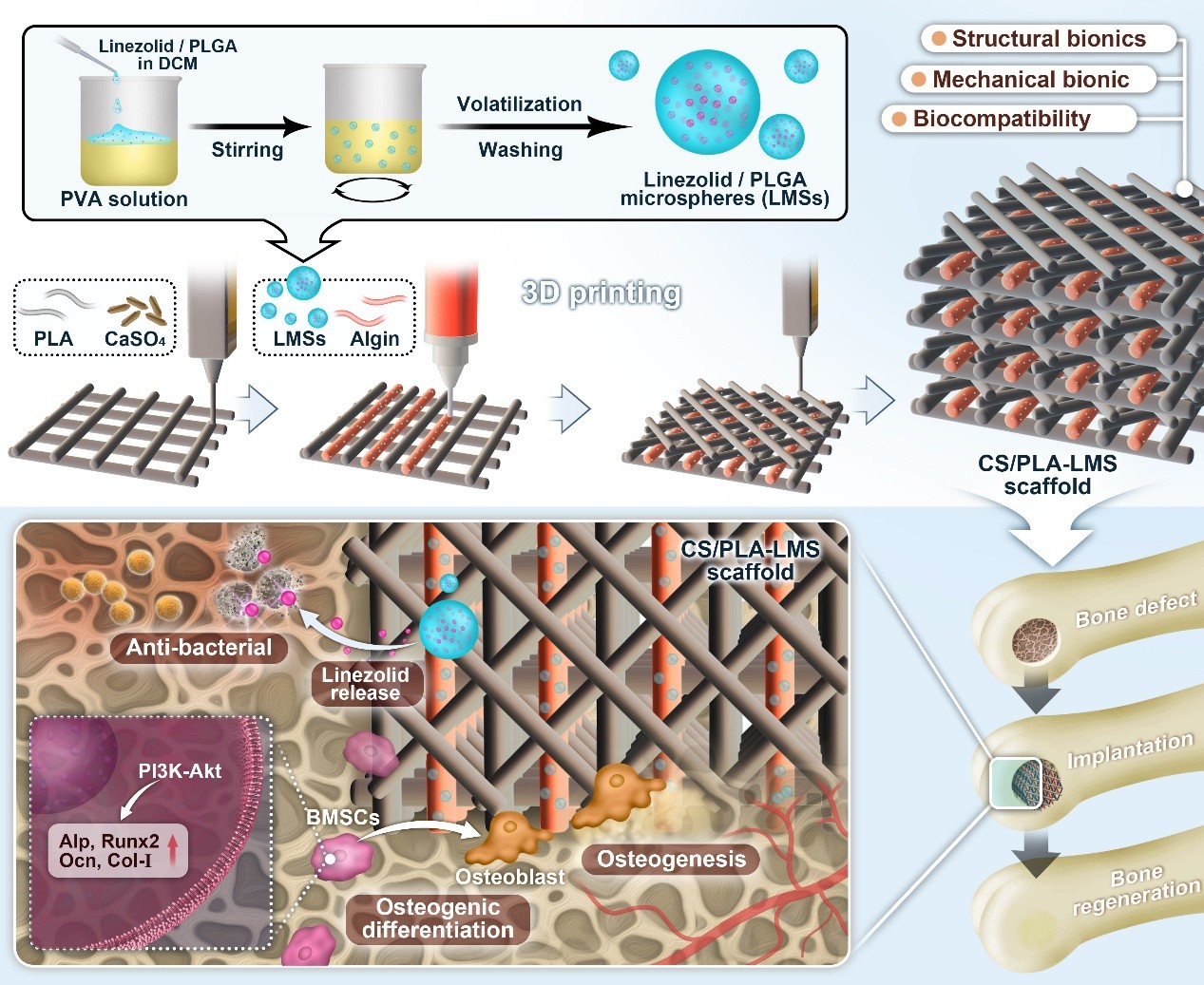
Large, complex bone defects pose a significant clinical challenge. Conventional bone grafting approaches cannot simultaneously achieve tissue regeneration and infection prevention, resulting in impaired healing outcomes and prolonged treatment cycles. Existing therapeutic strategies lack integrated solutions capable of concurrently providing infection prevention and osteogenesis promotion within a single platform. This study developed a novel multifunctional composite scaffold using dual-nozzle three-dimensional printing technology to simultaneously achieve infection prevention and accelerated bone regeneration. Linezolid-loaded polylactic-co-glycolic acid microspheres (LMS) were uniformly dispersed within the pores of calcium sulfate/polylactic acid (CS/PLA) scaffolds to successfully construct the composite scaffold. In vitro characterization revealed uniform distribution of microspheres within the scaffold pores, with the fabricated CS/PLA-LMS demonstrating excellent biocompatibility and mechanical properties, achieving an elastic modulus of 87 MPa. Furthermore, the composite scaffold effectively inhibited Staphylococcus aureus activity in vitro. In vivo rat femoral condyle defect model revealed that the composite scaffold significantly enhanced bone formation compared to blank controls. Additionally, bone volume fraction increased by 3.2 times, and trabecular spacing decreased by 50%, with mechanistic analysis indicating activation of the phosphoinositide 3-kinase-protein kinase B signaling pathway. The integrated design successfully prevented infection-related complications while promoting robust osteogenesis, offering a clinically relevant solution for treating complex bone defects where infection prevention and regenerative capacity are primary therapeutic concerns.
- Jihan M, Qi B, Bayaniahangar R, et al. Nanomaterials for bone tissue regeneration: updates and future perspectives. Nanomedicine (Lond). 2019;14(22):2987-3006. doi: 10.2217/nnm-2018-0445
- Spicer PP, Kretlow JD, Young S, Jansen JA, Kasper FK, Mikos AG. Evaluation of bone regeneration using the rat critical size calvarial defect. Nat Protoc. 2012;7(10): 1918-1929. doi: 10.1038/nprot.2012.113
- Gugala Z, Gogolewski S. Regeneration of segmental diaphyseal defects in sheep tibiae using resorbable polymeric membranes: a preliminary study. J Orthop Trauma. 1999;13(3):187-195. doi: 10.1097/00005131-199903000-00006
- Dimitriou R, Jones E, McGonagle D, Giannoudis PV. Bone regeneration: current concepts and future directions. BMC Med. 2011;9:66. doi: 10.1186/1741-7015-9-66
- Campana V, Milano G, Pagano E, et al. Bone substitutes in orthopaedic surgery: from basic science to clinical practice. J Mater Sci Mater Med. 2014;25(10):2445-2461. doi: 10.1007/s10856-014-5240-2
- Roberts TT, Rosenbaum AJ. Bone grafts, bone substitutes and orthobiologics: the bridge between basic science and clinical advancements in fracture healing. Organogenesis. 2012;8(4):114-124. doi: 10.4161/org.23306
- Nazeer MA, Onder OC, Sevgili I, Yilgor E, Kavakli IH, Yilgor I. 3D printed poly (lactic acid) scaffolds modified with chitosan and hydroxyapatite for bone repair applications. Mater Today Commun. 2020;25:101515. doi: 10.1016/j.mtcomm.2020.101515
- Khan SN, Cammisa FP Jr, Sandhu HS, Diwan AD, Girardi FP, Lane JM. The biology of bone grafting. J Am Acad Orthop Surg. 2005;13(1):77-86. doi: 10.5435/00124635-200501000-00009
- Chiarello E, Cadossi M, Tedesco G, et al. Autograft, allograft and bone substitutes in reconstructive orthopedic surgery. Aging Clin Exp Res. 2013;25(Suppl 1):S101-S103. doi: 10.1007/s40520-013-0088-8
- Stevenson S, Horowitz M. The response to bone allografts. J Bone Joint Surg Am. 1992;74(6):939-950. doi: 10.2106/00004623-199274060-00009
- Stevenson S. The immune response to osteochondral allografts in dogs. J Bone Joint Surg Am. 1987;69(4):573-582.
- Stevenson S, Li XQ, Martin B. The fate of cancellous and cortical bone after transplantation of fresh and frozen tissue-antigen-matched and mismatched osteochondral allografts in dogs. J Bone Joint Surg Am. 1991;73(8):1143-1156.
- Kavanagh N, Ryan EJ, Widaa A, et al. Staphylococcal osteomyelitis: disease progression, treatment challenges, and future directions. Clin Microbiol Rev. 2018;31(2): e00084-17. doi: 10.1128/CMR.00084-17
- Yıldırım A, Kapukaya A, Mertsoy Y, Yiğit Ş, Çaçan MA, Atiç R. Management of open fractures using a noncontact locking plate as an internal fixator. Indian J Orthop. 2017;51(3):312-317. doi: 10.4103/0019-5413.205686
- Masters EA, Ricciardi BF, Bentley KLdM, Moriarty TF, Schwarz EM, Muthukrishnan G. Skeletal infections: microbial pathogenesis, immunity and clinical management. Nat Rev Microbiol. 2022;20(7):385-400. doi: 10.1038/s41579-022-00686-0
- Vallet-Regí M, Lozano D, González B, Izquierdo-Barba I. Biomaterials against bone infection. Adv Healthc Mater. 2020;9(6):e1901647. doi: 10.1002/adhm.201901647
- Prince GE, Yang X, Fu J, et al. Yolk-porous shell biphasic bioceramic granules enhancing bone regeneration and repair beyond homogenous hybrid. Mater Sci Eng C Mater Biol Appl. 2019;100:433-444. doi: 10.1016/j.msec.2019.03.026
- Yang Y, Chu L, Yang S, et al. Dual-functional 3D-printed composite scaffold for inhibiting bacterial infection and promoting bone regeneration in infected bone defect models. Acta Biomater. 2018;79:265-275. doi: 10.1016/j.actbio.2018.08.015
- Zhang L, Yang G, Johnson BN, Jia X. Three-dimensional (3D) printed scaffold and material selection for bone repair. Acta Biomater. 2019;84:16-33. doi: 10.1016/j.actbio.2018.11.039
- Liu W, Li J, Cheng M, et al. A surface-engineered polyetheretherketone biomaterial implant with direct and immunoregulatory antibacterial activity against methicillin-resistant Staphylococcus aureus. Biomaterials. 2019;208:8-20. doi: 10.1016/j.biomaterials.2019.04.008
- El-Rashidy AA, Roether JA, Harhaus L, Kneser U, Boccaccini AR. Regenerating bone with bioactive glass scaffolds: a review of in vivo studies in bone defect models. Acta Biomater. 2017;62:1-28. doi: 10.1016/j.actbio.2017.08.030
- Lopes MS, Jardini A, Maciel Filho R. Poly (lactic acid) production for tissue engineering applications. Procedia Eng. 2012;42:1402-1413.
- Maadani AM, Salahinejad E. Performance comparison of PLA- and PLGA-coated porous bioceramic scaffolds: mechanical, biodegradability, bioactivity, delivery and biocompatibility assessments. J Control Release. 2022;351:1-7. doi: 10.1016/j.jconrel.2022.09.022
- Ma B, Han J, Zhang S, et al. Hydroxyapatite nanobelt/ polylactic acid Janus membrane with osteoinduction/barrier dual functions for precise bone defect repair. Acta Biomater. 2018;71:108-117. doi: 10.1016/j.actbio.2018.02.033
- Tyler B, Gullotti D, Mangraviti A, Utsuki T, Brem H. Polylactic acid (PLA) controlled delivery carriers for biomedical applications. Adv Drug Deliv Rev. 2016;107:163-175. doi: 10.1016/j.addr.2016.06.018
- Zwingenberger S, Nich C, Valladares RD, Yao Z, Stiehler M, Goodman SB. Recommendations and considerations for the use of biologics in orthopedic surgery. BioDrugs. 2012;26(4):245-256. doi: 10.2165/11631680-000000000-00000
- Kumar RA, Sivashanmugam A, Deepthi S, Bumgardner JD, Nair SV, Jayakumar R. Nano-fibrin stabilized CaSO4 crystals incorporated injectable chitin composite hydrogel for enhanced angiogenesis & osteogenesis. Carbohydrate Polym. 2016;140:144-153. doi: 10.1016/j.carbpol.2015.12.077
- Zhou Z, Buchanan F, Mitchell C, Dunne N. Printability of calcium phosphate: calcium sulfate powders for the application of tissue engineered bone scaffolds using the 3D printing technique. Mater Sci Eng C Mater Biol Appl. 2014;38:1-10. doi: 10.1016/j.msec.2014.01.033
- Aquino-Martínez R, Angelo AP, Pujol FV. Calcium-containing scaffolds induce bone regeneration by regulating mesenchymal stem cell differentiation and migration. Stem Cell Res Ther. 2017;8(1):265. doi: 10.1186/s13287-017-0713-0
- Mitragotri S, Burke PA, Langer R. Overcoming the challenges in administering biopharmaceuticals: formulation and delivery strategies. Nat Rev Drug Discov. 2014;13(9):655-672. doi: 10.1038/nrd4363
- Wright JC, Burgess DJ. Long Acting Injections and Implants. Springer Science & Business Media; 2011.
- Jain R, Shah NH, Malick AW, Rhodes CT. Controlled drug delivery by biodegradable poly (ester) devices: different preparative approaches. Drug Dev Ind Pharm. 1998;24(8):703-727. doi: 10.3109/03639049809047401
- Li J, Li K, Du Y, et al. Dual-nozzle 3D printed nano-hydroxyapatite scaffold loaded with vancomycin sustained-release microspheres for enhancing bone regeneration. Int J Nanomed. 2023;18:307-322. doi: 10.2147/ijn.S394366
- Wang L, Wang J, Zhou X, et al. A new self-healing hydrogel containing hucMSC-derived exosomes promotes bone regeneration. Front Bioeng Biotechnol. 2020;8:564731. doi: 10.3389/fbioe.2020.564731
- Wang L, Wei X, He X, et al. Osteoinductive dental pulp stem cell-derived extracellular vesicle-loaded multifunctional hydrogel for bone regeneration. ACS Nano. 2024;18(12):8777-8797. doi: 10.1021/acsnano.3c11542
- Davidson BL, Hilfinger JM, Beer SJ. Extended release of adenovirus from polymer microspheres: potential use in gene therapy for brain tumors. Adv Drug Deliv Rev. 1997;27(1):59-66.
- Rocha CV, Gonçalves V, da Silva MC, Bañobre-López M, Gallo J. PLGA-based composites for various biomedical applications. Int J Mol Sci. 2022;23(4):2034. doi: 10.3390/ijms23042034
- Wang Y, Qin B, Xia G, Choi SH. FDA’s poly (lactic-co-glycolic acid) research program and regulatory outcomes. AAPS J. 2021;23(4):92. doi: 10.1208/s12248-021-00611-y
- Wan B, Bao Q, Burgess D. Long-acting PLGA microspheres: advances in excipient and product analysis toward improved product understanding. Adv Drug Deliv Rev. 2023;198:114857. doi: 10.1016/j.addr.2023.114857
- Yuan Z, Wei P, Huang Y, et al. Injectable PLGA microspheres with tunable magnesium ion release for promoting bone regeneration. Acta Biomater. 2019;85:294-309. doi: 10.1016/j.actbio.2018.12.017
- Hickey T, Kreutzer D, Burgess DJ, Moussy F. Dexamethasone/ PLGA microspheres for continuous delivery of an anti-inflammatory drug for implantable medical devices. Biomaterials. 2002;23(7):1649-1656. doi: 10.1016/s0142-9612(01)00291-5
- Huiwen W, Shuai L, Jia X, et al. 3D-printed nanohydroxyapatite/methylacrylylated silk fibroin scaffold for repairing rat skull defects. J Biol Eng. 2024;18(1):22. doi: 10.1186/s13036-024-00416-5
- Sitthisang S, Hou X, Treetong A, et al. Nanomechanical mapping of PLA hydroxyapatite composite scaffolds links surface homogeneity to stem cell differentiation. Sci Rep. 2024;14(1):21097. doi: 10.1038/s41598-024-72073-z
- Wang W, Liu P, Zhang B, et al. Fused deposition modeling printed PLA/Nano β-TCP composite bone tissue engineering scaffolds for promoting osteogenic induction function. Int J Nanomedicine. 2023;18:5815-5830. doi: 10.2147/ijn.S416098
- Senatov F, Zimina A, Chubrik A, et al. Effect of recombinant BMP-2 and erythropoietin on osteogenic properties of biomimetic PLA/PCL/HA and PHB/HA scaffolds in critical-size cranial defects model. Mater Sci Eng C Mater Biol Appl. 2022;135:112680. doi: 10.1016/j.msec.2022.112680
- Gao S, Li J, Lei Q, et al. Calcium sulfate-Cu(2+) delivery system improves 3D-printed calcium silicate artificial bone to repair large bone defects. Front Bioeng Biotechnol. 2023;11:1224557. doi: 10.3389/fbioe.2023.1224557
- Liu S, Fu H, Lv Y, et al. α-Hemihydrate calcium sulfate/n-hydroxyapatite combined with metformin promotes osteogenesis in vitro and in vivo. Front Bioeng Biotechnol. 2022;10:899157. doi: 10.3389/fbioe.2022.899157
- Macedo J, Marques R, Vervaet C, Pinto JF. Production of bi-compartmental tablets by FDM 3D printing for the withdrawal of diazepam. Pharmaceutics. 2023;15(2):538. doi: 10.3390/pharmaceutics15020538
- Morata L, Cuesta M, Rojas JF, et al. Risk factors for a low linezolid trough plasma concentration in acute infections. Antimicrob Agents Chemother. 2013;57(4):1913-1917. doi: 10.1128/aac.01694-12
- Mandal S, Viraj, Nandi SK, Roy M. Effects of multiscale porosity and pore interconnectivity on in vitro and in vivo degradation and biocompatibility of Fe-Mn-Cu scaffolds. J Mater Chem B. 2021;9(21):4340-4354. doi: 10.1039/d1tb00641j
- Qin Y, Jing Z, Zou D, et al. A metamaterial scaffold beyond modulus limits: enhanced osteogenesis and angiogenesis of critical bone defects. Nat Commun. 2025;16(1):2180. doi: 10.1038/s41467-025-57609-9
- Wang Q, Chen Y, Ding H, et al. Optogenetic activation of mechanical nociceptions to enhance implant osseointegration. Nat Commun. 2025;16(1):3093. doi: 10.1038/s41467-025-58336-x
- Zhao SJ, Kong FQ, Jie J, et al. Macrophage MSR1 promotes BMSC osteogenic differentiation and M2-like polarization by activating PI3K/AKT/GSK3β/β-catenin pathway. Theranostics. 2020;10(1):17-35. doi: 10.7150/thno.36930