

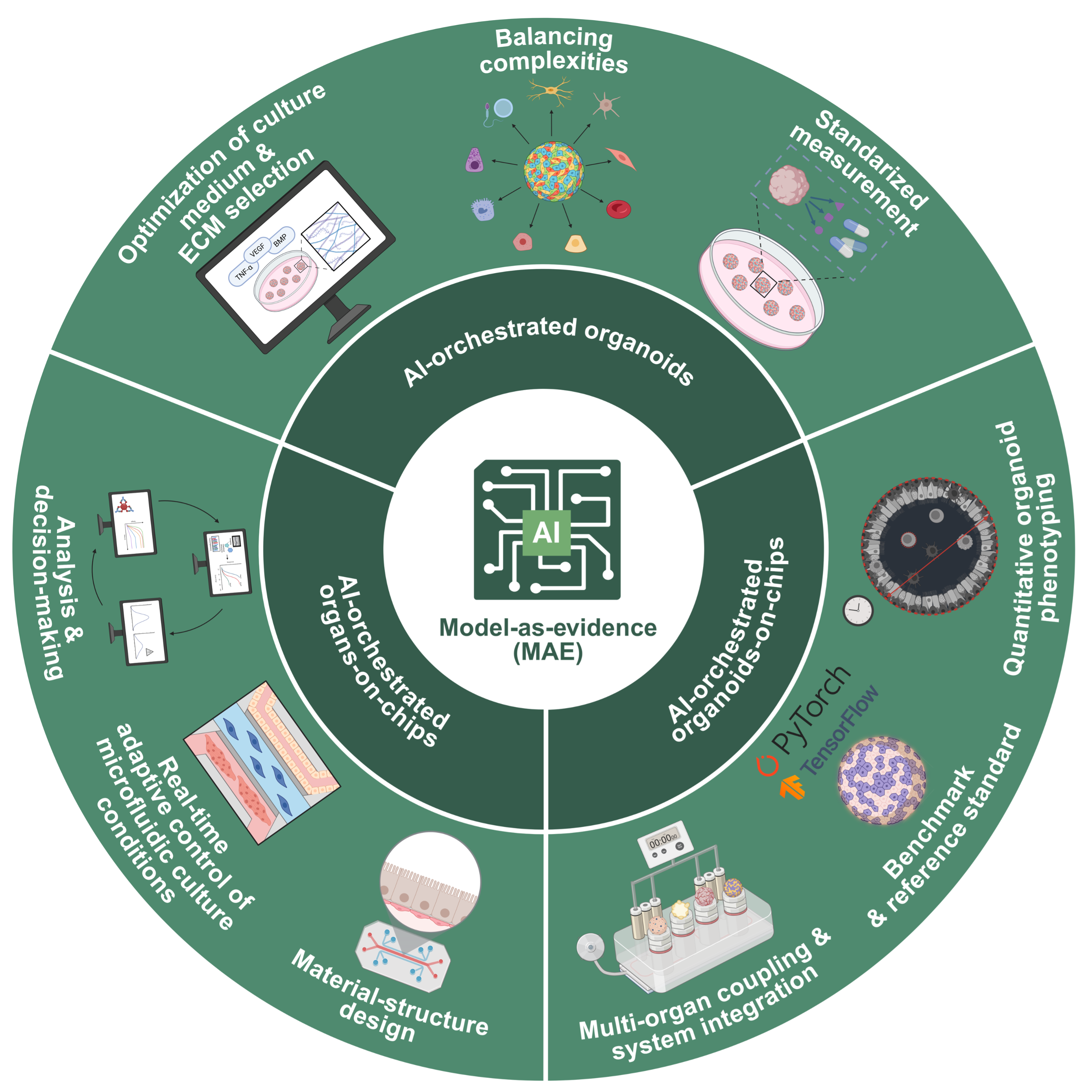
Model-as-evidence framework established using AI-integrated organoids and organs-on-chips systems

The predictive capability crisis in drug discovery and development remains a significant factor hindering medical progress. Among emerging solutions, organoids offer the ability to reflect the biological fidelity of tissues, whereas organs-on-chips (OoCs) demonstrate strengths in simulating dynamic tissue microenvironments and enabling multi‑organ interactions. Herein, we present a unifying artificial intelligence (AI)-orchestrated framework that incorporates both platforms into verifiable and regulatable “model-as-evidence” (MAE) pathways capable of self-optimization with researcher oversight. AI is employed as an indispensable computational integration layer addressing three bottlenecks that render conventional approaches infeasible: (i) combinatorial explosion in high-dimensional parameter spaces, (ii) multi-scale coupling between molecular interactions and tissue-scale level morphogenesis, and (iii) requirement for real-time adaptive control of dynamic microenvironments exceeding human cognitive bandwidth. At the organoid level, this integration enables multi‑objective optimization to improve reproducibility, reduce resource use, and achieve predictive modeling to capture extracellular matrix–cells interactions. For OoCs, active learning algorithms condense protracted design cycles into efficient, goal-directed experimentation, and deep reinforcement learning sustains physiological steady states through adaptive, real-time control. The convergence of these systems within an integrated platform enables standardized, cross-comparable phenotyping pipelines that transform heterogeneous experimental data into a universal quantitative language, and harmonized evaluation benchmarks align experimental outputs across laboratories. Together, this framework generates curated evidence packages that directly address regulatory concerns related to efficacy and safety. By systematizing experimental design, parameter control, and interpretive analytics, this AI-orchestrated approach elevates organoid and OoC technologies from specialized crafts to a robust engineering discipline. It establishes a virtuous cycle wherein AI accelerates iterative validation and optimization, freeing researchers to focus on higher-order hypothesis generation and translational strategy. Thus, through AI-orchestrated quantification of biological fidelity and engineering rigor, these platforms may mature into credible, scalable, and transformative toolboxes for next-generation biomedical translation.
- Parrish MC, Tan YJ, Grimes KV, Mochly-Rosen D. Surviving in the valley of death: opportunities and challenges in translating academic drug discoveries. Annu Rev Pharmacol. Toxicol. 2019;59(1):405-421. doi: 10.1146/annurev-pharmtox-010818-021625
- Begley CG, Ellis LM. Drug development: Raise standards for preclinical cancer research. Nature. 2012;483(7391):531-3. doi: 10.1038/483531a
- Clevers H. Modeling development and disease with organoids. Cell. 2016;165(7):1586-1597. doi: 10.1016/j.cell.2016.05.082
- Zhou L, Chen S, Liu J, et al. When artificial intelligence (AI) meets organoids and organs-on-chips (OoCs): Game-changer for drug discovery and development? Innov Life. 2025;3(1):100115. doi: 10.59717/j.xinn-life.2024.100115
- Sun D, Gao W, Hu H, Zhou S. Why 90% of clinical drug development fails and how to improve it? Acta Pharm Sin B. 2022;12(7):3049-3062. doi: 10.1016/j.apsb.2022.02.002
- Ioannidis JP. Why most published research findings are false. PLoS Med. 2005;2(8):e124. doi: 10.1371/journal.pmed.0020124
- Mullard A. Parsing clinical success rates. Nat Rev Drug Discov. 2016;15(7):447-448. doi: 10.1038/nrd.2016.136
- Prinz F, Schlange T, Asadullah K. Believe it or not: how much can we rely on published data on potential drug targets? Nat Rev Drug Discov. 2011;10(9):712-712. doi: 10.1038/nrd3439-c1
- Freedman LP, Cockburn IM, Simcoe TS. The economics of reproducibility in preclinical research. PLoS Biol. 2015;13(6):e1002165. doi: 10.1371/journal.pbio.1002165
- Baker M. 1,500 scientists lift the lid on reproducibility. Nature. 2016;533:452-454. doi: 10.1038/533452a
- Errington TM, Iorns E, Gunn W, Tan FE, Lomax J, Nosek BA. An open investigation of the reproducibility of cancer biology research. eLife. 2014;3:e04333. doi: 10.7554/eLife.04333
- Drost J, Clevers H. Organoids in cancer research. Nat Rev Cancer 2018;18(7):407-418. doi: 10.1038/s41568-018-0007-6
- Sato T, Vries RG, Snippert HJ, et al. Single Lgr5 stem cells build crypt-villus structures in vitro without a mesenchymal niche. Nature. 2009;459(7244):262-265. doi: 10.1038/nature07935
- Sato T, Stange DE, Ferrante M, et al. Long-term expansion of epithelial organoids from human colon, adenoma, adenocarcinoma, and Barrett’s epithelium. Gastroenterology. 2011;141(5):1762-1772. doi: 10.1053/j.gastro.2011.07.050
- Huh D, Matthews BD, Mammoto A, Montoya-Zavala M, Hsin HY, Ingber DE. Reconstituting organ-level lung functions on a chip. Science. 2010;328(5986):1662-1668. doi: 10.1126/science.1188302
- Bhatia SN, Ingber DE. Microfluidic organs-on-chips. Nat Biotechnol. 2014;32(8):760-72. doi: 10.1038/nbt.2989
- Ingber DE. Human organs-on-chips for disease modelling, drug development and personalized medicine. Nat Rev Genet. 2022;23(8):467-491. doi: 10.1038/s41576-022-00466-9
- Zhang B, Radisic M. Organ-on-a-chip devices advance to market. Lab Chip. 2017;17(14):2395-2420. doi: 10.1039/C6LC01554A
- Kandasamy K, Dasarathy G, Oliva J, Schneider J, Poczos B. Multi-fidelity gaussian process bandit optimisation. J Artif Intell Res. 2019;66:151-196. doi: 10.1613/jair.1.11288
- Shahriari B, Swersky K, Wang Z, Adams RP, De Freitas N. Taking the human out of the loop: A review of Bayesian optimization. Proc IEEE 2015;104(1):148-175. doi: 10.1109/JPROC.2015.2494218
- Engler AJ, Sen S, Sweeney HL, Discher DE. Matrix elasticity directs stem cell lineage specification. Cell. 2006;126(4):677- 689. doi: 10.1016/j.cell.2006.06.044
- Trappmann B, Gautrot JE, Connelly JT, et al. Extracellular-matrix tethering regulates stem-cell fate. Nat Mater. 2012;11(7):642-649. doi: 10.1038/nmat3339
- Quang Tran D, Bae S-H. Proximal Policy Optimization Through a Deep Reinforcement Learning Framework for Multiple Autonomous Vehicles at a Non-Signalized Intersection. Appl Sci. 2020;10(16):5722. doi: 10.3390/app10165722
- Schulman J, Wolski F, Dhariwal P, Radford A, Klimov O. Proximal policy optimization algorithms. arXiv. Preprint posted online 2017. doi: 10.48550/arXiv.1707.06347
- Greggio C, De Franceschi F, Figueiredo-Larsen M, et al. Artificial three-dimensional niches deconstruct pancreas development in vitro. Development. 2013;140(21):4452- 4462. doi: 10.1242/dev.096628
- Caliari SR, Burdick JA. A practical guide to hydrogels for cell culture. Nat Methods. 2016;13(5):405-14. doi: 10.1038/nmeth.3839
- Lundberg SM, Lee S-I. A unified approach to interpreting model predictions. Adv Neural Inf. Process. Syst. 2017;30. Accessed May 17, 2026. https://proceedings.neurips.cc/paper_files/paper/2017/file/8a20a8621978632d76c43dfd28b67767-Paper.pdf
- Driehuis E, Kolders S, Spelier S, et al. Oral mucosal organoids as a potential platform for personalized cancer therapy. Cancer Discov. 2019;9(7):852-871. doi: 10.1158/2159-8290.CD-18-1522
- Watson CL, Mahe MM, Múnera J, et al. An in vivo model of human small intestine using pluripotent stem cells. Nat Med. 2014;20(11):1310-1314. doi: 10.1038/nm.3737
- Camp JG, Badsha F, Florio M, et al. Human cerebral organoids recapitulate gene expression programs of fetal neocortex development. Proc Natl Acad Sci USA. 2015;112(51):15672-7. doi: 10.1073/pnas.1520760112
- Pollen AA, Bhaduri A, Andrews MG, et al. Establishing cerebral organoids as models of human-specific brain evolution. Cell. 2019;176(4):743-756. e17. doi: 10.1016/j.cell.2019.01.017
- Marks M, Israel U, Dilip R, et al. CellSAM: a foundation model for cell segmentation. Nat Methods. 2025. doi: 10.1038/s41592-025-02879-w
- Archit A, Freckmann L, Nair S, et al. Segment Anything for Microscopy. Nat Methods. 2025;22(3):579-591. doi: 10.1038/s41592-024-02580-4
- Ali M, Wu T, Hu H, et al. A review of the Segment Anything Model (SAM) for medical image analysis: Accomplishments and perspectives. Comput Med Imaging Graph. 2025;119:102473. doi: 10.1016/j.compmedimag.2024.102473
- Zhang Y, Shen Z, Jiao R. Segment anything model for medical image segmentation: Current applications and future directions. Comput Biol Med. 2024;171:108238. doi: 10.1016/j.compbiomed.2024.108238
- Zhang L, Deng X, Lu Y. Segment Anything Model (SAM) for Medical Image Segmentation: A Preliminary Review. IEEE BIBM 2023:4187-4194. doi: 10.1109/BIBM58861.2023.10386032
- Kundacina I, Kundacina O, Miskovic D, Radonic V. Advancing microfluidic design with machine learning: a Bayesian optimization approach. Lab Chip. 2025;25(4):657- 672. doi: 10.1039/D4LC00872C
- Miller JS, Stevens KR, Yang MT, et al. Rapid casting of patterned vascular networks for perfusable engineered three-dimensional tissues. Nat Mater. 2012;11(9):768-774. doi: 10.1038/nmat3357
- Fu C-Y, Tseng S-Y, Yang S-M, Hsu L, Liu C-H, Chang H-Y. A microfluidic chip with a U-shaped microstructure array for multicellular spheroid formation, culturing and analysis. Biofabrication. 2014;6(1):015009. doi: 10.1088/1758-5082/6/1/015009
- Bein A, Shin W, Jalili-Firoozinezhad S, et al. Microfluidic Organ-on-a-Chip Models of Human Intestine. Cell Mol Gastroenterol Hepatol. 2018;5(4):659-668. doi: 10.1016/j.jcmgh.2017.12.010
- Mosadegh B, Lockett MR, Minn KT, et al. A paper-based invasion assay: Assessing chemotaxis of cancer cells in gradients of oxygen. Biomaterials. 2015;52:262-271. doi: 10.1016/j.biomaterials.2015.02.012
- Kolesky DB, Truby RL, Gladman AS, Busbee TA, Homan KA, Lewis JA. 3D Bioprinting of Vascularized, Heterogeneous Cell-Laden Tissue Constructs. Adv Mater. 2014;26(19):3124-3130. doi: 10.1002/adma.201305506
- Gracioso Martins AM, Wilkins MD, Ligler FS, Daniele MA, Freytes DO. Microphysiological System for High- Throughput Computer Vision Measurement of Microtissue Contraction. ACS Sens. 2021;6(3):985-994. doi: 10.1021/acssensors.0c02172
- Wong CWT, Lee JZX, Jaeschke A, et al. Lung cancer intravasation-on-a-chip: Visualization and machine learning-assisted automatic quantification. Bioact Mater. 2025;51:858-875. doi: 10.1016/j.bioactmat.2025.06.028
- Shin W, Wu A, Massidda MW, et al. A Robust Longitudinal Co-culture of Obligate Anaerobic Gut Microbiome with Human Intestinal Epithelium in an Anoxic-Oxic Interface-on-a-Chip. Front Bioeng Biotechnol. 2019;7:13. doi: 10.3389/fbioe.2019.00013
- Montes-Olivas S, Legge D, Lund A, et al. In-silico and in-vitro morphometric analysis of intestinal organoids. PLoS Comput Biol. 2023;19(8):e1011386. doi: 10.1371/journal.pcbi.1011386
- Thompson J, Koe R, Le A, Goodman G, Brown DS, Kuntz A. Early Failure Detection in Autonomous Surgical Soft- Tissue Manipulation via Uncertainty Quantification. arXiv. Preprint posted online 2025. doi: 10.48550/arXiv.2501.10561
- van Leeuwen PJ, Chiu JC, Yang C-KK. Uncertainty quantification for deep learning. Environ Data Sci. 2024;4. doi: 10.1017/eds.2025.10027
- Zeevi T, Venkataraman R, Staib LH, Onofrey JA. Monte- Carlo Frequency Dropout for Predictive Uncertainty Estimation in Deep Learning. IEEE ISBI. 2024:1-5. doi: 10.1109/ISBI56570.2024.10635511
- Han DY. Artificial Intelligence in and Beyond Healthcare Psychology. J Clin Psychol Med Settings. 2025;32(4):600-607. doi: 10.1007/s10880-025-10101-4
- Chauhan SB, Gaur R, Akram A, Singh I. Artificial Intelligence Driven insights for Regulatory Intelligence in Medical Devices: Evaluating EMA, FDA and CDSCO Frameworks. Glob Clin Eng J. 2025;7:11-24. doi: 10.31354/globalce.v7i2.210
- Barker N, Huch M, Kujala P, et al. Lgr5(+ve) stem cells drive self-renewal in the stomach and build long-lived gastric units in vitro. Cell Stem Cell. 2010;6(1):25-36. doi: 10.1016/j.stem.2009.11.013
- Huch M, Bonfanti P, Boj SF, et al. Unlimited in vitro expansion of adult bi‐potent pancreas progenitors through the Lgr5/R‐spondin axis. EMBO J. 2013;32(20):2708-2721. doi: 10.1038/emboj.2013.204
- Conesa A, Madrigal P, Tarazona S, et al. A survey of best practices for RNA-seq data analysis. Genome Biol. 2016/01/26 2016;17(1):13.
- DOI:·10.1186/s13059-016-0881-8
- Sunyer R, Conte V, Escribano J, et al. Collective cell durotaxis emerges from long-range intercellular force transmission. Science. 2016;353(6304):1157-1161. doi: 10.1126/science.aaf7119
- Workman MJ, Mahe MM, Trisno S, et al. Engineered human pluripotent-stem-cell-derived intestinal tissues with a functional enteric nervous system. Nat Med. 2017;23(1):49- 59. doi: 10.1038/nm.4233
- Finkbeiner SR, Hill David R, Altheim Christopher H, et al. Transcriptome-wide Analysis Reveals Hallmarks of Human Intestine Development and Maturation In Vitro and In Vivo. Stem Cell Rep. 2015;4(6):1140-1155. doi: 10.1016/j.stemcr.2015.04.010
- Lindeboom RGH, van Voorthuijsen L, Oost KC, et al. Integrative multi‐omics analysis of intestinal organoid differentiation. Mol Syst Biol. 2018;14(6):MSB188227. doi: 10.15252/msb.20188227
- Sachs N, De Ligt J, Kopper O, et al. A living biobank of breast cancer organoids captures disease heterogeneity. Cell. 2018;172(1):373-386. e10. doi: 10.1016/j.cell.2017.11.010
- Vlachogiannis G, Hedayat S, Vatsiou A, et al. Patient-derived organoids model treatment response of metastatic gastrointestinal cancers. Science. 2018;359(6378):920-926. doi: 10.1126/science.aao2774
- Broutier L, Mastrogiovanni G, Verstegen MM, et al. Human primary liver cancer-derived organoid cultures for disease modeling and drug screening. Nat Med. 2017;23(12):1424- 1435. doi: 10.1038/nm.4438
- Cho J, Lee MJ, Park J, et al. Label-free, High-Resolution 3D Imaging and Machine Learning Analysis of Intestinal Organoids via Low-Coherence Holotomography. J Vis Exp. 2025;(222):e68529. doi: 10.3791/68529
- Wang B, Ganjee R, Khandaker I, et al. Deep learning based characterization of human organoids using optical coherence tomography. Biomed Opt Express. 2024;15(5):3112-3127. doi: 10.1364/BOE.515781
- Bao D, Wang L, Zhou X, Yang S, He K, Xu M. Automated detection and growth tracking of 3D bio-printed organoid clusters using optical coherence tomography with deep convolutional neural networks. Front Bioeng Biotechnol. 2023;11:1133090. doi: 10.3389/fbioe.2023.1133090
- Gu J, Liu F, Li L, Mao J. Advances and Challenges in Modeling Autosomal Dominant Polycystic Kidney Disease: A Focus on Kidney Organoids. Biomedicines. 2025;13(2):523. doi: 10.3390/biomedicines13020523
- Bukas C, Subramanian H, See F, et al. MultiOrg: a multi-rater organoid-detection dataset. In: Proceedings of the Advances in Neural Information Processing Systems 37. 2024; Vancouver, BC, Canada. doi: 10.52202/079017-3036
- Li Y, Zhang H, Xiang Z, Yuan Z. Predictive Modeling of Oxygen Gradient in Gut-on-a-Chip Using Machine Learning and Finite Element Simulation. Appl Sci. 2026;16(2):571. doi: 10.3390/app16020571
- Grigoryan B, Paulsen SJ, Corbett DC, et al. Multivascular networks and functional intravascular topologies within biocompatible hydrogels. Science. 2019;364(6439):458-464. doi: 10.1126/science.aav9750
- Grünewald TA, Liebi M, Wittig NK, et al. Mapping the 3D orientation of nanocrystals and nanostructures in human bone: Indications of novel structural features. Sci Adv. 2020;6(24):eaba4171. doi: 10.1126/sciadv.aba4171
- Shelat R, Bhatt LK, Paunipagar B, Kurian T, Khanna A, Chandra S. Regeneration of hyaline cartilage in osteochondral lesion model using L-lysine magnetic nanoparticles labeled mesenchymal stem cells and their in vivo imaging. J Tissue Eng Regen Med. 2020;14(11):1604- 1617. doi: 10.1002/term.3120
- Ates GC, Gorguluarslan RM. Two-stage convolutional encoder-decoder network to improve the performance and reliability of deep learning models for topology optimization. Struct Multidiscip Optim. 2021;63(4):1927-1950. doi: 10.1007/s00158-020-02788-w
- Jui E, Kingsley G, Phan HKT, et al. Shear Stress Induces a Time-Dependent Inflammatory Response in Human Monocyte-Derived Macrophages. Ann Biomed Eng. 2024;52(11):2932-2947. doi: 10.1007/s10439-024-03546-5
- Gao Z, Li Y. Enhancing single-cell biology through advanced AI-powered microfluidics. Biomicrofluidics. 2023;17(5):051301. doi: 10.1063/5.0170050
- Vela I, Chen Y. Prostate cancer organoids: a potential new tool for testing drug sensitivity. Expert Rev Anticancer Ther. 2015;15(3):261-3. doi: 10.1586/14737140.2015.1003046
- Biology ASfC. Molecular biology of the cell. vol 15. American Society for Cell Biology; 2004. Accessed May 17, 2026. https://books.google.com.hk/books/about/Molecular_Biology_of_the_Cell.html?id=Z-1FAQAAIAAJ&redir_ esc=y
- Gal Y, Ghahramani Z. Dropout as a Bayesian Approximation: Representing Model Uncertainty in Deep Learning. 2016; Proceedings of Machine Learning Research. Available from: https://proceedings.mlr.press/v48/gal16.pdf [Last accessed on May 17, 2026].
- Esch M, King T, Shuler M. The role of body-on-a-chip devices in drug and toxicity studies. Annu Rev Biomed Eng. 2011;13(1):55-72. doi: 10.1146/annurev-bioeng-071910-124629
- Kim HJ, Huh D, Hamilton G, Ingber DE. Human gut-on-a-chip inhabited by microbial flora that experiences intestinal peristalsis-like motions and flow. Lab Chip. 2012;12(12):2165-2174. doi: 10.1039/C2LC40074J
- Edabashi H, Elghadafi R, Rajkanth N, et al. A hybrid technique for measurement of intra/extracellular proteins. PLoS ONE. 2023;18(5):e0282948. doi: 10.1371/journal.pone.0282948
- Matano M, Date S, Shimokawa M, et al. Modeling colorectal cancer using CRISPR-Cas9-mediated engineering of human intestinal organoids. Nat Med. 2015;21(3):256-62. doi: 10.1038/nm.3802
- Drost J, van Boxtel R, Blokzijl F, et al. Use of CRISPR-modified human stem cell organoids to study the origin of mutational signatures in cancer. Science. 2017;358(6360):234-238. doi: 10.1126/science.aao3130
- Karra N, Fernandes J, James J, Swindle EJ, Morgan H. The effect of membrane properties on cell growth in an ‘Airway barrier on a chip’. Organs Chip. 2023;5:100025. doi: 10.1016/j.ooc.2022.100025
- Chen P, Zhang X, Ding R, et al. Patient-Derived Organoids Can Guide Personalized-Therapies for Patients with Advanced Breast Cancer. Adv Sci. 2021;8(22):e2101176. doi: 10.1002/advs.202101176
- Ju M, Qi A, Bi J, et al. A five-mRNA signature associated with post-translational modifications can better predict recurrence and survival in cervical cancer. J Cell Mol. Med. 2020;24(11):6283-6297. doi: 10.1111/jcmm.15270
- Abbas Y, van Wyk M, Sze H, et al. A primary human Gut/ Liver microphysiological system to estimate human oral bioavailability. Drug Metab. Dispos. 2025;53(9):100130. doi: 10.1016/j.dmd.2025.100130
- Chen R, Luo L, Zhang YZ, Liu Z, Liu AL, Zhang YW. Bayesian network-based survival prediction model for patients having undergone post-transjugular intrahepatic portosystemic shunt for portal hypertension. World J Gastroenterol. 2024;30(13):1859-1870. doi: 10.3748/wjg.v30.i13.1859
- Takebe T, Sekine K, Enomura M, et al. Vascularized and functional human liver from an iPSC-derived organ bud transplant. Nature. 2013;499(7459):481-484. doi: 10.1038/nature12271
- Takebe T, Sekine K, Kimura M, et al. Massive and Reproducible Production of Liver Buds Entirely from Human Pluripotent Stem Cells. Cell Rep. 2017;21(10):2661- 2670. doi: 10.1016/j.celrep.2017.11.005
- Camp JG, Sekine K, Gerber T, et al. Multilineage communication regulates human liver bud development from pluripotency. Nature. 2017;546(7659):533-538. doi: 10.1038/nature22796
- Jalili-Firoozinezhad S, Gazzaniga FS, Calamari EL, et al. A complex human gut microbiome cultured in an anaerobic intestine-on-a-chip. Nat Biomed Eng. 2019;3(7):520-531. doi: 10.1038/s41551-019-0397-0
- Duivenvoorden AAM, Claes BSR, van der Vloet L, et al. Lipidomic Phenotyping Of Human Small Intestinal Organoids Using Matrix-Assisted Laser Desorption/ Ionization Mass Spectrometry Imaging. Anal Chem. 2023;95(50):18443-18450. doi: 10.1021/acs.analchem.3c03543
- Chao CJ, Gu YR, Kumar W, et al. Foundation versus domain-specific models for left ventricular segmentation on cardiac ultrasound. NPJ Digit Med. 2025;8(1):341. doi: 10.1038/s41746-025-01730-y
- Tong L, Li X, Shu T, et al. Organfit: a multi-scale convolutional model with ellipse fitting for organoid identification. Complex Intell Syst. 2025;12(2):67. doi: 10.1007/s40747-025-02177-0
- Hafner M, Niepel M, Chung M, Sorger PK. Growth rate inhibition metrics correct for confounders in measuring sensitivity to cancer drugs. Nat Methods. 2016;13(6):521-7. doi: 10.1038/nmeth.3853
- Pozdeyev N, Yoo M, Mackie R, Schweppe RE, Tan AC, Haugen BR. Integrating heterogeneous drug sensitivity data from cancer pharmacogenomic studies. Oncotarget. 2016;7(32):51619-51625. doi: 10.18632/oncotarget.10010
- Herrera C. The Pre-clinical Toolbox of Pharmacokinetics and Pharmacodynamics: in vitro and ex vivo Models. Front Pharmacol. 2019;10:578. doi: 10.3389/fphar.2019.00578
- Chen Y, Zhang J, Zhang B, et al. Optimizing drug sensitivity assays in patient-derived tumor organoids: a comparison of IC50 estimation methods and experimental parameters. Biol Methods Protoc. 2025;10(1):bpaf012. doi: 10.1093/biomethods/bpaf012
- Peklaj K, Cerovšek M. Implementing laboratory computerized systems in pharmaceutical industry: regulatory compliance. Ventil. 2025;31(1):26-32. Accessed May 17, 2026. https://revija-ventil.si/wp-content/uploads/cerovsek_02-2025.pdf
- Amershi S, Weld D, Vorvoreanu M, et al. Guidelines for Human-AI Interaction. In: Proceedings of the 2019 CHI Conference on Human Factors in Computing Systems; 2019; Glasgow, Scotland UK. doi: 10.1145/3290605.3300233
- Borten MA, Bajikar SS, Sasaki N, Clevers H, Janes KA. Automated brightfield morphometry of 3D organoid populations by OrganoSeg. Sci Rep. 2018;8(1):5319. doi: 10.1038/s41598-017-18815-8
- Chang SY, Weber EJ, Ness KV, Eaton DL, Kelly EJ. Liver and Kidney on Chips: Microphysiological Models to Understand Transporter Function. Clin Pharmacol Ther. 2016;100(5):464-478. doi: 10.1002/cpt.436
- U.S. Food and Drug Administration (FDA). Guidance for Industry Estimating the Maximum Safe Starting Dose in Initial Clinical Trials for Therapeutics in Adult Healthy Volunteers. July 2005. Pharmacol Toxicol. 2020. Accessed May 17, 2026. https://api.semanticscholar.org/ CorpusID:17535338
- Kim R, Sung JH. Microfluidic gut-axis-on-a-chip models for pharmacokinetic-based disease models. Biomicrofluidics. 2024;18(3):031507. doi: 10.1063/5.0206271
- Waring MJ, Arrowsmith J, Leach AR, et al. An analysis of the attrition of drug candidates from four major pharmaceutical companies. Nat Rev Drug Discov. 2015;14(7):475-486. doi: 10.1038/nrd4609
- DiMasi JA, Grabowski HG, Hansen RW. Innovation in the pharmaceutical industry: New estimates of R&D costs. J Health Econ. 2016;47:20-33. doi: 10.1016/j.jhealeco.2016.01.012
- Anand O, Pepin XJ, Kolhatkar V, Seo P. The Use of Physiologically Based Pharmacokinetic Analyses---in Biopharmaceutics Applications-Regulatory and Industry Perspectives: Anand et al. Pharm Res. 2022;39(8):1681-1700. doi: 10.1007/s11095-022-03280-4
- Quaid K, Xing X, Chen Y-H, et al. iPSCs and iPSC-derived cells as a model of human genetic and epigenetic variation. Nat Commun. 2025;16(1):1750. doi: 10.1038/s41467-025-56569-4
- Discher DE, Mooney DJ, Zandstra PW. Growth factors, matrices, and forces combine and control stem cells. Science. 2009;324(5935):1673-1677. doi: 10.1126/science.1171643
- Wei X, Feng T, Huang Q, Chen Q, Zuo C, Ma H. Deep learning-powered biomedical photoacoustic imaging. Neurocomputing. 2024;573:127207. doi: 10.1016/j.neucom.2023.127207
- Muhyi HA, Sukmadewi R, Chan A, Kahfi AA. Organizational Readiness for Artificial Intelligence with Systematic Mapping Study in Public and Private Sectors. Sosiohumaniora. 2024;26(3):483-494. doi: 10.24198/sosiohumaniora.v26i3.56493
- Topol E. Deep medicine: how artificial intelligence can make healthcare human again. Hachette UK; 2019. Accessed May 17, 2026. Available from: https://books.google.com.hk/books?hl=zh-CN&lr=&id=_EFlDwAAQBAJ&oi=fnd&pg=PT8&ots=BJ7DqZYxfP&sig=V_NOc8HR0TkycFhAm_2PpUpGJGM&redir_ esc=y#v=onepage&q&f=false [Last accessed on May 17, 2026].
- Christodoulou I, Goulielmaki M, Kritikos A, Zoumpourlis P, Koliakos G, Zoumpourlis V. Suitability of Human Mesenchymal Stem Cells Derived from Fetal Umbilical Cord (Wharton’s Jelly) as an Alternative In Vitro Model for Acute Drug Toxicity Screening. Cells. 2022;11(7). doi: 10.3390/cells11071102
- U.S. Food and Drug Administration. Drug development tool (DDT) qualification programs. FDA. Silver Spring, 2020. Accessed May 17, 2026. https://www.fda.gov/drugs/ development-approval-process-drugs/drug-development-tool-ddt-qualification-programs
- Cai ZQ, Si SB, Chen C, et al. Analysis of prognostic factors for survival after hepatectomy for hepatocellular carcinoma based on a bayesian network. PLoS ONE. 2015;10(3):e0120805. doi: 10.1371/journal.pone.0120805
- Clay I, Peerenboom N, Connors DE, et al. Reverse engineering of digital measures: inviting patients to the conversation. Digit Biomark. 2023;7(1):28-44. doi: 10.1159/000530413
- Bhavna, Ojha A, Bhargava S. Chapter 3 - International Council for Harmonisation (ICH) guidelines. In: Ali J, Baboota S, eds. Regulatory Affairs in the Pharmaceutical Industry. Academic Press. 2022:47-74. doi: 10.1016/B978-0-12-822211-9.00008-3
- Peroni S, Soiland-Reyes S, Sefton P, et al. Packaging research artefacts with RO-Crate. Data Sci. 2022;5(2):97-138. doi: 10.3233/ds-210053
- Yu M, Guo G, Huang L, et al. CD73 on cancer-associated fibroblasts enhanced by the A(2B)-mediated feedforward circuit enforces an immune checkpoint. Nat Commun. 2020;11(1):515. doi: 10.1038/s41467-019-14060-x
- B S, Bhargavi MS. Explainable AI for Pancreatic Cancer Prediction and Survival Prognosis: An Interpretable Deep Learning and Machine Learning Approach. Informatica. 2024;48(4):623-640. doi: 10.31449/inf.v48i4.5151
- Alharbi W, Alfayez AA. Explainable artificial intelligence in pancreatic cancer prediction: from transparency to clinical decision-making. Front Oncol. 2025;15. doi: 10.3389/fonc.2025.1720039
- Nakkiran P, Kaplun G, Bansal Y, Yang T, Barak B, Sutskever I. Deep double descent: Where bigger models and more data hurt. J Stat Mech. 2021;2021(12):124003. doi: 10.1088/1742-5468/ac3a74
- ICH harmonised tripartite guideline: Quality risk management Q9. Current Step 4 version. 2005. Accessed May 17, 2026. https://www.pharmatech.nl/pdf/part3/ quality_risk_management_en.pdf