

Ocular changes in Alzheimer’s disease

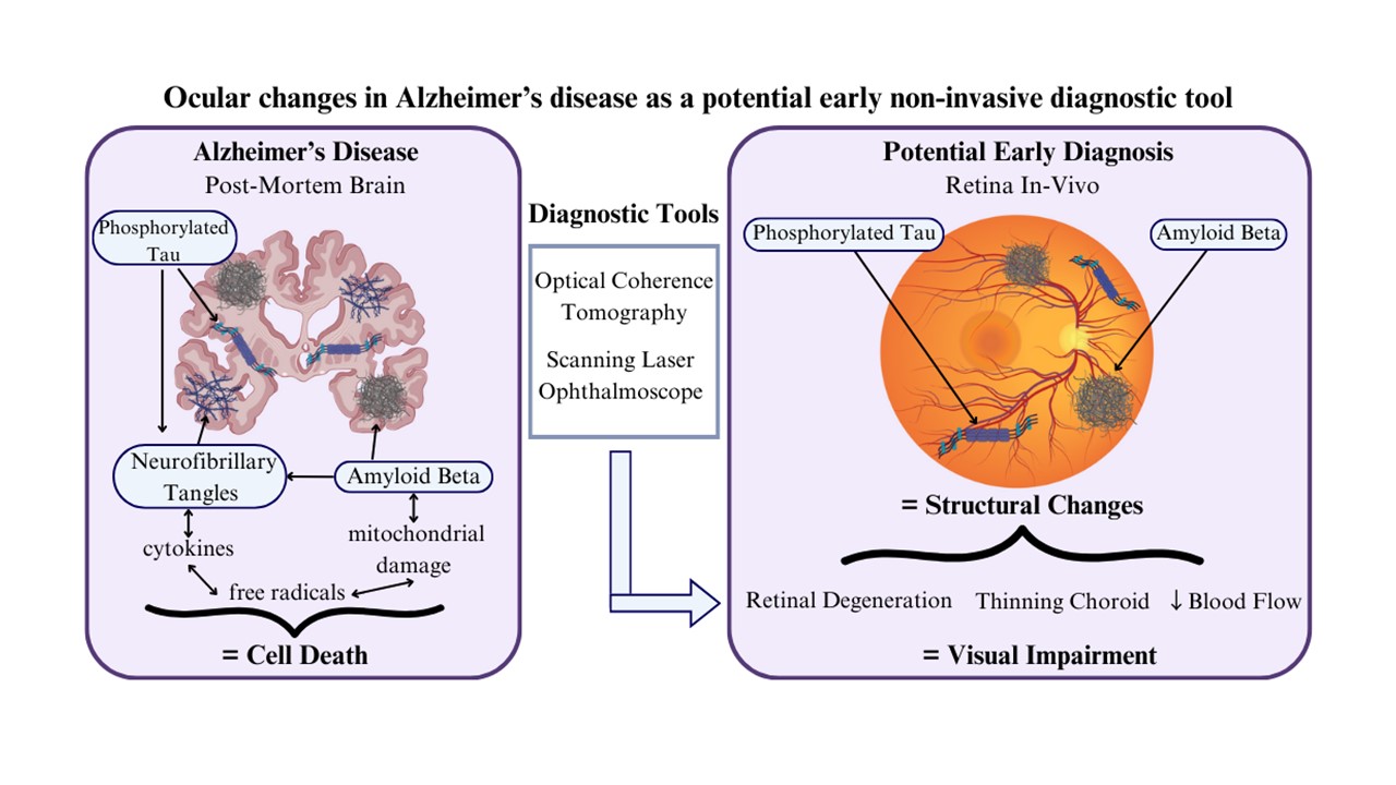
Alzheimer’s disease (AD) is an incurable, irreversible condition that bears a large global burden. An early diagnosis is imperative to allow the maximum time for treatment interventions. The eye’s involvement has been identified in AD, making it a promising option for a non-invasive early diagnostic and screening tool. The pathology of AD observed in the brain such as amyloid beta (Aβ) plaques and phosphorylated tau have been mirrored in the retina. There is also evidence that AD pathology in the retina precedes the onset of symptoms and the deposition of plaques in the brain. It is estimated that Aβ burden results in structural changes in the eye such as degeneration of the retinal nerve fiber layer, thinning of the macula, reduced blood flow rate, and thinning of the choroid. These structural changes can be observed using non-invasive imaging techniques such as optical coherence tomography. This review examines the existing literature on AD pathology in the retina and discusses the possibility of using retinal imaging techniques to screen for AD.
- Lane CA, Hardy J, Schott JM. Alzheimer’s disease. Eur J Neurol. 2018;25(1):59-70. doi: 10.1111/ene.13439
- World Health Organization. Dementia; 2023. Available from: https://www.who.int/news-room/facts-inpictures/ detail/dementia [Last accessed on 2024 Apr 01].
- Chen XQ, Mobley WC. Alzheimer disease pathogenesis: Insights from molecular and cellular biology studies of oligomeric Aβ and tau species. Front Neurosci. 2019;13:659. doi: 10.3389/fnins.2019.00659
- Hart NJ, Koronyo Y, Black, KL, Koronyo-Hamaoui M. Ocular indicators of Alzheimer’s: Exploring disease in the retina. Acta Neuropathol. 2016;132(6):767-787. doi: 10.1007/s00401-016-1613-6
- Ascaso FJ, Cruz N, Modrego PJ, et al. Retinal alterations in mild cognitive impairment and Alzheimer’s disease: An optical coherence tomography study. J Neurol. 2014;261:1522-1530. doi: 10.1007/s00415-014-7374-z
- Asanad S, Ross-Cisneros FN, Nassisi M, Barron E, Karanjia R, Sadun AA. The retina in Alzheimer’s disease: Histomorphometric analysis of an ophthalmologic biomarker. Invest Ophthalmol Vis Sci. 2019;60:1491-1500. doi: 10.1167/iovs.18-25966
- Zhang XX, Tian Y, Wang ZT, Ma YH, Tan L, Yu JT. The epidemiology of Alzheimer’s disease modifiable risk factors and prevention. J Prev Alzheimers Dis. 2021;8(3):313-321. doi: 10.14283/jpad.2021.15
- Public Health Agency of Canada. Dementia: Overview; 2024. Available from: https://www.canada.ca/en/publichealth/ services/diseases/dementia.html [Last accessed on 2024 Apr 18].
- Public Health Agency of Canada. Dementia in Canada; 2024. Available from: https://www.canada.ca/en/publichealth/ services/publications/diseases-conditions/dementia.html [Last accessed on 2024 Apr 18].
- Nichols E, Szoeke CEI, Vollset SE, et al. Global, regional, and national burden of Alzheimer’s disease and other dementias, 1990-2016: A systematic analysis for the Global Burden of Disease Study 2016. Lancet Neurol. 2019;18(1):88-106. doi: 10.1016/S1474-4422(18)30403-4
- Ashok A, Singh N, Chaudhary S, et al. Retinal degeneration and Alzheimer’s disease: An evolving link. Int J Mol Sci. 2020;21(19):7290. doi: 10.3390/ijms21197290
- Sultan S, Al-Hammadi M, Alshareef A, Al-Barakati AM, Al-Huthali RK, Al-Jahdali NH. An update on treatment of Alzheimer disease-a literature review. Eur J Pharm Med Res. 2018;205(7):8-18.
- Ewers M, Sperling RA, Klunk WE, Weiner MW, Hampel H. Neuroimaging markers for the prediction and early diagnosis of Alzheimer’s disease dementia. Trends Neurosci. 2011;34(8):430-442. doi: 10.1016/j.tins.2011.05.005
- Winblad B, Palmer K, Kivipelto M, et al. Mild cognitive impairment--beyond controversies, towards a consensus: Report of the international working group on mild cognitive impairment. J Intern Med. 2004;256(3):240-246. doi: 10.1111/j.1365-2796.2004.01380.x
- Weller J, Budson A. Current understanding of Alzheimer’s disease diagnosis and treatment. F1000Res. 2018;7:F1000 Faculty Rev-1161. doi: 10.12688/f1000research.14506.1
- Blennow K, Hampel H, Weiner M, Zetterberg H. Cerebrospinal fluid and plasma biomarkers in Alzheimer disease. Nat Rev Neurol. 2010;6(3):131-144. doi: 10.1038/nrneurol.2010.4
- Xia X, Qin Q, Peng Y, Wang M, Yin Y, Tang Y. Retinal examinations provides early warning of Alzheimer’s disease. J Alzheimers Dis. 2022;90(4):1341-1357. doi: 10.3233/JAD-220596
- Arai H, Morikawa Y, Higuchi M, et al. Cerebrospinal fluid tau levels in neurodegenerative diseases with distinct tau-related pathology. Biochem Biophys Res Commun. 1997;236(2):262-264. doi: 10.1006/bbrc.1997.6908
- Xie F, Peng F. Radiopharmaceuticals for assessment of altered metabolism and biometal fluxes in brain aging and Alzheimer’s disease with positron emission tomography. J Alzheimers Dis. 2017;59(2):527-536. doi: 10.3233/JAD-170280
- Surguchov A, McMahan B, Masliah E, Surgucheva F. Synucleins in ocular tissues. J Neurosci Res. 2001;65(1): 68-77. doi: 10.1002/jnr.1129
- Romaus-Sanjurjo D, Regueiro U, López-López M, et al. Alzheimer’s disease seen through the eye: Ocular alterations and neurodegeneration. Int J Mol Sci. 2022;23(5):2486. doi: 10.3390/ijms23052486
- Koronyo Y, Biggs D, Barron E, et al. Retinal amyloid pathology and proof-of-concept imaging trial in Alzheimer’s disease. JCI Insight. 2017;2(16):e93621. doi: 10.1172/jci.insight.93621
- Sidiqi A, Wahl D, Lee S, et al. In vivo retinal fluorescence imaging with curcumin in an Alzheimer mouse model. Front Neurosci. 2020;14:713. doi: 10.3389/fnins.2020.00713
- Aumann S, Donner S, Fischer J, Müller F. Optical Coherence Tomography (OCT): Principle and technical realization. In: Bille JF, editor. High Resolution Imaging in Microscopy and Ophthalmology: New Frontiers in Biomedical Optics. Cham, CH: Springer International Publishing; 2019. p. 59-85. doi: 10.1007/978-3- 030-16638-0_3
- Gharbiya M, Trebbastoni A, Parisi F, et al. Choroidal thinning as a new finding in Alzheimer’s disease: Evidence from enhanced depth imaging spectral domain optical coherence tomography. J Alzheimer Dis. 2014;40(4): 907-917. doi: 10.3233/JAD-132039
- Cunha JP, Proença R, Dias‐Santos A, et al. Choroidal thinning: Alzheimer’s disease and aging. Alzheimers Dement (Amst). 2017;8:11-17. doi: 10.1016/j.dadm.2017.03.004
- Podoleanu AG, Rosen RB. Combinations of techniques in imaging the retina with high resolution. Prog Retin Eye Res. 2008;27(4):464-499. doi: 10.1016/j.preteyeres.2008.03.002
- Puyo L, Paques M, Fink M, Sahel JA, Atlan M. In vivo laser Doppler holography of the human retina. Biomed Opt Express. 2018;9(9):4113. doi: 10.1364/BOE.9.004113
- Salobrar-Garcia E, Méndez-Hernández C, Hoz R, et al. Ocular vascular changes in mild Alzheimer’s disease patients: Foveal avascular zone, choroidal thickness, and ONH hemoglobin analysis. J Pers Med. 2020;10(4):231. doi: 10.3390/jpm10040231
- Berisha F, Feke GT, Trempe CL, McMeel JW, Schepens CL. Retinal abnormalities in early Alzheimer’s disease. Invest Ophthalmol Vis Sci. 2007;48(5):2285-2289. doi: 10.1167/iovs.06-1029
- Koronyo-Hamaoui M, Koronyo Y, Ljubimov AV, et al. Identification of amyloid plaques in retinas from Alzheimer’s patients and noninvasive in vivo optical imaging of retinal plaques in a mouse model. Neuroimage. 2011;54:S204-S217. doi: 10.1016/j.neuroimage.2010.06.020
- Tadokoro K, Yamashita T, Kimura S, et al. Retinal amyloid imaging for screening Alzheimer’s disease. J Alzheimer Dis. 2021;83(2):927-934. doi: 10.3233/JAD-210327
- Lee S, Jiang K, McIlmoyle B, et al. Amyloid beta immunoreactivity in the retinal ganglion cell layer of the Alzheimer’s eye. Front Neurosci. 2020;14:758. doi: 10.3389/fnins.2020.00758
- Kitazawa M, Medeiros R, LaFerla F. Transgenic mouse models of Alzheimer disease: Developing a better model as a tool for therapeutic interventions. Curr Pharm Des. 2012;18(8):1131-1147. doi: 10.2174/138161212799315786
- Koronyo Y, Rentsendorj A, Mirzaei N, et al. Retinal pathological features and proteome signatures of Alzheimer’s disease. Acta Neuropathol. 2023;145(4):409-438. doi: 10.1007/s00401-023-02548-2
- Tsai Y, Lu B, Ljubimov AV, et al. Ocular changes in TgF344-AD rat model of Alzheimer’s disease. Invest Opthalmol Vis Sci. 2014;55(1):523-534. doi: 10.1167/iovs.13-12888
- Schön C, Hoffmann NA, Ochs SM, et al. Long-term in vivo imaging of fibrillar tau in the retina of P301S transgenic mice. PLoS One. 2012;7(12):e5347. doi: 10.1371/journal.pone.0053547
- Ho C, Troncoso JC, Knox D, Stark W, Eberhart CG. Beta‐amyloid, phospho‐tau and alpha‐synuclein deposits similar to those in the brain are not identified in the eyes of Alzheimer’s and Parkinson’s disease patients. Brain Pathol. 2014;24(1):25-32. doi: 10.1111/bpa.12070
- Den Haan J, Morrema THJ, Verbraak FD, et al. Amyloid-beta and phosphorylated tau in post-mortem Alzheimer’s disease retinas. Acta Neuropathol Commun. 2018;6(1):147. doi: 10.1186/s40478-018-0650-x
- Chiasseu M, Alarcon-Martinez L, Belforte N, et al. Tau accumulation in the retina promotes early neuronal dysfunction and precedes brain pathology in a mouse model of Alzheimer’s disease. Mol Neurodegener. 2017;12(1):58. doi: 10.1186/s13024-017-0199-3
- Hart de Ruyter FJ, Morrema THJ, den Haan J, et al. Phosphorylated tau in the retina correlates with tau pathology in the brain in Alzheimer’s disease and primary tauopathies. Acta Neuropathol. 2023;145(2):197-218. doi: 10.1007/s00401-022-02525-1
- Ballatore C, Lee VMY, Trojanowski JQ. Tau-mediated neurodegeneration in Alzheimer’s disease and related disorders. Nat Rev Neurosci. 2007;8(9):663-672. doi: 10.1038/nrn2194
- Paquet C, Boissonnot M, Roger F, Dighiero P, Gil R, Hugon J. Abnormal retinal thickness in patients with mild cognitive impairment and Alzheimer’s disease. Neurosci Lett. 2007;420(2):97-99. doi: 10.1016/j.neulet.2007.02.090
- He XF, Liu YT, Peng C, Zhang F, Zhuang S, Zhang JS. Optical coherence tomography assessed retinal nerve fiber layer thickness in patients with Alzheimer’s disease: A meta-analysis. Int J Ophthalmol. 2012;5(3):401-405. doi: 10.3980/j.issn.2222-3959.2012.03.30
- Gao L, Liu Y, Li X, Bai Q, Liu P. Abnormal retinal nerve fiber layer thickness and macula lutea in patients with mild cognitive impairment and Alzheimer’s disease. Arch Gerontol Geriatr. 2015;60(1):162-167. doi: 10.1016/j.archger.2014.10.011
- Garcia‐Martin E, Bambo MP, Marques ML, et al. Ganglion cell layer measurements correlate with disease severity in patients with Alzheimer’s disease. Acta Ophthalmol. 2016;94(6):e454-e459. doi: 10.1111/aos.12977
- Chiquita S, Campos EJ, Castelhano J, et al. Retinal thinning of inner sub-layers is associated with cortical atrophy in a mouse model of Alzheimer’s disease: A longitudinal multimodal in vivo study. Alzheimers Res Ther. 2019;11:90. doi: 10.1186/s13195-019-0542-8
- Bevan RJ, Hughes TR, Williams PA, Good MA, Morgan BP, Morgan JE. Retinal ganglion cell degeneration correlates with hippocampal spine loss in experimental Alzheimer’s disease. Acta Neuropathol Commun. 2020;8(1):216. doi: 10.1186/s40478-020-01094-2
- Kesler A, Vakhapova V, Korczyn AD, Naftaliev E, Neudorfer M. Retinal thickness in patients with mild cognitive impairment and Alzheimer’s disease. Clin Neurol Neurosurg. 2011;113(7):523-526. doi: 10.1016/j.clineuro.2011.02.014
- Parisi V, Restuccia R, Fattapposta F, Mina C, Bucci MG, Pierelli F. Morphological and functional retinal impairment in Alzheimer’s disease patients. Clin Neurophysiol. 2001;112(10):1860-1867. doi: 10.1016/S1388-2457(01)00620-4
- Georgevsky D, Retsas S, Raoufi N, Shimoni O, Golzan SM. A longitudinal assessment of retinal function and structure in the APP/PS1 transgenic mouse model of Alzheimer’s disease. Transl Neurodegener. 2019;8:30. doi: 10.1186/s40035-019-0170-z
- Mutlu U, Colijn JM, Ikram MA, et al. Association of retinal neurodegeneration on optical coherence tomography with dementia: A population-based study. JAMA Neurol. 2018;75(10):1256-1263. doi: 10.1001/jamaneurol.2018.1563
- Iseri PK, Altinaş Ö, Tokay T, Yüksel N. Relationship between cognitive impairment and retinal morphological and visual functional abnormalities in Alzheimer disease. J Neuroophthalmol. 2006;26(1):18-24. doi: 10.1097/01.wno.0000204645.56873.26
- Fujino Y, DeLucia MW, Davies P, Dickson DW. Ballooned neurones in the limbic lobe are associated with Alzheimer type pathology and lack diagnostic specificity. Neuropathol Appl Neurobiol. 2004;30(6):676-682. doi: 10.1111/j.1365-2990.2004.00593.x
- Reichenbach A, Wurm A, Pannicke T, Iandiev I, Wiedemann P, Bringmann A. Müller cells as players in retinal degeneration and edema. Graefes Arch Clin Exp Ophthalmol. 2007;245(5):627-636. doi: 10.1007/s00417-006-0516-y
- Patton N, Aslam T, MacGillivray T, Pattie A, Deary IJ, Dhillon B. Retinal vascular image analysis as a potential screening tool for cerebrovascular disease: A rationale based on homology between cerebral and retinal microvasculatures. J Anat. 2005;206(4):319-348. doi: 10.1111/j.1469-7580.2005.00395.x
- He JT, Zhao X, Xu L, Mao CY. Vascular risk factors and Alzheimer’s disease: Blood-brain barrier disruption, metabolic syndromes, and molecular links. J Alzheimers Dis. 2020;73(1):39-58. doi: 10.3233/JAD-190764
- Farkas E, Luiten PGM. Cerebral microvascular pathology in aging and Alzheimer’s disease. Prog Neurobiol. 2001;64(6):575-611. doi: 10.1016/S0301-0082(00)00068-X
- Fischer VW, Siddiqi A, Yusufaly Y. Altered angioarchitecture in selected areas of brains with Alzheimer’s disease. Acta Neuropathol. 1990;79(6):672-679. doi: 10.1007/BF00294246
- Dorr A, Sahota B, Chinta LV, et al. Amyloid-β-dependent compromise of microvascular structure and function in a model of Alzheimer’s disease. Brain. 2012;135(Pt 10): 3039-3050. doi: 10.1093/brain/aws243
- Ma Q, Zhao Z, Sagare AP, et al. Blood-brain barrier-associated pericytes internalize and clear aggregated amyloid-β42 by LRP1-dependent apolipoprotein E isoform-specific mechanism. Mol Neurodegener. 2018;13(1):57. doi: 10.1186/s13024-018-0286-0
- Halliday MR, Rege SV, Ma Q, et al. Accelerated pericyte degeneration and blood-brain barrier breakdown in apolipoprotein E4 carriers with Alzheimer’s disease. J Cereb Blood Flow Metab. 2016;36(1):216-227. doi: 10.1038/jcbfm.2015.44
- Shibata M, Yamada S, Kumar SR, et al. Clearance of Alzheimer’s amyloid-ss(1-40) peptide from brain by LDL receptor-related protein-1 at the blood-brain barrier. J Clin Invest. 2000;106(12):1489-1499. doi: 10.1172/JCI10498
- Cheung CY, Ong YT, Ikram MK, et al. Microvascular network alterations in the retina of patients with Alzheimer’s disease. Alzheimers Dement. 2014;10(2):135-142. doi: 10.1016/j.jalz.2013.06.009
- Shi H, Koronyo Y, Fuchs DT, et al. Retinal capillary degeneration and blood-retinal barrier disruption in murine models of Alzheimer’s disease. Acta Neuropathol Commun. 2020;8(1):202. doi: 10.1186/s40478-020-01076-4
- Nazari HK, Karimaghaei C, van der Merwe R, et al. Age dependence of retinal vascular plexus attenuation in the triple transgenic mouse model of Alzheimer’s disease. Exp Eye Res. 2022;214:108879. doi: 10.1016/j.exer.2021.108879
- Frost S, Kanagasingam Y, Sohrabi H, et al. Retinal vascular biomarkers for early detection and monitoring of Alzheimer’s disease. Transl Psychiatry. 2013;3(2):e233. doi: 10.1038/tp.2012.150
- Shi H, Koronyo Y, Rentsendorj A, et al. Identification of early pericyte loss and vascular amyloidosis in Alzheimer’s disease retina. Acta Neuropathol. 2020;139:813-836. doi: 10.1007/s00401-020-02134-w
- Sagare AP, Sweeney MD, Makshanoff J, Zlokovic BV. Shedding of soluble platelet-derived growth factor receptor-β from human brain pericytes. Neurosci Lett. 2015;607:97-101. doi: 10.1016/j.neulet.2015.09.025
- Dollery CT, Bulpitt CJ, Kohner EM. Oxygen supply to the retina from the retinal and choroidal circulations at normal and increased arterial oxygen tensions. Invest Ophthalmol. 1969;8(6):588-594.
- Laviers H, Zambarakji H. Enhanced depth imaging-OCT of the choroid: A review of the current literature. Graefes Arch Clin Exp Ophthalmol. 2014;252(12):1871-1883. doi: 10.1007/s00417-014-2840-y
- Salobrar-García E, de Hoz R, Ramírez AI, et al. Changes in visual function and retinal structure in the progression of Alzheimer’s disease. PLoS One. 2019;14(8):e0220535. doi: 10.1371/journal.pone.0220535
- Choi W, Mohler KJ, Potsaid B, et al. Choriocapillaris and choroidal microvasculature imaging with ultrahigh speed OCT angiography. PLoS One. 2013;8(12):e81499. doi: 10.1371/journal.pone.0081499
- Bayhan H, Bayhan S, Celikbilek A, et al. Evaluation of the chorioretinal thickness changes in Alzheimer’s disease using spectral‐domain optical coherence tomography. Clin Exp Ophthalmol. 2015;43(2):145-151. doi: 10.1111/ceo.12386
- Bulut M, Yaman A, Erol MK, et al. Choroidal thickness in patients with mild cognitive impairment and Alzheimer’s type dementia. J Ophthalmol. 2016;2016:7291257. doi: 10.1155/2016/7291257
- Den Haan J, van de Kreeke JA, van Berckel BN, et al. Is retinal vasculature a biomarker in amyloid proven Alzheimer’s disease? Alzheimers Dement (Amst). 2019;11:383-391. doi: 10.1016/j.dadm.2019.03.006
- Ramrattan RS, Van Der Schaft TL, Mooy CM, de Bruijn WC, Mulder PG, de Jong PT. Morphometric analysis of Bruch’s membrane, the choriocapillaris, and the choroid in aging. Invest Ophthalmol Vis Sci. 1994;35(6):2857-2864.
- Ning A, Cui J, To E, Ashe KH, Matsubara J. Amyloid-beta deposits lead to retinal degeneration in a mouse model of Alzheimer disease. Invest Ophthalmol Vis Sci. 2008;49(11):5136-5143. doi: 10.1167/iovs.08-1849
- Jirarattanasopa P, Ooto S, Nakata I, et al. Choroidal thickness, vascular hyperpermeability, and complement factor H in age-related macular degeneration and polypoidal choroidal vasculopathy. Invest Ophthalmol Vis Sci. 2012;53(7): 3663-3672. doi: 10.1167/iovs.12-9619
- Yoonessi A, Yoonessi A. Functional assessment of magno, parvo and konio-cellular pathways; Current state and future clinical applications. J Ophthalmic Vis Res. 2011;6(2): 119-126.
- Rizzo M, Anderson SW, Dawson J, Nawrot M. Vision and cognition in Alzheimer’s disease. Neuropsychologia. 2000;38(8):1157-1169. doi: 10.1016/S0028-3932(00)00023-3
- Tippett WJ, Sergio LE. Visuomotor integration is impaired in early stage Alzheimer’s disease. Brain Res. 2006;1102(1): 92-102. doi: 10.1016/j.brainres.2006.04.049
- Polo V, Rodrigo MJ, Garcia-Martin E, et al. Visual dysfunction and its correlation with retinal changes in patients with Alzheimer’s disease. Eye (Lond). 2017;31(7):1034-1041. doi: 10.1038/eye.2017.23
- Stein JD, Khawaja AP, Weizer JS. Glaucoma in adults-screening, diagnosis, and management: A review. JAMA. 2021;325(2):164-174. doi: 10.1001/jama.2020.21899