

Diagnosis of COVID-19 from computed tomography slices using flower pollination algorithm, k-nearest neighbor, and support vector machine classifiers

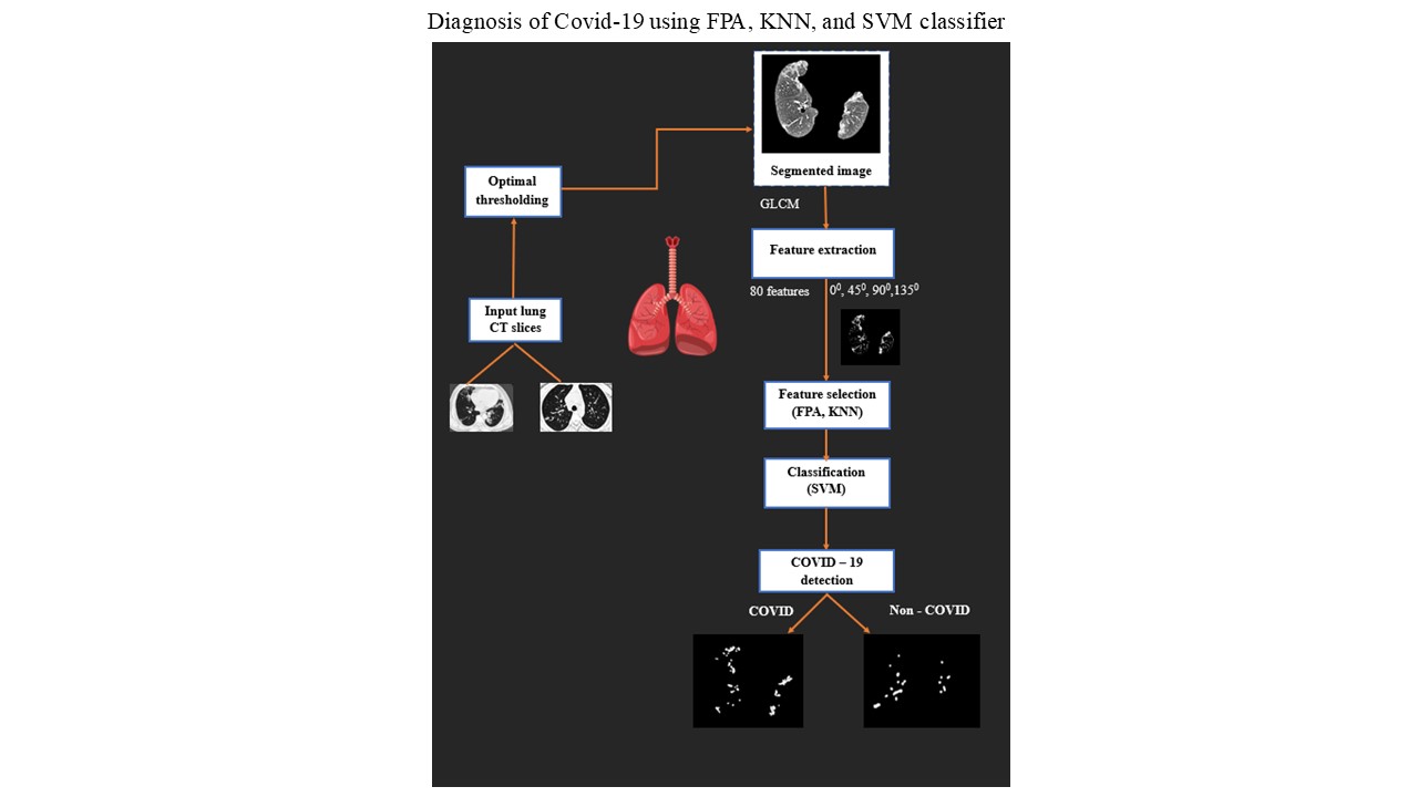
Coronavirus disease 19 (COVID-19), caused by the severe acute respiratory syndrome-coronavirus-2 virus, is commonly diagnosed through imaging techniques such as computed tomography (CT) scans, which reveal characteristic lung lesions. In this study, we propose a computer-aided diagnosis (CAD) system to assist in the early detection of COVID-19 from CT lung slices, leveraging advanced machine-learning algorithms for precise and efficient analysis. To achieve this, we developed a CAD system that diagnoses COVID-19 from CT lung slices. An adaptive Wiener filter was applied to remove noise from the CT images. The chest tissues were then segmented using an optimal thresholding method to extract regions of interest, which represent the COVID-19 lesions under investigation. The feature vectors were divided into training and testing with an 80/20 ratio. A wrapper-based flower pollination algorithm was employed alongside the k-nearest neighbor classifier to select the optimal feature set. These selected features were subsequently used to train a support vector machine (SVM) classifier. With feature selection, the SVM achieved an accuracy of 91.30% on a real-time dataset, outperforming seven other machine learning classifiers (radial basis function-SVM, k nearest neighbor, linear discriminant analysis, random forest, naïve Bayes, AdaBoost, extreme gradient boosting) and four deep learning classifiers (convolutional neural network, recurrent neural network, long short term memory, Bidirectional long short term memory). For the publicly available COVID-19 CT dataset, an accuracy of 88.18% was achieved. In conclusion, our COVID-19 CAD system improves diagnostic accuracy, with future work aimed at enhancing efficiency and expanding to covariant detection and severity assessment.
- Fuller HW. Lea HC, editor. On Diseases of the Lungs and Air- Passages: Their Pathology, Physical Diagnosis, Symptoms, and Treatment. Kissingert: Publishing; 1867.
- Tomashefski JF Jr, Farver CF. Anatomy and Histology of the Lung. In: Dail and Hammar’s Pulmonary Pathology. Springer New York; 2008:20-48. doi: 10.1007/978-0-387-68792-6_2
- Demedts M, Wells AU, Antó JM, et al. Interstitial lung diseases: An epidemiological overview. Eur Respir J. 2001;18(32 suppl):2S-16S. doi: 10.1183/09031936.01.18s320002
- Schwarz T, Johnson V. Lungs and Bronchi. Veterinary Computed Tomography. Wiley; 2011:261-277. doi: 10.1002/9781118785676.ch26
- Fan DP, Zhou T, Ji GP, et al. Inf-Net: Automatic COVID-19 Lung Infection Segmentation From CT Images. IEEE Trans Med Imaging. 2020;39(8):2626-2637. doi: 10.1109/tmi.2020.2996645
- Carotti M, Salaffi F, Sarzi-Puttini P, et al. Chest CT features of coronavirus disease 2019 (COVID-19) pneumonia: key points for radiologists. Radiol Med. 2020;125(7):636-646. doi: 10.1007/s11547-020-01237-4
- Fang Y, Zhang H, Xie J, et al. Sensitivity of chest CT for COVID-19: Comparison to RT-PCR. Radiology. 2020;296(2):E115-E117. doi: 10.1148/radiol.2020200432
- Ng MY, Lee EY, Yang J, et al. Imaging profile of the COVID-19 infection: Radiologic findings and literature review. Radiol Cardiothorac Imaging. 2020;2(1):e200034. doi: 10.1148/ryct.2020200034
- Ai T, Yang Z, Hou H, et al. Correlation of Chest CT and RT-PCR Testing for Coronavirus Disease 2019 (COVID- 19) in China: A Report of 1014 Cases. Radiology. 2020;296(2):E32-E40. doi: 10.1148/radiol.2020200642
- Pal NR, Pal SK. A review on image segmentation techniques. Pattern Recognit. 1993;26(9):1277-1294. doi: 10.1016/0031-3203(93)90135-J
- Norouzi A, Rahim MS, Altameem A, et al. Medical image segmentation methods, algorithms, and applications. IETE Tech Rev. 2014;31(3):199-213. doi: 10.1080/02564602.2014.906861
- Sitanggang S, Sonang S, Yuhandri Y, Setiawan A. Image transformation with lung image thresholding and segmentation method. J RESTI (Rekayasa Sist Teknol Inform). 2023;7(2):278-285. doi: 10.29207/resti.v7i2.4321
- Yu T, Huang L. An Adaptive Thresholding Method for Automatic Lung Segmentation in CT Images. In: IEEE AFRICON Conference; 2009:1-5.
- Sweetlin JD, Nehemiah HK, Kannan A. Feature selection using ant colony optimization with tandem-run recruitment to diagnose bronchitis from CT scan images. Comput Methods Programs Biomed. 2017;145:115-125. doi: 10.1016/j.cmpb.2017.04.009
- Sweetlin JD, Nehemiah HK, Kannan A. Computer aided diagnosis of drug sensitive pulmonary tuberculosis with cavities, consolidations and nodular manifestations on lung CT images. Int J Bio Inspired Comput. 2019;13(2):71-85. doi: 10.1504/IJBIC.2019.098405
- Dehmeshki J, Amin H, Valdivieso M, Ye X. Segmentation of pulmonary nodules in thoracic CT scans: A region growing approach. IEEE Trans Med Imaging. 2008;27(4):467-480. doi: 10.1109/TMI.2007.907555
- Nabipour S, Khorshidi A, Noorian B. Lung tumor segmentation using improved region growing algorithm. Nucl Eng Technol. 2020;52(10):2313-2319. doi: 10.1016/j.net.2020.03.011
- Prabin A, Veerappan J. Automatic segmentation of lung CT images by CC based region growing. J Theor Appl Inform Technol. 2014;68(1):63-69.
- Avinash S, Manjunath K, Kumar SS. An improved image processing analysis for the detection of lung cancer using Gabor filters and watershed segmentation technique. In: 2016 International Conference on Inventive Computation Technologies (ICICT). IEEE; 2016:1-6. doi: 10.1109/inventive.2016.7830084
- Kumar SL, Swathy M, Sathish S, Sivaraman J, Rajasekar M. Identification of lung cancer cell using watershed segmentation on CT images. Indian J Sci Technol. 2016;9:1-4. doi: 10.17485/ijst/2016/v9i1/85765
- Shojaii R, Alirezaie J, Babyn P. Automatic Lung Segmentation in CT Images Using Watershed Transform. In: IEEE International Conference on Image Processing; 2005.
- Nithila EE, Kumar SS. Segmentation of lung from CT using various active contour models. Biomed Signal Process Control. 2019;47:57-62.
- Kasinathan G, Jayakumar S, Gandomi AH, et al. Automated 3-D lung tumor detection and classification by an active contour model and CNN classifier. Expert Syst Appl. 2019;15(134):112-119. doi: 10.1016/j.eswa.2019.05.041
- Sangamithraa PB, Govindaraju S. Lung tumour detection and classification using EK-Mean clustering. In: 2016 International Conference on Wireless Communications, Signal Processing and Networking (WiSPNET). IEEE; 2016:2201- 2206. doi: 10.1109/wispnet.2016.7566533
- Joon P, Bajaj SB, Jatain A. Segmentation and Detection of Lung Cancer Using Image Processing and Clustering Techniques. In: Advances in Intelligent Systems and Computing. Springer Singapore; 2018:13-23. doi: 10.1007/978-981-13-1708-8_2
- Xu M, Qi S, Yue Y, et al. Segmentation of lung parenchyma in CT images using CNN trained with the clustering algorithm generated dataset. Biomed Eng Online. 2019;18:2. doi: 10.1186/s12938-018-0619-9
- Farag AA, Munim HE, Graham JH, Farag AA. A novel approach for lung nodules segmentation in chest CT using level sets. IEEE Trans Image Process. 2013;22:5202-5213. doi: 10.1109/TIP.2013.2282899
- Swierczynski P, Papież BW, Schnabel JA, Macdonald C. A level-set approach to joint image segmentation and registration with application to CT lung imaging. Comput Med Imaging Graph. 2018;65:58-68. doi: 10.1016/j.compmedimag.2017.06.003
- Wei J, Deihui X, Zhang B, Wang L, Kopriva I, Chen X. Random walk and graph cut for co-segmentation of lung tumor on PET-CT images. IEEE Trans Image Process. 2015;24(12):5854-5867. doi: 10.1109/TIP.2015.2488902
- Ali AM, Farag AA. Automatic Lung Segmentation of Volumetric Low-Dose CT Scans Using Graph Cuts. In: Advances in Visual Computing (Lecture Notes in Computer Science). Springer Berlin Heidelberg; 2008:258-267. doi: 10.1007/978-3-540-89639-5_25
- Bhuvaneswari P, Therese BA. Detection of cancer in lung with k-NN classification using genetic algorithm. Procedia Mater Sci. 2015;10:433-440. doi: 10.1016/j.mspro.2015.06.077
- Filho DC, Silva AO, Paiva AC, Nunes RA, Gattass M. Computer-aided diagnosis system for lung nodules based on computed tomography using shape analysis, a genetic algorithm, and SVM. Med Biol Eng Comput. 2017;55:1129-1146. doi: 10.1007/s11517-016-1577-7
- Herrmann P, Busana M, Cressoni M, et al. Using artificial intelligence for automatic segmentation of CT lung images in acute respiratory distress syndrome. Front Physiol. 2021;12:76118. doi: 10.3389/fphys.2021.676118
- Shi F, Wang J, Shi J, et al. Review of artificial intelligence techniques in imaging data acquisition, segmentation, and diagnosis for COVID-19. IEEE Rev Biomed Eng. 2021;14:4-15. doi: 10.1109/RBME.2020.2987975
- Van EM, Hoop D, Viergever MA, Prokop M, Ginneken BV. Automatic lung segmentation from thoracic computed tomography scans using a hybrid approach with error detection. Med Phys. 2009;36:2934-2947. doi: 10.1118/1.3147146
- Doi K. Computer-aided diagnosis in medical imaging: Historical review, current status, and future potential. Comput Med Imaging Graph. 2007;31(5):198-211. doi: 10.1016/j.compmedimag.2007.02.002
- Choi YJ, Baek JH, Park HS, et al. A computer-aided diagnosis system using artificial intelligence for the diagnosis and characterization of thyroid nodules on ultrasound: Initial clinical assessment. Thyroid. 2017;27(4):546-552. doi: 10.1089/thy.2016.0372
- Isaac A, Nehemiah HK, Dunston DS, Christo VRE, Kannan A. Feature selection using competitive coevolution of bio-inspired algorithms for the diagnosis of pulmonary emphysema. Biomed Signal Process Control. 2022;72:103340. doi: 10.1016/j.bspc.2021.103340
- Khin Y, Maneerat N, Sreng S, Hamamoto K. Ensemble deep learning for the detection of COVID-19 in unbalanced chest X-ray dataset. Appl Sci. 2021;11(22):10528. doi: 10.3390/app112210528
- Rajinikanth V, Kadry S, Thanaraj KP, Kamalanand K, Seo S. Firefly-Algorithm Supported Scheme to Detect COVID-19 Lesion in Lung CT Scan Images using Shannon Entropy and Markov-Random-Field. arXiv. Preprint posted online 2020. doi: 10.48550/arXiv.2004.09239
- Satapathy SC, Hemanth DJ, Kadry S, Manogaran G, Hannon NMS, Rajinikanth V. Segmentation and evaluation of COVID-19 lesion from CT scan slices - A study with Kapur/ Otsu Function and Cuckoo Search Algorithm. Research Square. Preprint posted online July 12, 2020. doi: 10.21203/rs.3.rs-40148/v1
- Mohammed S, Alkinani F, Hassan Y. Automatic computer aided diagnostic for COVID-19 based on chest X-ray image and particle swarm intelligence. Int J Intell Eng Syst. 2020;13(5):63-73. doi: 10.22266/ijies2020.1031.07
- Bhargava A, Bansal A, Goyal V. Machine learning-based automatic detection of novel coronavirus (COVID-19) disease. Multimed Tools Appl. 2022;81(10):13731-13750. doi: 10.1007/s11042-022-12508-9
- Shankar K, Mohanty SN, Yadav K, Gopalakrishnan T, Elmisery AM. Automated COVID-19 diagnosis and classification using convolutional neural network with fusion based feature extraction model. Cogn Neurodyn. 2021;10:1-4. doi: 10.1007/s11571-021-09712-y
- Kadry S, Rajinikanth V, Rho S, Raja NSM, Rao VS, Thanaraj KP. Development of a Machine-Learning System to Classify Lung CT Scan Images into Normal/COVID-19 Class. arXiv. Preprint posted online 2020. doi: 10.48550/arXiv.2004.13122
- Wu G, Zhou S, Wang Y, et al. A prediction model of outcome of SARS-CoV-2 pneumonia based on laboratory findings. Sci Rep. 2020;10(1):14042. doi: 10.1038/s41598-020-71114-7
- Banerjee A, Ray S, Vorselaars B, et al. Use of machine learning and artificial intelligence to predict SARS-CoV-2 infection from full blood counts in a population. Int Immunopharmacol. 2020;86:106705. doi: 10.1016/j.intimp.2020.106705
- Moutaz A, Awajan A, Mesleh A, Abraham A, Jatana V, Alhyari S. COVID-19 prediction and detection using deep learning. Int J Comput Inf Syst Ind Manag Appl. 2020;12:168- 181.
- Feng C, Wang L, Chen X, et al. A novel artificial intelligence-assisted triage tool to aid in the diagnosis of suspected COVID-19 pneumonia cases in fever clinics. Ann Transl Med. 2021;9(3):201-201. doi: 10.21037/atm-20-3073
- Najjar FH, Kadhim KA, Kareem MH, et al. Classification of COVID-19 from X-ray images using GLCM features and machine learning. Malays J Fundam Appl Sci. 2023;19(6):389-398. doi: 10.11113/mjfas.v19n3.2911
- Maryam A, Ahmad I, Imtiaz A, Mohammed A. Ensemble learning model for diagnosing COVID-19 from routine blood tests. Inform Med Unlocked. 2020;21:100449. doi: 10.1016/j.imu.2020.100449
- Atta A, Sultan K, Naseer I, et al. Supervised machine learning-based prediction of COVID-19. Comput Mater Contin. 2021;69(1):21-34. doi: 10.32604/cmc.2021.013453
- Rajinikanth V, Dey N, Raj ANJ, Hassanien AE, Santosh KC, Raja NSM. Harmony-Search and Otsu based System for Coronavirus Disease (COVID-19) Detection using Lung CT Scan Images. arXiv. Preprint posted online 2020. doi: 10.48550/arXiv.2004.03431
- Zhou T, Canu S, Ruan S. An automatic COVID-19 CT segmentation network using spatial and channel attention mechanism. arXiv. Preprint posted online 2020. doi: 10.48550/ARXIV.2004.06673
- Mobiny A, Cicalese PA, Zare S, et al. Radiologist-Level COVID-19 Detection Using CT Scans with Detail-Oriented Capsule Networks. arXiv. Preprint posted online 2020. doi: 10.48550/ARXIV.2004.07407
- Hasoon JN, Fadel AH, Hameed RS, et al. COVID-19 anomaly detection and classification method based on supervised machine learning of chest X-ray images. Results in Physics. 2021;31:105045. doi: 10.1016/j.rinp.2021.105045
- Mahdy LN, Ezzat KA, Elmousalami HH, Ella HA, Hassanien AE. Automatic X-ray COVID-19 Lung Image Classification System based on Multi-Level Thresholding and Support Vector Machine. medRxiv. Preprint posted online April 6, 2020. doi: 10.1101/2020.03.30.20047787
- Elizabeth DS, Nehemiah HK, Raj CSR, Kannan A. A novel segmentation approach for improving diagnostic accuracy of CAD systems for detecting lung cancer from chest computed tomography images. J Data and Information Quality. 2012;3(2):1-16. doi: 10.1145/2184442.2184444
- Rachel RB, Nehemiah HK, Marishanjunath CS, Manoharan RM. Diagnosis of pulmonary edema and COVID-19 from CT slices using squirrel search algorithm, support vector machine and back propagation neural network. J Intell Fuzzy Syst. 2023;44:1-4. doi: 10.3233/JIFS-222564
- Rachel RB, Nehemiah HK, Singh VK, Manoharan RM. Diagnosis of COVID-19 from CT slices using whale optimization algorithm, support vector machine and multi-layer perceptron. J Xray Sci Technol. 2023;32:253-269. doi: 10.3233/XST-230196
- Anisha I, Nehemiah HK, Anubha I, Kannan A. Computer- Aided Diagnosis system for diagnosis of pulmonary emphysema using bio-inspired algorithms. Comput Biol Med. 2020;124:103940. doi: 10.1016/j.compbiomed.2020.103940
- Glover B. Understanding Flowers and Flowering: An Integrated Approach. Oxford: Oxford University Press; 2007.
- Shifali Kalra, Sankalap Arora. Firefly Algorithm Hybridized with Flower Pollination Algorithm for Multimodal Functions. In: Advances in Intelligent Systems and Computing. Springer Singapore; 2016:207-219. doi: 10.1007/978-981-10-0767-5_23
- Yang XS. Flower Pollination Algorithm for Global Optimization. In: Unconventional Computation and Natural Computation (Lecture Notes in Computer Science). Springer Berlin Heidelberg; 2012:240-249. doi: 10.1007/978-3-642-32894-7_27
- Pavlyukevich I. Lévy flights, non-local search and simulated annealing. J Comput Phys. 2007;226(2):1830-1844. doi: 10.1016/j.jcp.2007.06.008
- Jain DK, Dubey SB, Choubey RK, et al. An approach for hyperspectral image classification by optimizing SVM using self organizing map. Journal of Computational Science. 2018;25:252-259. doi:10.1016/j.jocs.2017.07.016
- He B, Zhao W, Pi JY, et al. A biomarker basing on radiomics for the prediction of overall survival in non-small cell lung cancer patients. Respir Res. 2018;19:199. doi: 10.1186/s12931-018-0887-8
- Isaac A, Nehemiah HK, Kannan A. Computer-aided diagnosis system for diagnosis of cavitary and miliary tuberculosis using improved artificial bee colony optimization. IETE J Res. 2021;69:1-20. doi: 10.1080/03772063.2021.1946440
- Yang X, He X, Zhao J, Zhang Y, Zhang S, Xie P. COVID-CT-Dataset: A CT Scan Dataset about COVID-19. arXiv. Preprint posted online 2020. doi: 10.48550/arXiv.2003.13865
- Polsinelli M, Cinque L, Placidi G. A light CNN for detecting COVID-19 from CT scans of the chest. Pattern Recognit Lett. 2020;140:95-100. doi: 10.1016/j.patrec.2020.10.001
- Ali AE, Assadi TA. GLCMs based multi-inputs 1D CNN deep learning neural network for COVID-19 texture feature extraction and classification. Karbala Int J Mod Sci. 2022;8(1):28-39. doi: 10.33640/2405-609X.3201
- Pedro S, Luz E, Silva G, et al. COVID-19 detection in CT images with deep learning: A voting-based scheme and cross-datasets analysis. Inform Med Unlocked. 2020;20:100427. doi: 10.1016/j.imu.2020.100427