

Computational identification of immune-suppressive gene signatures associated with tumor progression and immunotherapy resistance

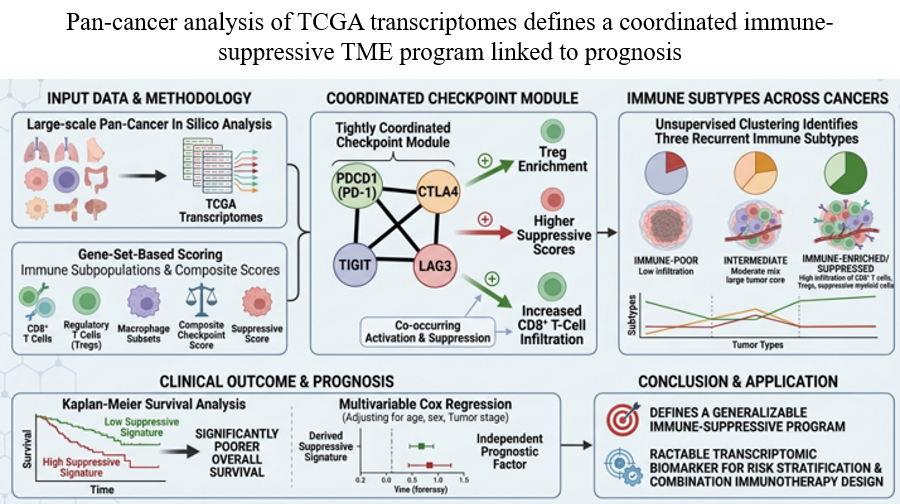
The tumor microenvironment integrates immune‑activating and immune‑suppressive cues that critically shape cancer progression and response to immunotherapy. Here, we performed a pan‑cancer in silico analysis of transcriptome data from The Cancer Genome Atlas to define an immune‑suppressive gene signature and associated immune states linked to checkpoint activity and clinical outcome. Gene set-based scoring was used to quantify CD8+ T cells, regulatory T cells, macrophage subsets, and composite checkpoint and suppressive immune signatures across tumor types. Correlation and network analyses revealed that PDCD1, CTLA4, TIGIT, and LAG3 form a tightly coordinated checkpoint module strongly associated with Treg enrichment, higher suppressive immune signatures, and, notably, increased CD8+ T‑cell infiltration, indicating co‑occurring immune activation and suppression rather than mutually exclusive states. Unsupervised clustering of immune scores identified three recurrent immune subtypes, immune‑poor, intermediate, and immune‑enriched/suppressed, spanning multiple cancers and capturing distinct immunological configurations. The derived suppressive signature stratified patients into high- and low-risk groups, with elevated suppressive immune signature scores associated with poorer overall survival in Kaplan–Meier analyses. Multivariable Cox regression confirmed that the suppressive signature is an independent prognostic factor after adjusting for age, sex, and tumor stage, suggesting that it encodes biological information not captured by standard clinicopathologic variables. Together, these findings define a coordinated immune‑suppressive program that transcends tissue of origin and provides a tractable transcriptomic biomarker for risk stratification and rational design of combination immunotherapeutic strategies.
- Hanahan D, Weinberg RA. Hallmarks of cancer: the next generation. Cell. 2011;144(5):646-674. doi: 10.1016/j.cell.2011.02.013
- Fridman WH, Pagès F, Sautès-Fridman C, Galon J. The immune contexture in human tumours: impact on clinical outcome. Nat Rev Cancer. 2012;12(4):298-306. doi: 10.1038/nrc3245
- Topalian SL, Drake CG, Pardoll DM. Immune checkpoint blockade: A common denominator Approach to Cancer Therapy. Cancer Cell. 2015;27(4):450-461. doi: 10.1016/j.ccell.2015.03.001
- Sharma P, Allison JP. The future of immune checkpoint therapy. Science. 2015;348(6230):56-61. doi: 10.1126/science.aaa8172
- Joyce JA, Fearon DT. T cell exclusion, immune privilege, and the tumor microenvironment. Science. 2015;348(6230):74- 80. doi: 10.1126/science.aaa6204
- Ribas A, Wolchok JD. Cancer immunotherapy using checkpoint blockade. Science. 2018;359(6382):1350-1355. doi: 10.1126/science.aar4060
- Hu Q, Zhu Y, Mei J, Liu Y, Zhou G. Extracellular matrix dynamics in tumor immunoregulation: from tumor microenvironment to immunotherapy. J Hematol Oncol. 2025;18(1):65. doi: 10.1186/s13045-025-01717-y
- de Visser KE, Joyce JA. The evolving tumor microenvironment: From cancer initiation to metastatic outgrowth. Cancer Cell. 2023;41(3):374-403. doi: 10.1016/j.ccell.2023.02.016
- Raskov H, Orhan A, Gaggar S, Gögenur I. Cancer-Associated Fibroblasts and Tumor-Associated Macrophages in Cancer and Cancer Immunotherapy. Front Oncol. 2021;11:668731. doi: 10.3389/fonc.2021.668731
- Zhang W, Xiang Y, Lu C, et al. The extracellular matrix in inflammation and cancer. Mol Biomed. 2026;7(1):21. doi: 10.1186/s43556-026-00415-6
- Weinstein JN, Collisson EA, Mills GB, et al. The Cancer Genome Atlas Pan-Cancer analysis project. Nat Genet. 2013;45(10):1113–1120. doi: 10.1038/ng.2764
- Gentles A, Newman A, Liu C, et al. The prognostic landscape of genes and infiltrating immune cells across human cancers. Nat Med. 2015;21(8):938–945. doi: 10.1038/nm.3909
- Newman A, Liu C, Green M, et al. Robust enumeration of cell subsets from tissue expression profiles. Nat Methods. 2015;12(5):453–457. doi: 10.1038/nmeth.3337
- Li B, Severson E, Pignon JC, et al. Comprehensive analyses of tumor immunity: implications for cancer immunotherapy. Genome Biol. 2016;17(1):174. doi: 10.1186/s13059-016-1028-7
- Rooney MS, Shukla SA, Wu CJ, Getz G, Hacohen N. Molecular and genetic properties of tumors associated with local immune cytolytic activity. Cell. 2015;160(1-2):48-61. doi: 10.1016/j.cell.2014.12.033
- Tumeh P, Harview C, Yearley J, et al. PD-1 blockade induces responses by inhibiting adaptive immune resistance. Nature. 2014;515(7528):568–571. doi: 10.1038/nature13954
- Munn DH, Mellor AL. IDO in the Tumor Microenvironment: Inflammation, Counter-Regulation, and Tolerance. Trends Immunol. 2016;37(3):193-207. doi: 10.1016/j.it.2016.01.002
- Mariathasan S, Turley S, Nickles D, et al. TGFβ attenuates tumour response to PD-L1 blockade by contributing to exclusion of T cells. Nature. 2018;554(7693):544–548. doi: 10.1038/nature25501
- Cristescu R, Mogg R, Ayers M, et al. Pan-tumor genomic biomarkers for PD-1 checkpoint blockade-based immunotherapy. Science. 2018;362(6411):eaar3593. doi: 10.1126/science.aar3593
- Thorsson V, Gibbs DL, Brown SD, et al. The Immune Landscape of Cancer. Immunity. 2018;48(4):812-830.e14. doi: 10.1016/j.immuni.2018.03.023
- Ayers M, Lunceford J, Nebozhyn M, et al. IFN-γ-related mRNA profile predicts clinical response to PD-1 blockade. J Clin Invest. 2017;127(8):2930-2940. doi: 10.1172/JCI91190
- Jiang P, Gu S, Pan D, et al. Signatures of T cell dysfunction and exclusion predict cancer immunotherapy response. Nat Med. 2018;24(10):1550–1558. doi: 10.1038/s41591-018-0136-1
- Li S, Zhang Y, Tong H, et al. Metabolic regulation of immunity in the tumor microenvironment. Cell Rep. 2025;44(11):116463. doi: 10.1016/j.celrep.2025.116463
- R Core Team. R: A language and environment for statistical computing. R Foundation for Statistical Computing. 2025. https://www.r-project.org/.
- Hänzelmann S, Castelo R, Guinney J. GSVA: Gene set variation analysis for microarray and RNA-seq data. BMC Bioinform. 2013;14(1):7. doi: 10.1186/1471-2105-14-7
- Terry M. Therneau. A package for survival analysis in R (Version 3.8-3). 2024. Available from: https://CRAN.R-project.org/package=survival.
- Alboukadel Kassambara, Marcin Kosinski, Przemyslaw Biecek. survminer: Drawing survival curves using ggplot2 (Version 0.5.2). 2026. doi: 10.32614/CRAN.package.survminer
- Lánczky A, Győrffy B. Web-Based Survival Analysis Tool Tailored for Medical Research (KMplot): Development and Implementation. J Med Internet Res. 2021;23(7):e27633. doi: 10.2196/27633
- D’Angelo A, Kilili H, Chapman R, et al. Immune-related pan-cancer gene expression signatures of patient survival revealed by NanoString-based analyses. PLoS ONE. 2023;18(1):e0280364. doi: 10.1371/journal.pone.0280364
- Turan T, Kongpachith S, Halliwill, K, et al. A balance score between immune stimulatory and suppressive microenvironments identifies mediators of tumour immunity and predicts pan-cancer survival. Br J Cancer 2021;124(4):760–769. doi: 10.1038/s41416-020-01145-4
- Anderson AC, Joller N, Kuchroo VK. Lag-3, Tim-3, and TIGIT: Co-inhibitory Receptors with Specialized Functions in Immune Regulation. Immunity. 2016;44(5):989–1004. doi: 10.1016/j.immuni.2016.05.001
- Yang ZZ, Kim HJ, Wu H, et al. TIGIT Expression Is Associated with T-cell Suppression and Exhaustion and Predicts Clinical Outcome and Anti-PD-1 Response in Follicular Lymphoma. Clin Cancer Res. 2020;26(19):5217– 5231. doi: 10.1158/1078-0432.CCR-20-0558
- Aliazis K, Christofides A, Shah R, et al. The tumor microenvironment’s role in the response to immune checkpoint blockade. Nature Cancer. 2025;6(6):924–937. doi: 10.1038/s43018-025-00986-3
- Danaher P, Warren S, Lu R, et al. Pan-cancer adaptive immune resistance as defined by the Tumor Inflammation Signature (TIS): results from The Cancer Genome Atlas (TCGA). J Immunother Cancer 2018;6(1):63. doi: 10.1186/s40425-018-0367-1
- Wherry EJ, Kurachi M. Molecular and cellular insights into T cell exhaustion. Nature reviews. Immunology. 2015;15(8):486–499. doi: 10.1038/nri3862
- Blank CU, Haining WN, Held W, et al. Defining ‘T cell exhaustion.’ Nat Rev Immunol. 2019;19(11):665-674. doi: 10.1038/s41577-019-0221-9
- Jiang Y, Li Y, Zhu B. T-cell exhaustion in the tumor microenvironment. Cell Death Dis. 2015;6(6):e1792. doi: 10.1038/cddis.2015.162
- Joller N, Kuchroo VK. Tim-3, Lag-3, and TIGIT. Curr Top Microbiol Immunol. 2017;410:127–156. doi: 10.1007/82_2017_62
- Venkatachalapathy M. Targeting intratumoral Tregs: The promise of CD25×TIGIT bispecific antibodies in solid tumor therapy. Mol Ther. 2024;32(11):3758–3760. doi: 10.1016/j.ymthe.2024.10.013
- Ge Z, Zhou G, Campos Carrascosa L, et al. TIGIT and PD1 Co-blockade Restores ex vivo Functions of Human Tumor- Infiltrating CD8+ T Cells in Hepatocellular Carcinoma. Cell Mol Gastroenterol Hepatol. 2021;12(2):443–464. doi: 10.1016/j.jcmgh.2021.03.003
- Zhou L, Velegraki M, Wang Y, et al. Spatial and functional targeting of intratumoral Tregs reverses CD8+ T cell exhaustion and promotes cancer immunotherapy. J Clin Investig. 2024;134(14):e180080. doi: 10.1172/JCI180080
- Nutsch K, Banta KL, Wu TD, et al. TIGIT and PD-L1 co-blockade promotes clonal expansion of multipotent, non-exhausted antitumor T cells by facilitating co-stimulation. Nature Cancer. 2024;5(12):1834–1851. doi: 10.1038/s43018-024-00870-6
- Ma J, Yan S, Zhao Y, et al. Blockade of PD-1 and LAG-3 expression on CD8+ T cells promotes the tumoricidal effects of CD8+ T cells. Front Immunol. 2023;14:1265255. doi: 10.3389/fimmu.2023.1265255
- Zheng S, Wang W, Shen L, Yao Y, Xia W, Ni C. Tumor battlefield within inflamed, excluded or desert immune phenotypes: the mechanisms and strategies. Exp Hematol Oncol. 2024;13(1):80. doi: 10.1186/s40164-024-00543-1
- Tiwari A, Oravecz T, Dillon LA, et al. Towards a consensus definition of immune exclusion in cancer. Front Immunol. 2023;14:1084887. doi: 10.3389/fimmu.2023.1084887
- Kather JN, Suarez-Carmona M, Charoentong P, et al. Topography of cancer-associated immune cells in human solid tumors. eLife. 2018;7:e36967. doi: 10.7554/eLife.36967
- Chen DS, Mellman I. Elements of cancer immunity and the cancer-immune set point. Nature. 2017;541(7637):321–330. doi: 10.1038/nature21349
- Lindskrog SV, Prip F, Lamy P, et al. An integrated multi-omics analysis identifies prognostic molecular subtypes of non-muscle-invasive bladder cancer. Nature Commun. 2021;12(1):2301. doi: 10.1038/s41467-021-22465-w
- Mezheyeuski A, Backman M, Mattsson J, et al. An immune score reflecting pro- and anti-tumoural balance of tumour microenvironment has major prognostic impact and predicts immunotherapy response in solid cancers. eBioMedicine. 2023;88:104452. doi: 10.1016/j.ebiom.2023.104452
- Yang X, Luo B, Tian J, et al. Biomarkers and ImmuneScores in lung cancer: predictive insights for immunotherapy and combination treatment strategies. Biol Proced Online. 2025;27(1):25. doi: 10.1186/s12575-025-00287-0
- Teng F, Meng X, Wang X, et al. Expressions of CD8+TILs, PD-L1 and Foxp3+TILs in stage I NSCLC guiding adjuvant chemotherapy decisions. Oncotarget. 2016;7(39):64318– 64329. doi: 10.18632/oncotarget.11793
- Simon S, Labarriere N. PD-1 expression on tumor-specific T cells: Friend or foe for immunotherapy? Oncoimmunology. 2017;7(1):e1364828. doi: 10.1080/2162402X.2017.1364828
- Que Y, Xiao W, Guan YX, et al. PD-L1 Expression Is Associated with FOXP3+ Regulatory T-Cell Infiltration of Soft Tissue Sarcoma and Poor Patient Prognosis. J Cancer. 2017;8(11):2018–2025. doi: 10.7150/jca.18683
- Damotte D, Warren S, Arrondeau J, et al. The tumor inflammation signature (TIS) is associated with anti-PD-1 treatment benefit in the CERTIM pan-cancer cohort. J Transl Med. 2019;17(1):357. doi: 10.1186/s12967-019-2100-3
- Sahin I, Turen S, Santapuram P, Sahin IH. The tumor microenvironment of pancreatic adenocarcinoma and immune checkpoint inhibitor resistance: a perplex relationship. Cancer Drug Resist. 2020;3(4):699–709. doi: 10.20517/cdr.2020.48
- Finan JM, Guo Y, Goodyear SM, Brody J.R. Challenges and Opportunities in Targeting the Complex Pancreatic Tumor Microenvironment. JCO Oncol Adv. 2024;1:e2400050. doi: 10.1200/OA-24-00050
- Lindau D, Gielen P, Kroesen M, Wesseling P, Adema GJ. The immunosuppressive tumour network: myeloid-derived suppressor cells, regulatory T cells and natural killer T cells. Immunology. 2013;138(2):105–115. doi: 10.1111/imm.12036
- Haist M, Stege H, Grabbe S, Bros M. The Functional Crosstalk between Myeloid-Derived Suppressor Cells and Regulatory T Cells within the Immunosuppressive Tumor Microenvironment. Cancers. 2021;13(2):210. doi: 10.3390/cancers13020210
- Tiwari JK, Negi S, Kashyap M, Nizamuddin S, Singh A, Khattri A. Pan-Cancer Analysis Shows Enrichment of Macrophages, Overexpression of Checkpoint Molecules, Inhibitory Cytokines, and Immune Exhaustion Signatures in EMT-High Tumors. Front Oncol. 2022;11:793881. doi: 10.3389/fonc.2021.793881
- Nadjafikhah M, Nasiri M. A comparative study of manifold learning methods for scRNA-seq with a trajectory-aware metric. Sci Rep. 2025;15(1):28923. doi: 10.1038/s41598-025-14301-8
- Li W, Cerise JE, Yang Y, Han H. Application of t-SNE to human genetic data. J Bioinform Comput Biol. 2017;15(04):1750017. doi: 10.1142/S0219720017500172
- Cieslak MC, Castelfranco AM, Roncalli V, Lenz PH, Hartline D.K. t-Distributed Stochastic Neighbor Embedding (t-SNE): A tool for eco-physiological transcriptomic analysis. Mar Genom. 2020;51:100723. doi: 10.1016/j.margen.2019.100723
- Silva R, Melo-Pinto P. t-SNE: A study on reducing the dimensionality of hyperspectral data for the regression problem of estimating oenological parameters. Artif Intell Agric. 2023;7:58–68. doi: 10.1016/j.aiia.2023.02.003