

Jawbone periosteal cell spheroids exhibit early osteogenic responses through F-actin/NF-κB–mediated mechanotransduction

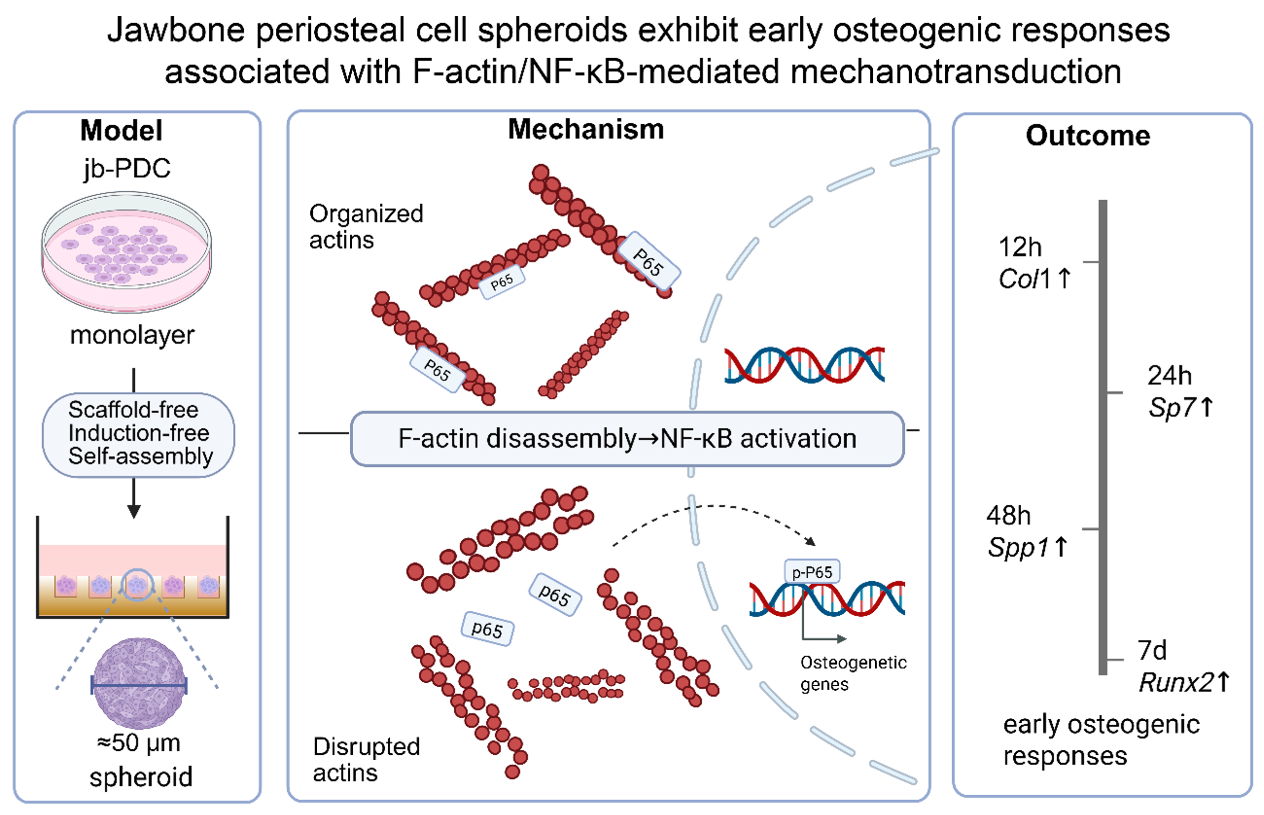
Three-dimensional (3D) spheroids have emerged as a powerful approach for studying ossification and bone regeneration. However, it remains unclear whether a 3D self-assembly process can initiate early osteogenic responses under physiologically relevant, induction-free conditions. In this study, we established a scaffold-free, self-assembling spheroid model using mouse jawbone periosteum-derived cells (jb-PDCs) without exogenous osteogenic inducers. Compared with monolayer cultures, spheroid formation induced the sequential upregulation of osteogenic markers, including Col1a2 at 12 h, Sp7 at 24 h, Spp1 at 48 h, and Runx2 after seven days. Transcriptomic profiling revealed significant enrichment of nuclear factor κ-light-chain-enhancer of activated B cells (NF-κB) signaling. Increased phosphorylation of the p65 subunit further confirmed NF-κB pathway activation. Pharmacological inhibition of NF-κB abolished the spheroid-associated upregulation of Col1a2, Sp7, and Spp1, whereas direct NF-κB activation in conventional monolayer cultures recapitulated this osteogenic gene expression program. Furthermore, spheroid formation was accompanied by marked F-actin disassembly. Pharmacological stabilization of actin filaments reduced both p65 phosphorylation and osteogenic gene expression. Together, these findings identify an F-actin/NF-κB mechanotransduction axis through which 3D self-assembly promotes early osteogenic gene expression in jb-PDCs. Our results suggest that cellular aggregation can intrinsically couple cytoskeletal remodeling to initiate early osteogenic responses. The inducer-free jb-PDC spheroid system also provides a useful platform for investigating the mechanobiology of early osteogenic responses and may inform the development of biomimetic strategies for craniofacial bone regeneration.
- Singh V, Bhagol A, Ashwin V. Controversies in the dental implant treatment planning for anterior maxillary aesthetic zone – A review. Natl J Maxillofac Surg. 2023;14(1):3-8. doi:10.4103/njms.njms_59_20
- Dimitriou R, Jones E, McGonagle D, Giannoudis PV. Bone regeneration: current concepts and future directions. BMC Med. 2011;9(1). doi:10.1186/1741-7015-9-66
- Kawecki F, Clafshenkel WP, Fortin M, Auger FA, Fradette J. Biomimetic Tissue‐Engineered Bone Substitutes for Maxillofacial and Craniofacial Repair: The Potential of Cell Sheet Technologies. Adv Healthc Mater. 2017;7(6). doi:10.1002/adhm.201700919
- Debnath S, Yallowitz AR, McCormick J, et al. Discovery of a periosteal stem cell mediating intramembranous bone formation. Nature. 2018;562(7725):133-139. doi:10.1038/s41586-018-0554-8
- Zhang S, Zhu J, Jin S, Sun W, Ji W, Chen Z. Jawbone periosteum‐derived cells with high osteogenic potential controlled by R‐spondin 3. FASEB J. 2024;38(19). doi:10.1096/fj.202400988rr
- Jin S, Zhang C, Ji W. Danxibao cexu jiexi xiahegu yu zhiya fayu yizhixing. [Single-cell Sequencing Resolves Heterogeneity in Mandible and Limb Bud Development]. Journal of Oral Research. 2025;41(10):849-855. [In Chinese]. doi:10.13701/j.cnki.kqyxyj.2025.10.004
- Duchamp de Lageneste O, Julien A, Abou-Khalil R, et al. Periosteum contains skeletal stem cells with high bone regenerative potential controlled by Periostin. Nat Commun. 2018;9(1). doi:10.1038/s41467-018-03124-z
- de Souza Castro G, de Souza W, Lima TSM, et al. The Effects of Titanium Dioxide Nanoparticles on Osteoblasts Mineralization: A Comparison between 2D and 3D Cell Culture Models. Nanomaterials. 2023;13(3):425. doi:10.3390/nano13030425
- Logovskaya LV, Bukharova TB, Volkov AV, Vikhrova EB, Makhnach OV, Goldshtein DV. Induction of Osteogenic Differentiation of Multipotent Mesenchymal Stromal Cells from Human Adipose Tissue. Bull Exp Biol Med. 2013;155(1):145-150. doi:10.1007/s10517-013-2100-x
- Ru X, Cai P, Tan M, Zheng L, Lu Z, Zhao J. Temporal transcriptome highlights the involvement of cytokine/JAK/STAT3 signaling pathway in the osteoinduction of BMSCs. J Orthop Surg Res. 2023;18(1). doi:10.1186/s13018-023-03767-9
- Clevers H. Modeling Development and Disease with Organoids. Cell. 2016;165(7):1586-1597. doi:10.1016/j.cell.2016.05.082
- Bai J, Leng S, Peng L, Svitina H, Papantoniou I. The rise of callus organoids for skeletal repair: Embedding developmental biology principles in technology-based tissue engineering. Organoid Res. 2026;2(2):026090011. doi:10.36922/or026090011
- Li C, Zhang Y, Du Y, et al. A Review of Advanced Biomaterials and Cells for the Production of Bone Organoid. Small Sci. 2023;3(8). doi:10.1002/smsc.202300027
- Park Y, Franz CK, Ryu H, et al. Three-dimensional, multifunctional neural interfaces for cortical spheroids and engineered assembloids. Sci Adv. 2021;7(12). doi:10.1126/sciadv.abf9153
- Zhu J, Zhang S, Jin S, et al. Endochondral Repair of Jawbone Defects Using Periosteal Cell Spheroids. J Dent Res. 2023;103(1):31-41. doi:10.1177/00220345231205273
- Zhang X, Jiang W, Wu X, et al. Divide-and-conquer strategy with engineered ossification center organoids for rapid bone healing through developmental cell recruitment. Nat Commun. 2025;16(1). doi:10.1038/s41467-025-61619-y
- Juhásová J, Juhás S, Klíma J, Strnádel J, Holubová M, Motlík J. Osteogenic differentiation of miniature pig mesenchymal stem cells in 2D and 3D environment. Physiol Res. 2011:559-571. doi:10.33549/physiolres.932028
- Jiang J, Zhu J, Lin H, Jin S, He Q, Ji W. High-Throughput Preosteoblastic Spheroids Elevate Fibroblast Growth Factor 23 via Parathyroid Hormone Signaling Pathway. Tissue Eng Part C Methods. 2024;30(9):402-413. doi:10.1089/ten.tec.2024.0195
- Schindelin J, Arganda-Carreras I, Frise E, et al. Fiji: an open-source platform for biological-image analysis. Nat Methods. 2012;9(7):676-682. doi:10.1038/nmeth.2019
- Gulati GS, Murphy MP, Marecic O, et al. Isolation and functional assessment of mouse skeletal stem cell lineage. Nat Protoc. 2018;13(6):1294-1309. doi:10.1038/nprot.2018.041
- Feher K, Kirsch J, Radbruch A, Chang HD, Kaiser T. Cell population identification using fluorescence-minus-one controls with a one-class classifying algorithm. Bioinformatics. 2014;30(23):3372-3378. doi:10.1093/bioinformatics/btu575
- Langmead B, Salzberg SL. Fast gapped-read alignment with Bowtie 2. Nat Methods. 2012;9(4):357-359. doi:10.1038/nmeth.1923
- Li B, Dewey CN. RSEM: accurate transcript quantification from RNA-Seq data with or without a reference genome. BMC Bioinform. 2011;12(1). doi:10.1186/1471-2105-12-323
- Love MI, Huber W, Anders S. Moderated estimation of fold change and dispersion for RNA-seq data with DESeq2. Genome Biol. 2014;15(12). doi:10.1186/s13059-014-0550-8
- Kanehisa M, Araki M, Goto S, et al. KEGG for linking genomes to life and the environment. Nucleic Acids Res. 2007;36(Database):D480-D484. doi:10.1093/nar/gkm882
- Ashburner M, Ball CA, Blake JA, et al. Gene Ontology: tool for the unification of biology. Nat Genet. 2000;25(1):25-29. doi:10.1038/75556
- Subramanian A, Tamayo P, Mootha VK, et al. Gene set enrichment analysis: A knowledge-based approach for interpreting genome-wide expression profiles. Proc Natl Acad Sci USA. 2005;102(43):15545-15550. doi:10.1073/pnas.0506580102
- Qing J, Guo Q, Lv L, et al. Organoid Culture Development for Skeletal Systems. Tissue Eng Part B Rev. 2023;29(5):545-557. doi:10.1089/ten.teb.2023.0022
- Wang L, Ruan M, Bu Q, Zhao C. Signaling Pathways Driving MSC Osteogenesis: Mechanisms, Regulation, and Translational Applications. Int J Mol Sci. 2025;26(3):1311. doi:10.3390/ijms26031311
- Huang W. Signaling and transcriptional regulation in osteoblast commitment and differentiation. Front Biosci. 2007;12(8-12):3068. doi:10.2741/2296
- Chen Y, Yang S, Lovisa S, et al. Type-I collagen produced by distinct fibroblast lineages reveals specific function during embryogenesis and Osteogenesis Imperfecta. Nat Commun. 2021;12(1). doi:10.1038/s41467-021-27563-3
- Zhu XX, Yan YW, Chen D, et al. Long non-coding RNA HoxA-AS3 interacts with EZH2 to regulate lineage commitment of mesenchymal stem cells. Oncotarget. 2016;7(39):63561-63570. doi:10.18632/oncotarget.11538
- Nakashima K, Zhou X, Kunkel G, et al. The Novel Zinc Finger-Containing Transcription Factor Osterix Is Required for Osteoblast Differentiation and Bone Formation. Cell. 2002;108(1):17-29. doi:10.1016/s0092-8674(01)00622-5
- Hoshiba T, Kawazoe N, Tateishi T, Chen G. Development of Stepwise Osteogenesis-mimicking Matrices for the Regulation of Mesenchymal Stem Cell Functions. J Biol Chem. 2009;284(45):31164-31173. doi:10.1074/jbc.m109.054676
- Bruderer M, Richards R, Alini M, Stoddart M. Role and regulation of RUNX2 in osteogenesis. eCM. 2014;28:269-286. doi:10.22203/ecm.v028a19
- Mussbacher M, Salzmann M, Brostjan C, et al. Cell Type-Specific Roles of NF-κB Linking Inflammation and Thrombosis. Front Immunol. 2019;10. doi:10.3389/fimmu.2019.00085
- Radi S, EzEldeen M, Végvári Á, et al. The proteome of osteoblasts in a 3D culture perfusion bioreactor model compared with static conditions. Sci Rep. 2025;15(1). doi:10.1038/s41598-025-96632-0
- Yu H, Lin L, Zhang Z, Zhang H, Hu H. Targeting NF-κB pathway for the therapy of diseases: mechanism and clinical study. Sig Transduct Target Ther. 2020;5(1). doi:10.1038/s41392-020-00312-6
- Swarnkar G, Zhang K, Mbalaviele G, Long F, Abu-Amer Y. Constitutive Activation of IKK2/NF-κB Impairs Osteogenesis and Skeletal Development. PLoS ONE. 2014;9(3):e91421. doi:10.1371/journal.pone.0091421
- Sandra F, Sudiono J, Chouw A, Celinna M, Dewi NM, Djamil MS. Inhibition of lipopolysaccharide-induced NF-κB maintains osteogenesis of dental pulp and periodontal ligament stem cells. Braz Oral Res. 2024;38. doi:10.1590/1807-3107bor-2024.vol38.0037
- Jimi E, Takakura N, Hiura F, Nakamura I, Hirata-Tsuchiya S. The Role of NF-κB in Physiological Bone Development and Inflammatory Bone Diseases: Is NF-κB Inhibition “Killing Two Birds with One Stone”? Cells. 2019;8(12):1636. doi:10.3390/cells8121636
- Xu Y, Wang Y, Pang X, et al. Potassium dihydrogen phosphate promotes the proliferation and differentiation of human periodontal ligament stem cells via nuclear factor kappa B pathway. Exp Cell Res. 2019;384(1):111593. doi:10.1016/j.yexcr.2019.111593
- Boyce BF, Li J, Yao Z, Xing L. Nuclear Factor-Kappa B Regulation of Osteoclastogenesis and Osteoblastogenesis. Endocrinol Metab. 2023;38(5):504-521. doi:10.3803/enm.2023.501
- Are AF, Galkin VE, Pospelova TV, Pinaev GP. The p65/RelA Subunit of NF-κB Interacts with Actin-Containing Structures. Exp Cell Res. 2000;256(2):533-544. doi:10.1006/excr.2000.4830
- Németh ZH, Deitch EA, Davidson MT, Szabó C, Vizi ES, Haskó G. Disruption of the actin cytoskeleton results in nuclear factor‐κB activation and inflammatory mediator production in cultured human intestinal epithelial cells. J Cell Physiol. 2004;200(1):71-81. doi:10.1002/jcp.10477
- Tiskratok W, Yamada M, Watanabe J, Kartikasari N, Kimura T, Egusa H. Substrate stiffness controls proinflammatory responses in human gingival fibroblasts. Sci Rep. 2023;13(1). doi:10.1038/s41598-023-28541-z
- Woodley JP, Lambert DW, Asencio IO. Understanding Fibroblast Behavior in 3D Biomaterials. Tissue Eng Part B Rev. 2022;28(3):569-578. doi:10.1089/ten.teb.2021.0010
- Saraswathibhatla A, Indana D, Chaudhuri O. Cell–extracellular matrix mechanotransduction in 3D. Nat Rev Mol Cell Biol. 2023;24(7):495-516. doi:10.1038/s41580-023-00583-1
- Case LB, Waterman CM. Integration of actin dynamics and cell adhesion by a three-dimensional, mechanosensitive molecular clutch. Nat Cell Biol. 2015;17(8):955-963. doi:10.1038/ncb3191
- Debruyne AC, Okkelman IA, Heymans N, et al. Live Microscopy of Multicellular Spheroids with the Multimodal Near-Infrared Nanoparticles Reveals Differences in Oxygenation Gradients. ACS Nano. 2024;18(19):12168-12186. doi:10.1021/acsnano.3c12539
- Pollard TD, Cooper JA. Actin, a Central Player in Cell Shape and Movement. Science. 2009;326(5957):1208-1212. doi:10.1126/science.1175862