

The efficacy and safety of acupuncture combined with ranibizumab in the treatment of macular edema secondary to retinal vein occlusion

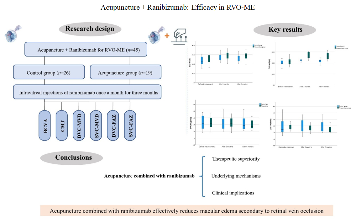
Background: Macular edema secondary to retinal vein occlusion (RVO-ME) impairs vision. Intravitreal ranibizumab is commonly used, but the adjunctive value of acupuncture remains unclear. Objective: To evaluate the clinical efficacy and safety of acupuncture combined with intravitreal ranibizumab injection for RVO-ME. Methods: Patients with RVO-ME (n = 45) were randomized into a control group (ranibizumab monotherapy) and an acupuncture group (ranibizumab and acupuncture). Both groups received monthly intravitreal ranibizumab (0.5 mg/0.05 mL) for 3 months, with a total follow-up of 6 months. Best-corrected visual acuity (BCVA), central macular thickness (CMT), macular vessel density (MVD) of superficial vascular complex (SVC), and deep vascular complex (DVC), foveal avascular zone (FAZ) area, and safety outcomes were assessed. Results: At 3 and 6 months post-treatment, BCVA, CMT, SVC-MVD, and DVC-MVD improved significantly in both groups (p<0.05). The acupuncture group showed significant reductions in SVC-FAZ and DVC-FAZ at 6 months (p<0.05), whereas the control group showed no such changes. Between-group differences at 6 months were significant for BCVA, CMT, DVC-MVD, and DVC-FAZ (p<0.05), with DVC-MVD differing significantly at 3 months (p<0.05). Adverse events (subconjunctival hemorrhage, elevated intraocular pressure, subcutaneous hemorrhage) were mild and comparable between groups (p>0.05). Conclusion: Acupuncture combined with ranibizumab effectively reduces RVO-ME, improves the microvascular structure of the macula, and is safe and reliable, with no serious adverse reactions. Relevance for patients: Patients with vision loss from RVO-ME may benefit from this combined treatment, which improves visual acuity, reduces retinal edema, and supports better long-term macular health with a favorable safety profile.
- Costa JV, Moura-Coelho N, Abreu AC, Neves P, Ornelas M, Furtado MJ. Macular edema secondary to retinal vein occlusion in a real-life setting: A multicenter, nationwide, 3-year follow-up study. Graefes Arch Clin Exp Ophthalmol. 2021;259(3):343-350. doi: 10.1007/s00417-020-04932-0
- Hirano T, Toriyama Y, Iesato Y, et al. Effect of leaking perifoveal microaneurysms on resolution of diabetic macular edema treated by combination therapy using anti-vascular endothelial growth factor and short pulse focal/ grid laser photocoagulation. JPN J Ophthalmol. 2017; 61(1):51-60. doi: 10.1007/s10384-016-0483-8
- Li XR, Zhou HY. Special attention to the cause, treatment and prevention of macular edema. RAO. 2019;39(7): 601-605. doi: 10.13389/j.cnki.rao,2019.0139
- Schmidt-Erfurth U, Garcia-Arumi J, Gerendas BS, et al. Guidelines for the management of retinal vein occlusion by the European society of retina specialists (EURETINA). Ophthalmologica. 2019;242(3):123-162. doi: 10.1159/000502041
- Chen XD, Bu JP. Observation of differentiation of syndrome on ischemic retinal vein occlusion after laser photocoagulation. IES. 2011;11(3):461-463. doi: CNKI: SUN:GJYK.0.2011-03-033
- Li X, Zhuang ZY, Bai M. Thinking mode of disease, etiology, syndrome type and treatment of macular edema in retinal vein occlusion. Chin J Ophthalmol. 2020;30(2):135-139. doi: 10.13444/j.cnki.zgzyykzz.2020.02.014
- Sierpina VS, Frenkel MA. Acupuncture: A clinical review. South Med J. 2005;98(3):330-337. doi: 10.1097/01.SMJ.0000140834.30654.0F
- Zhuang Y, Xing JJ, Li J, Zeng BY, Liang FR. History of acupuncture research. Int Rev Neurobiol. 2013;111:1-23. doi: 10.1016/B978-0-12-411545-3.00001-8
- Wang Y, Kong DN. Clinical efficacy of acupuncture plus compound xueshuantong for retinal vein occlusion and its effect on the expressions of VEGF and ET-1 in peripheral blood. Shanghai J Acu-Mox. 2019;38(8):883-887. doi: 10.13460/j.issn.1005-0957.2019.08.0883
- Zhao J, Bi XD, Si YF, Zhou L. Clinical trial of ranibizumab injection in the treatment of macular edema in non-ischemic branch retinal vein occlusion. Chin J Clin Pharmacol. 2021;37(11):1330-1332. doi: 10.13699/j.cnki.1001-6821.2021.11.007
- Pieramiei DJ, Avery RL. Ranibizumab: Treatment in patients with neovascular age-related macular degeneration. Expert Opin Bio Ther. 2006;6(11):1237-1245. doi: 10.1517/14712598.6.11.1237
- Liu W. Advances in the treatment of macular edema secondary to retinal vein occlusion. Chin J Ophthalmol Otorhinolaryngol. 2015;15(4):236-239. doi: 10.14166/j.issn.1671-2420.2015.04.004
- Wan SS, Yang YN, Xing YQ, Man ZH, Rao ZQ. Efficacy and safety of Ranibizumab for macular edema due to retinal vein occlusion: A systematic review. China Med Herald. 2013;10(27):62-64,67. doi: 10.3969/j.issn.1673-7210.2013.27.022
- Gao W, Yi XL. Meta-analysis of efficacy of ranibizumab and laser treatment for diabetic macular edema. J Xinjiang Med Univ. 2014;37(10):1321-1325. doi: 10.3969/j.issn.1009-5551.2014.10.020
- Khalil GF, Iafe NA, Jean-Pierre H, Irena T, Sadda SR, David S. Optical coherence tomography angiography analysis of the foveal avascular zone and macular vessel density after anti-VEGF therapy in eyes with diabetic macular edema and retinal vein occlusion. IOVS. 2017;58(1):30-34. doi: 10.1167/iovs.16-20579
- Feucht N, Schnbach EM, Lanzl I, Kotliar K, Lohmann CP, Maier M. Changes in the foveal microstructure after intravitreal bevacizumab application in patients with retinal vascular disease. Clin Ophthalmol. 2013;7(1):173-178. doi: 10.2147/OPTH.S37544
- Balaratnasingam C, Inoue M, Ahn S, Mccann J, Dhrami-Gavazi E, Yannuzzi LA. Visual acuity is correlated with the area of the foveal avascular zone in diabetic retinopathy and retinal vein occlusion. Ophthalmology. 2016;123(11):2352-2367. doi: 10.1016/j.ophtha.2016.07.008
- Leszczynska A, Ramm L, Spoerl E, Pillunat LE, Naim T. The short-term effect of acupuncture on different ocular blood flow parameters in patients with primary open-angle glaucoma: A randomized, clinical study. Clin Ophthalmol. 2018;12(1):1285-1291. doi: 10.2147/OPTH.S170396
- Giuffre C, Cicinelli MV, Marchese A, Coppola M, Parodi MB, Bandello F. Simultaneous intravitreal dexamethasone and aflibercept for refractory macular edema secondary to retinal vein occlusion. Graefes Arch Clin Exp Opthalmol. 2020;258(4):787-793. doi: 10.1007/s00417-019-04577-8
- Naruse S, Mori K, Kurihara M, et al. Chorioretinal blood flow changes following acupuncture between thumb and forefinger. JPN J Ophthalmol. 2001;45(2):205. doi: 10.1016/S0021-5155(00)00371-3
- Chu TC, Potter DE. Ocular hypotension induced by electroacupuncture. J Ocul Pharmacol Ther. 2002;18(4): 293-305. doi: 10.1089/10807680260218461
- Wang TH. Clinical Efficacy of Acupuncture Combined with Laser in the Treatment of Retinal Vein Obstruction Macular Edema. Jinan: Shandong University of Traditional Chinese Medicine; 2024. doi: 10.27282/d.cnki.gsdzu.2024.000442
- Chen CL, Bojikian KD, Wen JC, et al. Peripapillary retinal nerve fiber layer vascular microcirculation in eyes with glaucoma and single-hemifield visual field loss. JAMA Ophthalmol. 2017;135(5):461-468. doi: 10.1001/jamaophthalmol.2017.0261
- Wang AL, Li YJ, Hou XL, et al. Effects of acupuncture therapy on macular blood flow and structure in glaucoma. CJTCMP. 2023;38(3):1374-1378.
- Akagi T, Iida Y, Nakanishi H, et al. Microvascular density in glaucomatous eyes with hemifield visual field defects: An optical coherence tomography angiography study. Am J Ophthalmol. 2016;168:237-249. doi: 10.1016/j.ajo.2016.06.009
- Seknazi D, Coscas F, Sellam A, et al. Optical coherence tomography angiography in retinal vein occlusion: Correlations between macular vascular density, visual acuity, and peripheral nonperfusion area on fluorescein angiography. Retina. 2017;38(8):1562-1570. doi: 10.1097/IAE.0000000000001737
- Wemer JU, Bohm F, Lang GE, Dreyhaupt J, Lang GK, Enders C. Comparison of foveal avascular zone between optical coherence tomography angiography and fluorescein angiography in patients with retinal vein occlusion. PLoS One. 2019;14(6):e0217849. doi: 10.1371/journal.pone.0217849
- Ouedemi M, Assi H, Nefaa F, et al. Anatomo-functional study in branch retinal vein occIusion using swept source optical coherence tomography angiography. J Fr Ophtalmol. 2019;42(3):255-261. doi: 10.1016/j.jfo.2018.09.010
- Taku W, Tatsuhiko S, Chikako HU, et al. Retinal microvasculature and visual acuity in eyes with branch retinal vein occlusion: Imaging analysis by optical coherence tomography angiography. Invest Ophthalmol Vis Sci. 2017;58(4):2087-2094. doi: 10.1167/iovs.16-21208
- Deng Y, Zhong QW, Zhang AQ, et al. Microvascular changes after conbercept therapy in central retinal vein occlusion analyzed by optical coherence tomography angiography. Int J Ophthalmol. 2019;12(5):802-808. doi: 10.18240/ijo.2019.05.16
- Qin WX, Zhao YR, Xu JF, Wang PF, Yang T. Clinical observation of pressing while removing filiform needles to reduce adverse reactions. Shanghai J Acu-Mox. 2024;43(11):1269-1274. doi: 10.13460/j.issn.1005-0957.2024.11.1269
- Yang CX, Wang YY, Ma Y. Causative analysis of a case with buccinator diastem hematocele caused by retrobulbar injection and her nursing care. Chin Nurs Res. 2007;21(13):1219. doi: 10.3969/j.issn.1009-6493.2007.13.041
- Zhang J, Zhang R. Two cases of accidental bleeding induced by acupuncture near eyes. Zhongguo Zhen Jiu. 2014;34(2):186-188. doi: 10.13703/j.0255-2930.2014.02.024
- Gross A, Cestari DM. Optic neuropathy following retrobulbar injection: A review. Semin Ophthalmol. 2014;29(5-6):434-439. doi: 10.3109/08820538.2014.959191
- Kim CH, Kim US. Large exotropia after retrobulbar anesthesia. Indian J Ophthalmol. 2016;64(1):91-92. doi: 10.4103/0301-4738.178148
- Dikci S, Yılmaz T, Gök ZE, Demirel S, Genc O. Choroidal neovascularization secondary to ocular penetration during retrobulbar anesthesia and its treatment. Oman J Ophthalmol. 2017;10(1):44-46. doi: 10.4103/0974-620X.200695
- Mimouni M, Abualhasan H, Mtanes K, Mazzawi F, Barak Y. Patients’ experience of anxiety and pain during retrobulbar injections prior to vitrectomy. J Ophthalmol. 2019;2019:8098765. doi: 10.1155/2019/8098765