

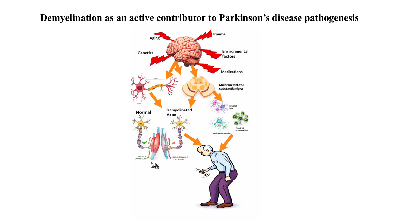
Demyelination in Parkinson’s disease: Not just a secondary bystander, but also as an active contributor to pathogenesis

Parkinson’s disease (PD) primarily affects the gray matter, with a gradual loss of dopaminergic neurons in the substantia nigra and the buildup of intraneuronal α-synuclein. Recent neuroimaging and neuropathological studies have indicated widespread alterations in white matter microstructure, raising a vital question: Is demyelination merely a consequence of axonal degeneration, or does it actively drive neurodegeneration? This review discusses the role of demyelination in PD and highlights future research directions. A literature search was conducted using keywords such as “myelin,”“demyelination,” “bystander,” “remyelination,” “impact of demyelination,” “causes of Parkinson’s,” Parkinson’s disease,” “myelination,” “functional magnetic resonance imaging,” “neurodegeneration,” “factors affecting Parkinson’s,” “PD,” “α-synuclein,” “synucleinopathy,” and “aging” across Scopus, Web of Science, OpenGrey, PubMed, ResearchGate, Cochrane Library, and Google Scholar. These terms were systematically combined using Boolean operators and linked to Parkinsonism. The review prioritized peer-reviewed studies published in the last 10 to 15 years to highlight recent findings, with a few older key studies included. The collected evidence presents a complex picture: the “bystander” hypothesis, which posits that neuronal death triggers Wallerian degeneration and myelin breakdown, is supported; however, multiple lines of evidence also suggest an early, active role for oligodendrocytes. α-synuclein buildup disrupts cellular metabolic support, leading to energy deficits in axons and heightened oxidative stress before neurons are visibly lost. Furthermore, neuroinflammatory responses associated with early demyelination exacerbate the toxic environment, accelerating dopaminergic neuron loss. In conclusion, demyelination is both a consequence and an active contributor to PD progression. Recognizing these dynamics offers new opportunities for therapies that protect myelin, potentially transforming the course of PD.
- Ascherio A, Schwarzschild MA. The epidemiology of Parkinson’s disease: risk factors and prevention. Lancet Neurol. 2016;15(12):1257-1272. doi: 10.1016/S1474-4422(16)30230-7
- Salat D, Noyce AJ, Schrag A, Tolosa E. Challenges of modifying disease progression in prediagnostic Parkinson’s disease. Lancet Neurol. 2016;15(6):637-648. doi: 10.1016/S1474-4422(16)00060-0
- Collier TJ, Kanaan NM, Kordower JH. Aging and Parkinson’s disease: Different sides of the same coin? Mov Disord. 2017;32(7):983-990. doi: 10.1002/mds.27037.
- Gepshtein S, Li X, Snider J, Plank M, Lee D, Poizner H. Dopamine function and the efficiency of human movement. J Cogn Neurosci. 2014;26(3):645-657. doi: 10.1162/JOCN_A_00503
- Lamptey RNL, Chaulagain B, Trivedi R, Gothwal A, Layek B, Singh J. A Review of the Common Neurodegenerative Disorders: Current Therapeutic Approaches and the Potential Role of Nanotherapeutics. Int J Mol Sci. 2022;23(3):1851. doi: 10.3390/IJMS23031851
- Kline EM, Houser MC, Herrick MK, Seibler P, Klein C, West A, Tansey MG. Genetic and Environmental Factors in Parkinson’s Disease Converge on Immune Function and Inflammation. Mov Disord. 2021;36(1):25-36. doi: 10.1002/mds.28411.
- Ball N, Teo WP, Chandra S, Chapman J. Parkinson’s Disease and the Environment. Front Neurol. 2019;10:218. doi: 10.3389/fneur.2019.00218.
- Khatri DK, Choudhary M, Sood A, Singh SB. Anxiety: An ignored aspect of Parkinson’s disease, lacking attention. Biomedicine and Pharmacotherapy. 2020:110776. doi: 10.1016/J.BIOPHA.2020.110776
- Chaudhuri KR, Healy DG, Schapira AH. Non-motor symptoms of Parkinson’s disease: Diagnosis and management. Lancet Neurol. 2006;5(3):235–45. doi: 10.1016/S1474-4422(06)70373-8.
- Armstrong MJ, Okun MS. Diagnosis and Treatment of Parkinson’s Disease: A Review. JAMA. 2020;323(6):548-560. doi: 10.1001/JAMA.2019.22360
- Ben-Shlomo Y, Darweesh S, Llibre-Guerra J, Marras C, San Luciano M, Tanner C. The epidemiology of Parkinson’s disease. Lancet. 2024;403(10423):283-292. doi: 10.1016/S0140-6736(23)01419-8
- Kulisevsky J. Pharmacological management of Parkinson’s disease motor symptoms: update and recommendations from an expert. Rev Neurol. 2022;75:S1-S10. doi: 10.33588/RN.75S04.2022217
- Postuma RB, Berg D, Stern M, Poewe W, Olanow CW, Oertel W, et al. MDS clinical diagnostic criteria for Parkinson’s disease. Mov Disord. 2015;30(12):1591-601. doi: 10.1002/mds.26424.
- Mack J, Marsh L. Parkinson’s Disease: Cognitive Impairment. Focus. 2017;15(1):42-54. doi: 10.1176/APPI.FOCUS.20160043
- Bohnen NI, Albin RL. White matter lesions in Parkinson’s disease. Nat Rev Neurol. 2011;7(4):229-236. doi: 10.1038/NRNEUROL.2011.21
- Hughes RC. Parkinson’s Disease and Its Management. BMJ. 1994;308(6923):281. doi: 10.1136/BMJ.308.6923.281
- Jankovic J, Aguilar LG. Current approaches to the treatment of Parkinson’s disease. Neuropsychiatr Dis Treat. 2008;4(4):743-757. doi: 10.2147/NDT.S2006
- Jankovic J, Goodman I, Safirstein B, et al. Safety and Tolerability of Multiple Ascending Doses of PRX002/ RG7935, an Anti-Synuclein Monoclonal Antibody, in Patients with Parkinson’s Disease: A Randomised Clinical Trial. JAMA Neurol. 2018;75(10):1206-1214. doi: 10.1001/JAMANEUROL.2018.1487
- Feigin VL, Nichols E, Alam T, et al. Global, regional, and national burden of neurological disorders, 1990–2016: a systematic analysis for the Global Burden of Disease Study 2016. Lancet Neurol. 2019;18(5):459-480. doi: 10.1016/S1474-4422(18)30499-X
- Naghavi M, Ong KL, Aali A, et al. Global burden of 288 causes of death and life expectancy decomposition in 204 countries and territories and 811 subnational locations, 1990–2021: a systematic analysis for the Global Burden of Disease Study 2021. Lancet. 2024;403(10440):2100-2132. doi: 10.1016/S0140-6736(24)00367-2
- Coleman C, Martin I. Unravelling Parkinson’s Disease Neurodegeneration: Does Ageing Hold the Clues? J Parkinsons Dis. 2022;12(8):2321-2338. doi: 10.3233/JPD-223363
- Aarsland D. Cognitive impairment in Parkinson’s disease and dementia with Lewy bodies. Parkinsonism Relat Disord. 2016;22:S144-S148. doi: 10.1016/j.parkreldis.2015.09.034
- Aarsland D, Batzu L, Halliday GM, et al. Parkinson’s disease-associated cognitive impairment. Nat Rev Dis Primers. 2021;7(1). doi: 10.1038/S41572-021-00280-3
- Lang AE, Melamed E, Poewe W, Rascol O. Trial designs used to study neuroprotective therapy in Parkinson’s disease. Movement Disorders. 2013;28(1):86-95. doi: 10.1002/MDS.24997
- Ye H, Robak LA, Yu M, Cykowski M, Shulman JM. Genetics and Pathogenesis of Parkinson’s Syndrome. Annu Rev Pathol. 2023;18:95-121. doi: 10.1146/annurev-pathmechdis-031521-034145.
- Barker S, Paul BD, Pieper AA. Increased Risk of Aging- Related Neurodegenerative Disease after Traumatic Brain Injury. Biomedicines. 2023;11(4):1154. doi: 10.3390/biomedicines11041154.
- Beheshti I. Exploring Risk and Protective Factors in Parkinson’s Disease. Cells. 2025;14(10):710. doi: 10.3390/cells14100710.
- Sturrock RR. Myelination of the Mouse Corpus Callosum. Neuropathol Appl Neurobiol. 1980;6(6):415-420. doi: 10.1111/J.1365-2990.1980.TB00219.X
- Fields RD. A new mechanism of nervous system plasticity: Activity-dependent myelination. Nat Rev Neurosci. 2015;16(12):756-767. doi: 10.1038/NRN4023
- Yeung MSY, Zdunek S, Bergmann O, et al. Dynamics of oligodendrocyte generation and myelination in the human brain. Cell. 2014;159(4):766-774. doi: 10.1016/J.CELL.2014.10.011
- Stadelmann C, Timmler S, Barrantes-Freer A, Simons M. Myelin in the Central Nervous System: Structure, Function, and Pathology. Physiol Rev. 2019;99(3):1381-1431. doi: 10.1152/PHYSREV.00031.2018
- Cohen CCH, Popovic MA, Klooster J, Weil MT, Möbius W, et al. Saltatory Conduction along Myelinated Axons Involves a Periaxonal Nanocircuit. Cell. 2020;180(2):311-322.e15. doi: 10.1016/j.cell.2019.11.039.
- Harris JJ, Attwell D. The energetics of CNS white matter. J Neurosci. 2012;32(1):356-71. doi: 10.1523/JNEUROSCI.3430-11.2012.
- Grydeland H, Vértes PE, Váša F, et al. Waves of Maturation and Senescence in Micro-structural MRI Markers of Human Cortical Myelination over the Lifespan. Cerebral Cortex. 2019;29(3):1369-1381. doi: 10.1093/CERCOR/BHY330
- Abu-Rub M, Miller RH. Emerging Cellular and Molecular Strategies for Enhancing Central Nervous System (CNS) Remyelination. Brain Sci. 2018;8(6):111. doi: 10.3390/BRAINSCI8060111
- Pi KS, Sang Y, Straus SK. Viral Proteins with PxxP and PY Motifs May Play a Role in Multiple Sclerosis. Viruses. 2022;14(2):281. doi: 10.3390/V14020281
- Uchewa OO, Alobu EJ, Ikechukwu FC, et al. The role of glia cell in the neural mechanism of memory formation, storage, and motor control: A review. Neuroscience. 2025;588:174- 192. doi: 10.1016/J.NEUROSCIENCE.2025.10.015
- Keirstead HS, Blakemore WF. The role of oligodendrocytes and oligodendrocyte progenitors in CNS remyelination. Adv Exp Med Biol. 2000;468:183-197. doi: 10.1007/978-1-4615-4685-6_15
- Arancibia-Cárcamo IL, Ford MC, Cossell L, Ishida K, Tohyama K, Attwell D. Node of Ranvier length as a potential regulator of myelinated axon conduction speed. eLife. 2017;6. doi: 10.7554/ELIFE.23329
- Philips T, Rothstein JD. Oligodendroglia: metabolic supporters of neurons. J Clin Invest. 2017;127(9):3271-3280. doi: 10.1172/JCI90610
- Chang KJ, Redmond SA, Chan JR. Remodelling myelination: Implications for mechanisms of neural plasticity. Nat Neurosci. 2016;19(2):190-197. doi: 10.1038/NN.4200
- Nave KA, Werner HB. Myelination of the nervous system: Mechanisms and functions. Annu Rev Cell Dev Biol. 2014;30:503-533. doi: 10.1146/ANNUREV-CELLBIO-100913-013101/CITE/REFWORKS
- Oliveira JT, Yanick C, Wein N, Gomez Limia CE. Neuron- Schwann cell interactions in peripheral nervous system homeostasis, disease, and preclinical treatment. Front Cell Neurosci. 2023;17:1248922. doi: 10.3389/fncel.2023.1248922
- Braak H, Del Tredici K. Neuropathological Staging of Brain Pathology in Sporadic Parkinson’s disease: Separating the Wheat from the Chaff. J Parkinsons Dis. 2017;7(s1):S73-S87. doi: 10.3233/JPD-179001
- Fields RD, Dutta DJ, Belgrad J, Robnett M. Cholinergic signalling in myelination. Glia. 2017;65(5):687-698. doi: 10.1002/GLIA.23101
- Zhang Y, Burock MA. Diffusion Tensor Imaging in Parkinson’s Disease and Parkinsonian Syndrome: A Systematic Review. Front Neurol. 2020;11:531993. doi: 10.3389/fneur.2020.531993
- Chen Y, Xia Y. Iterative sparse and deep learning for accurate diagnosis of Alzheimer’s disease. Pattern Recognit. 2021;116:107944. doi: 10.1016/J.PATCOG.2021.107944
- Smith LJ, Lee CY, Menozzi E, Schapira AHV. Genetic variations in GBA1 and LRRK2 genes: Biochemical and clinical consequences in Parkinson’s disease. Front Neurol. 2022;13:971252. doi: 10.3389/fneur.2022.971252.
- Chen W, Hu Y, Ju D. Gene therapy for neurodegenerative disorders: advances, insights, and prospects. Acta Pharm Sin B. 2020;10(8):1347-1359. doi: 10.1016/J.APSB.2020.01.015
- Parrilla GE, Gupta V, Wall R Vander, et al. The role of myelin in neurodegeneration: implications for drug targets and neuroprotection strategies. Rev Neurosci. 2023;35(3):271- 292. doi: 10.1515/REVNEURO-2023-0081
- Srinivasan E, Chandrasekhar G, Chandrasekar P, et al. Alpha-Synuclein Aggregation in Parkinson’s Disease. Front Med. 2021;8:736978. doi: 10.3389/fmed.2021.736978
- Breydo L, Wu JW, Uversky VN. α-Synuclein misfolding and Parkinson’s disease. Biochimica et Biophysica Acta (BBA) - Molecular Basis of Disease. 2012;1822(2):261-285. doi: 10.1016/j.bbadis.2011.10.002
- Johnson JT, Awosiminiala FW, Anumudu CK. Exploring Protein Misfolding and Aggregate Pathology in Neurodegenerative Diseases: From Molecular Mechanisms to Clinical Interventions. Appl Sci.2025;15(18). doi: 10.3390/app151810285
- Coggan JS, Bittner S, Stiefel KM, Meuth SG, Prescott SA. Physiological Dynamics in Demyelinating Diseases: Unraveling Complex Relationships through Computer Modeling. Int J Mol Sci. 2015;16(9):21215-36. doi: 10.3390/ijms160921215.
- Todorich B, Pasquini JM, Garcia CI, Paez PM, Connor JR. Oligodendrocytes and myelination: the role of iron. Glia. 2009;57(5):467-478. doi: 10.1002/glia.20784
- Ettle B, Schlachetzki JCM, Winkler J. Oligodendroglia and Myelin in Neurodegenerative Diseases: More Than Just Bystanders? Mol Neurobiol. 2016;53(5):3046-3062. doi: 10.1007/S12035-015-9205-3
- Bokulic Panichi L, Stanca S, Dolciotti C, Bongioanni P. The Role of Oligodendrocytes in Neurodegenerative Diseases: Unwrapping the Layers. Int J Mol Sci. 2025;26(10):4623. doi: 10.3390/ijms26104623
- Chen, J.-F., Wang, F., Huang, N.-X., Xiao, L., & Mei, F. Oligodendrocytes and myelin: Active players in Neurodegenerative brains? Dev Neurobiol. 2022; 82:160– 174. doi: 10.1002/dneu.22867
- Shi H, Hu X, Leak RK, Shi Y, An C, Suenaga J, Chen J, Gao Y. Demyelination as a rational therapeutic target for ischemic or traumatic brain injury. Exp Neurol. 2015;272:17-25. doi: 10.1016/j.expneurol.2015.03.017.
- Ebrahimi-Fakhari D., Wahlster L., McLean P.J. Protein Degradation Pathways in Parkinson’s Disease: Curse or Blessing. Acta Neuropathol. 2012;124:153–172. doi: 10.1007/s00401-012-1004-6.
- Kira J., Yamasaki R., Ogata H. Anti-Neurofascin Autoantibody and Demyelination. Neurochem. Int. 2019;130:104360. doi: 10.1016/j.neuint.2018.12.011
- Desplats P, Lee HJ, Bae EJ, Patrick C, Rockenstein E, Crews L, Spencer B, Masliah E, Lee SJ. Inclusion formation and neuronal cell death through neuron-to-neuron transmission of alpha-synuclein. Proc Natl Acad Sci USA. 2009;106(31):13010-5. doi: 10.1073/pnas.0903691106.
- Abeliovich A, Schmitz Y, Fariñas I, et al. Mice lacking α-synuclein display functional deficits in the nigrostriatal dopamine system. Neuron. 2000;25(1):239-252. doi: 10.1016/S0896-6273(00)80886-7
- Isik S, Yeman Kiyak B, Akbayir R, Seyhali R, Arpaci T. Microglia Mediated Neuroinflammation in Parkinson’s Disease. Cells. 2023;12(7):1012. doi: 10.3390/cells12071012.
- Tansey M.G., Wallings R.L., Houser M.C., Herrick M.K., Keating C.E., Joers V. Inflammation and Immune Dysfunction in Parkinson’s Disease. Nat Rev Immunol. 2022;22:657–673. doi: 10.1038/s41577-022-00684-6.
- Guo J, Huang X, Dou L, et al. Ageing and ageing-related diseases: from molecular mechanisms to interventions and treatments. Signal Transduct Target Ther. 2022;7(1). doi: 10.1038/S41392-022-01251-0
- Milber JM, Noorigian J V., Morley JF, et al. Lewy pathology is not the first sign of degeneration in vulnerable neurons in parkinson disease. Neurology. 2012;79(24):2307-2314. doi: 10.1212/WNL.0B013E318278FE32
- McKeith IG, Boeve BF, Dickson DW, et al. Diagnosis and management of dementia with Lewy bodies. Neurology. 2017;89(1):88-100. doi: 10.1212/WNL.0000000000004058
- Stiefel K.M., Torben-Nielsen B., Coggan J.S. Proposed evolutionary changes in the role of myelin. Front Neurosci. 2013;8.doi: 10.3389/fnins.2013.00202.
- Maitre M, Jeltsch-David H, Okechukwu NG, Klein C, Patte- Mensah C, Mensah-Nyagan AG. Myelin in Alzheimer’s disease: culprit or bystander? Acta Neuropathol Commun. 2023;11(1). doi: 10.1186/S40478-023-01554-5
- Bezzola L, Mérillat S, Gaser C, Jäncke L. Training-induced neural plasticity in golf novices. J Neurosci. 2011;31(35):12444-12448. doi: 10.1523/JNEUROSCI.1996-11.2011
- Scholz J, Klein MC, Behrens TEJ, Johansen-Berg H. Training induces changes in white-matter architecture. Nat Neurosci. 2009;12(11):1370-1371. doi: 10.1038/NN.2412
- Kiuchi K, Kitamura S, Taoka T, et al. Gray and white matter changes in subjective cognitive impairment, amnestic mild cognitive impairment and Alzheimer’s disease: A voxel-based analysis study. PLoS ONE. 2014;9(8):e104007. doi: 10.1371/JOURNAL.PONE.0104007
- Khazaee A, Ebrahimzadeh A, Babajani-Feremi A. Identifying patients with Alzheimer’s disease using resting-state fMRI and graph theory. Clin Neurophysiol. 2015;126(11):2132- 2141. doi: 10.1016/J.CLINPH.2015.02.060
- Kassa RM, Sechi E, Flanagan EP, et al. Onset of progressive motor impairment in patients with critical central nervous system demyelinating lesions. Mult Scler J. 2021;27(6):895- 902. doi: 10.1177/1352458520940983
- Carvalho de Abreu DC, Pieruccini-Faria F, Son S, Montero- Odasso M, Camicioli R. Is white matter hyperintensity burden associated with cognitive and motor impairment in patients with parkinson’s disease? A systematic review and meta-analysis. Neurosci Biobehav Rev. 2024;161:105677. doi: 10.1016/J.NEUBIOREV.2024.105677
- Yang W, Xu S, Zhou M, Chan P. Aging-related biomarkers for the diagnosis of Parkinson’s disease based on bioinformatics analysis and machine learning. Aging. 2024;16(17):12191- 12208. doi: 10.18632/aging. 205954
- Zhou C, Cheng O. Associations of the Life’s Essential 8 with Parkinson’s disease: a population-based study. Front Ageing Neurosci. 2025;17:1510411. doi: 10.3389/fnagi.2025.1510411
- Decramer T, Demaerel P, Van Loon J, Thijs V. Wallerian degeneration of the superior cerebellar peduncle. JAMA Neurol. 2015;72(10):1206-1208. doi: 10.1001/JAMANEUROL.2015.1170
- DeFrancesco-Lisowitz A, Lindborg JA, Niemi JP, Zigmond RE. The neuroimmunology of degeneration and regeneration in the peripheral nervous system. Neuroscience. 2015;302:174-203. doi: 10.1016/j.neuroscience.2014.09.027.
- Lunn E.R., Perry V.H., Brown M.C., Rosen H., Gordon S. Absence of Wallerian degeneration does not hinder regeneration in peripheral nerve. Eur. J Neurosci. 1989;1(1):27–33. doi: 10.1111/j.1460-9568.1989.tb00771.x.
- Cobb SR, Mehringer CM. Wallerian degeneration in a patient with Schilder disease: MR imaging demonstration. Radiology. 1987;162(2):521-522. doi: 10.1148/RADIOLOGY.162.2.3797667
- Matsusue E, Sugihara S, Fujii S, Kinoshita T, Ohama E, Ogawa T. Wallerian degeneration of the corticospinal tracts: Postmortem MR-pathologic correlations. Acta Radiol. 2007;48(6):690-694. doi: 10.1080/02841850701342112
- Lyon MF, Ogukolade BW, Brown MC, Atherton DJ, Perry VH. A gene affecting Wallerian nerve degeneration maps distally on mouse chromosome 4. Proc Natl Acad Sci USA. 1993;90(20):9717-9720. doi: 10.1073/PNAS.90.20.9717
- Tian R, Zhou Y, Ren Y, Zhang Y, Tang W. Wallerian degeneration: From mechanism to disease to imaging. Heliyon. 2024;11(1):e40729. doi: 10.1016/j.heliyon.2024.e40729.
- Rozo JA, Martínez-Gallego I, Rodríguez-Moreno A. Cajal, the neuronal theory and the idea of brain plasticity. Front Neuroanat. 2024;18:1331666. doi: 10.3389/fnana.2024.1331666.
- Hickey WF, Vass K, Lassmann H. Bone marrow-derived elements in the central nervous system: An immunohistochemical and ultrastructural survey of rat chimeras. J Neuropathol Exp Neurol. 1992;51(3):246-256. doi: 10.1097/00005072-199205000-00002
- Figlewicz DA, Hofteig JH, Druse MJ. Maternal deficiency of protein or protein and calories during lactation: Effect upon CNS myelin subfraction formation in rat offspring. Life Sci. 1978;23(21):2163-2172. doi: 10.1016/0024-3205(78)90190-X
- Ohara S, Ikuta F. On the occurrence of the fenestrated vessels in Wallerian degeneration of the peripheral nerve. Acta Neuropathol. 1985;68(3):259–262. doi: 10.1007/BF00690205.
- Lindholm D, Heumann R, Meyer M, Thoenen H. Interleukin-1 regulates synthesis of nerve growth factor in non-neuronal cells of rat sciatic nerve. Nature. 1987;330(6149):658–659. doi: 10.1038/330658a0.
- Zhang JD, Zhong ZA, Xing WY. Environmental enrichment for neuropathic pain via modulation of neuroinflammation. Front Mol Neurosci. 2025;18. doi: 10.3389/fnmol. 2025.1547647.
- Harboe M, Torvund-Jensen J, Kjaer-Sorensen K, Laursen LS. Ephrin-A1-EphA4 signalling negatively regulates myelination in the central nervous system. Glia. 2018;66(5):934-950. doi: 10.1002/GLIA.23293
- Quarles RH. Comparison of CNS and PNS myelin proteins in the pathology of myelin disorders. J Neurol Sci. 2005;228(2):187-189. doi: 10.1016/j.jns.2004.10.005
- Yiu G, He Z. Glial inhibition of CNS axon regeneration. Nat Rev Neurosci. 2006;7(8):617-627. doi: 10.1038/NRN1956
- Woodruff RH, Fruttiger M, Richardson WD, Franklin RJM. Platelet-derived growth factor regulates oligodendrocyte progenitor numbers in adult CNS and their response following CNS demyelination. Mol Cell Neurosci. 2004;25(2):252-262. doi: 10.1016/j.mcn.2003.10.014
- Coleman M. Axon degeneration mechanisms: commonality amid diversity. Nat Rev Neurosci. 2005;6:889–898. doi: 10.1038/nrn1788.
- Saxena S, Caroni P. Mechanisms of axon degeneration: From development to disease. Prog Neurobiol. 2007;83(3):174-191. doi: 10.1016/j.pneurobio.2007.07.007
- Ahdab R, Kikano R, Saade H, Riachi N. Early corticospinal tract Wallerian degeneration versus mesencephalic substantia nigra degeneration secondary to striatal stroke. Clin Neurol Neurosurg. 2014;118:101-102. doi: 10.1016/j.clineuro.2013.12.005
- Mishra B, Carson R, Hume RI, Collins CA. Sodium and potassium currents influence Wallerian degeneration of injured Drosophila axons. J Neurosci. 2013;33(48):18728-39. doi: 10.1523/JNEUROSCI.1007-13.2013.
- Goldner R, Yaron A. TIR Axons Apart: Unpredicted NADase Controls Axonal Degeneration. Neuron. 2017;93(6):1239- 1241. doi: 10.1016/j.neuron.2017.03.005
- Vargas ME, Barres BA. Why is Wallerian degeneration in the CNS so slow? Annu Rev Neurosci. 2007;30(1):153-179. doi: 10.1146/ANNUREV.NEURO.30.051606.094354
- Salazar Campos JM, Burbulla LF, Jäkel S. Are oligodendrocytes bystanders or drivers of Parkinson’s disease pathology? PLoS Biol. 2025;23(1):e3002977. doi: 10.1371/JOURNAL.PBIO.3002977
- de Lau LM, Breteler MM. Epidemiology of Parkinson’s disease. Lancet Neurology. 2006;5(6):525-535. doi: 10.1016/S1474-4422(06)70471-9
- Nussbaum RL, Ellis CE. Alzheimer’s Disease and Parkinson’s Disease. NN Engl. J. Med. 2003;348(14):1356-1364. doi: 10.1056/NEJM2003RA020003
- Grotewold N, Albin RL. Update: Protective and risk factors for Parkinson’s disease. Parkinsonism Relat Disord. 2024;125:107026. doi: 10.1016/j.parkreldis.2024.107026.
- Pang SY, Ho PW, Liu HF, Leung CT, Li L, Chang EES, Ramsden DB, Ho SL. The interplay of aging, genetics, and environmental factors in the pathogenesis of Parkinson’s disease. Transl Neurodegener. 2019 Aug 16;8:23. doi: 10.1186/s40035-019-0165-9.
- López-Otín C, Blasco MA, Partridge L, Serrano M, Kroemer G. The hallmarks of aging. Cell. 2013;153(6):1194. doi: 10.1016/J.CELL.2013.05.039
- Leng J, Goldstein DR. Impact of aging on viral infections. Microbes Infect. 2010;12(14-15):1120-1124. doi: 10.1016/J.MICINF.2010.08.009
- Li XJ, Li S. Proteasomal dysfunction in aging and Huntington disease. Neurobiol Dis. 2011;43(1):4-8. doi: 10.1016/J.NBD.2010.11.018
- Hirsch EC, Graybiel AM, Duyckaerts C, Javoy-Agid F. Neuronal loss in the pedunculopontine tegmental nucleus in Parkinson’s disease and in progressive supranuclear palsy. Proc Natl Acad Sci USA. 1987;84(16):5976-5980. doi: 10.1073/PNAS.84.16.5976
- Gonzalez-Rodriguez P, Zampese E, Surmeier DJ. Selective neuronal vulnerability in Parkinson’s disease. In: Progress in Brain Research. Elsevier. 2020:61-89. doi: 10.1016/bs.pbr.2020.02.005
- Surmeier J, Zampese E, Galtieri D, Schumacker PT. Life on the Edge: Determinants of Selective Neuronal Vulnerability in Parkinson’s Disease. In: Mitochondrial Dysfunction in Neurodegenerative Disorders. Springer International Publishing. 2016:141-173. doi: 10.1007/978-3-319-28637-2_6
- Lin MW, Lin CC, Chen YH, Yang H Bin, Hung SY. Celastrol inhibits dopaminergic neuronal death of Parkinson’S disease through activating mitophagy. Antioxidants. 2019;9(1):37. doi: 10.3390/ANTIOX9010037
- Martin LJ, Pan Y, Price AC, et al. Parkinson’s disease α-synuclein transgenic mice develop neuronal mitochondrial degeneration and cell death. J Neurosci. 2006;26(1):41-50. doi: 10.1523/JNEUROSCI.4308-05.2006
- Lee AY, Choi KT, Chang MC. Prediction of muscle loss after stroke by analysis of corticospinal tract. Transl Neurosci. 2020;11(1):328-333. doi: 10.1515/TNSCI-2020-0114
- Sulzer D. Multiple hit hypotheses for dopamine neuron loss in Parkinson’s disease. Trends Neurosci. 2007;30(5):244-250. doi: 10.1016/J.TINS.2007.03.009
- Kastner A, Hirsch EC, Lejeune O, Javoy‐Agid F, Rascol O, Agid Y. Is the Vulnerability of Neurons in the Substantia Nigra of Patients with Parkinson’s Disease Related to Their Neuromelanin Content? J Neurochem. 1992;59(3):1080- 1089. doi: 10.1111/J.1471-4159.1992.TB08350.X
- Lax NZ, Hepplewhite PD, Reeve AK, et al. Cerebellar ataxia in patients with mitochondrial DNA disease: A molecular clinicopathological study. J Neuropathol Exp Neurol. 2012;71(2):148-161. doi: 10.1097/NEN.0B013E318244477D
- Reeve A, Simcox E, Turnbull D. Ageing and Parkinson’s disease: Why is advancing age the biggest risk factor? Ageing Res Rev. 2014;14:19-30. doi: 10.1016/J.ARR.2014.01.004
- Vila M. Neuromelanin, ageing, and neuronal vulnerability in Parkinson’s disease. Movement Disorders. 2019;34(10):1440- 1451. doi: 10.1002/MDS.27776
- Elstner M, Müller SK, Leidolt L, et al. Neuromelanin, neurotransmitter status, and brainstem location determine the differential vulnerability of catecholaminergic neurons to mitochondrial DNA deletions. Mol Brain. 2011;4(1). doi: 10.1186/1756-6606-4-43
- Pandya VA, Patani R. Region-specific vulnerability in neurodegeneration: lessons from normal ageing. Ageing Res Rev. 2021;67. doi: 10.1016/J.ARR.2021.101311
- Pirko I, Noseworthy JH. Demyelinating Disorders of the Central Nervous System. TTextb Clin Neurol. 2007:1103. doi: 10.1016/B978-141603618-0.10048-7
- Chen D, Huang Y, Shi Z, et al. Demyelinating processes in aging and stroke in the central nervous system and the prospect of a treatment strategy. CNS Neurosci Ther. 2020;26(12):1219-1229. doi: 10.1111/cns.13497
- Murayama R, Cai Y, Nakamura H, Hashimoto K. Demyelination in psychiatric and neurological disorders: Mechanisms, clinical impact, and novel therapeutic strategies. Neurosci Biobehav Rev. 2025;174:106209. doi: 10.1016/J.NEUBIOREV.2025.106209
- Yang K, Wu Z, Long J, et al. White matter changes in Parkinson’s disease. Npj Parkinson’s Disease. 2023;9(1). doi: 10.1038/s41531-023-00592-z
- Poewe W, Seppi K, Tanner CM, et al. Parkinson’s disease. Nat Rev Dis Prim. 2017;3(1). doi: 10.1038/nrdp.2017.13
- Schapira AHV, Chaudhuri KR, Jenner P. Non-motor features of Parkinson’s disease. Nat Rev Neurosci. 2017;18(7):435- 450. doi: 10.1038/nrn.2017.62
- Chen CZ, Neumann B, Förster S, Franklin RJM. Schwann cell remyelination of the central nervous system: why does it happen, and what are the benefits? Open Biol. 2021;11(1). doi: 10.1098/rsob.200352
- Akram AS, Grezenko H, Singh P, Ahmed M, Hassan BD, Hagenahalli Anand V, Elashry AA, Nazir F, Khan R. Advancing the Frontier: Neuroimaging Techniques in the Early Detection and Management of Neurodegenerative Diseases. Cureus. 2024;16(5):e61335. doi: 10.7759/cureus.61335.
- Saab AS, Nave KA. Myelin dynamics: protecting and shaping neuronal functions. Curr Opin Neurobiol. 2017;47:104-112. doi: 10.1016/j.conb.2017.09.013
- Jiang J, Sun Y, Ma Y, Xu C, Zhao X, Fu H. Advances in Therapeutics Research for Demyelinating Diseases. Pharmaceuticals. 2025;18(12):1835. doi: 10.3390/ph18121835
- Flores-León M, Outeiro TF. Expanding our understanding of synucleinopathies: proteinopathy, proteinopenia, and lipidopathy. FEBS J. 2025;292(18):4774. doi: 10.1111/febs.70011
- Kouli A, Torsney KM, Kuan WL. Parkinson’s Disease: Aetiology, Neuropathology, and Pathogenesis. Parkinson’s Dis Pathog Clin Asp. 2018:3-26. doi: 10.15586/codonpublications.parkinsonsdisease.2018.ch1
- Kim WS, Kagedal K, Halliday GM. Alpha-synuclein biology in Lewy body diseases. Alzheimer’s Res Ther. 2014;6(5-8). doi: 10.1186/s13195-014-0073-2
- Iranzo A, Tolosa E, Gelpi E, et al. Neurodegenerative disease status and post-mortem pathology in idiopathic rapid-eye-movement sleep behavior disorder: An observational cohort study. Lancet Neurol. 2013;12(5):443-453. doi: 10.1016/S1474-4422(13)70056-5
- Zhang W, Xiao D, Mao Q, Xia H. Role of neuroinflammation in neurodegeneration development. Signal Transduction and Targeted Therapy. 2023;8(1). doi: 10.1038/s41392-023-01486-5
- Ford B. Pain in Parkinson’s disease. Mov Disord. 2010;25(S1). doi: 10.1002/mds. 22716
- Truong DD, Bhidayasiri R, Wolters E. Management of non-motor symptoms in advanced Parkinson’s disease. J Neurol Sci. 2008;266(1–2):216-228. doi: 10.1016/j.jns.2007.08.015
- Solari N, Bonito-Oliva A, Fisone G, Brambilla R. Understanding cognitive deficits in Parkinson’s disease: lessons from preclinical animal models. Learn Mem. 2013;20(10):592-600. doi: 10.1101/lm.032029.113
- Xie S, Yang J, Huang S, et al. Disrupted myelination network in the cingulate cortex of Parkinson’s disease. IET Syst Biol. 2022;16(3-4):98-119. doi: 10.1049/syb2.12043
- Chauhan A, Jeans AF. Is Parkinson’s disease truly a prion-like disorder? An appraisal of current evidence. Neurol Res Int. 2015;2015:1-8. doi: 10.1155/2015/345285
- Festa LK, Grinspan JB, Jordan-Sciutto KL. White matter injury across neurodegenerative diseases. Trends Neurosci. 2024;47(1):47-57. doi: 10.1016/J.TINS.2023.11.003
- Huang P, Zhang M. Magnetic Resonance Imaging Studies of Neurodegenerative Disease: From Methods to Translational Research. Neurosci Bull. 2023;39(1):99-112. doi: 10.1007/S12264-022-00905-X
- Jones DK, Cercignani M. Twenty-five pitfalls in the analysis of diffusion MRI data. NMR Biomed. 2010;23(7):803-820. doi: 10.1002/NBM.1543
- Boespflug EL, Storrs J, Sadat-Hossieny S, et al. Full diffusion characterisation implicates regionally disparate neuropathology in Mild Cognitive Impairment. Brain Struct Funct. 2014;219(1):367-379. doi: 10.1007/S00429-013-0506-X
- Berardi A, Brown JA, Jackson BS, et al. White Matter, Cognition, and Electrophysiological Variables in Bipolar Disorder: Using Multimodal Integration of Biomarker Variables Associated with Bipolar Disorder to Elucidate Deficits. Bipolar Disord. 2025;27(3):205-216. doi: 10.1111/BDI.70010
- Cedres N, Diaz-Galvan P, Diaz-Flores L, et al. The interplay between grey- and white-matter neurodegeneration in subjective cognitive decline. Aging. 2021;13(16):19963- 19977. doi: 10.18632/AGING.203467
- Maiti P, Manna J, Dunbar GL, Maiti P, Dunbar GL. Current understanding of the molecular mechanisms in Parkinson’s disease: Targets for potential treatments. Transl Neurodegener. 2017;6(1). doi: 10.1186/s40035-017-0099-z
- Berg D. Biomarkers for the early detection of Parkinson’s and Alzheimer’s disease. Neurodegener Dis. 2008;5(3–4):133- 136. doi: 10.1159/000113682
- De Santis S, Drakesmith M, Bells S, Assaf Y, Jones DK. Why diffusion tensor MRI does well only some of the time: Variance and covariance of white matter tissue microstructure attributes in the living human brain. Neuroimage. 2014;89:35-44. doi: 10.1016/J.NEUROIMAGE.2013.12.003
- Groh J, Simons M. White matter ageing and its impact on brain function. Neuron. 2025;113(1):127-139. doi: 10.1016/J.NEURON.2024.10.019
- Jiang Z, He M, Young C, et al. Dopaminergic Neurons in Zona Incerta Drive Appetitive Self-Grooming. Advanced Science. 2024;11(36). doi: 10.1002/advs. 202308974
- Duda JE, Giasson BI, Gur TL, et al. Immunohistochemical and biochemical studies demonstrate a distinct profile of α-Synuclein permutations in multiple system atrophy. J Neuropathol Exp Neurol. 2000;59(9):830-841. doi: 10.1093/JNEN/59.9.830
- Tsao JW, Brown MC, Carden MJ, McLean WG, Perry VH. Loss of the Compound Action Potential: An Electrophysiological, Biochemical and Morphological Study of Early Events in Axonal Degeneration in the C57BL/Ola Mouse. Eur J Neurosci. 1994;6(4):516-524. doi: 10.1111/J.1460-9568.1994.TB00295.X
- Choonara YE, Pillay V, Du Toit LC, et al. Trends in the molecular pathogenesis and clinical therapeutics of common neurodegenerative disorders. Int J Mol Sci. 2009;10(6):2510- 2557. doi: 10.3390/IJMS10062510
- Orr CF, Rowe DB, Mizuno Y, Mori H, Halliday GM. A possible role for humoral immunity in the pathogenesis of Parkinson’s disease. Brain. 2005;128(11):2665-2674. doi: 10.1093/BRAIN/AWH625
- Klein C, Westenberger A. Genetics of Parkinson’s disease. Cold Spring Harb Perspect Med. 2012;2(1):a008888. doi: 10.1101/cshperspect.a008888
- Bertram L, Tanzi RE. The genetic epidemiology of neurodegenerative disease. J Clin Invest. 2005;115(6):1449-1457. doi: 10.1172/JCI24761
- Hernandez DG, Reed X, Singleton AB. Genetics in Parkinson disease: Mendelian versus non-Mendelian inheritance. J Neurochem. 2016;139(s1):59-74. doi: 10.1111/jnc.13593
- Bonifati V, Rizzu P, Van Baren MJ, et al. Mutations in the DJ-1 gene are associated with autosomal recessive early-onset Parkinsonism. Science. 2003;299(5604):256-259. doi: 10.1126/science.1077209
- Ramirez A, Heimbach A, Gründemann J, et al. Hereditary Parkinsonism with dementia is caused by mutations in ATP13A2, encoding a lysosomal type 5 P-type ATPase. Nat Genet. 2006;38(10):1184-1191. doi: 10.1038/ng1884
- Martin I, Dawson VL, Dawson TM. Recent advances in the genetics of parkinson’s disease. Annu Rev Genomics Hum Genet. 2011;12(1):301-325. doi: 10.1146/annurev-genom-082410-101440
- Kim HJ, DiBernardo AB, Sloane JA, et al. WAVE1 is required for oligodendrocyte morphogenesis and normal CNS myelination. J Neurosci. 2006;26(21):5849-5859. doi: 10.1523/JNEUROSCI.4921-05.2006
- García-Domínguez M. Neuroinflammation: Mechanisms, Dual Roles, and Therapeutic Strategies in Neurological Disorders. Curr Issues Mol Biol. 2025; 47(6):417. doi: 10.3390/cimb47060417
166 Double KL. Neuronal vulnerability in parkinson’s disease. Parkinsonism Relat Disord. 2012;18:S6. doi: 10.1016/s1353-8020(11)70098-0
- Kamath T, Abdulraouf A, Burris SJ, et al. Single-cell genomic profiling of human dopamine neurons identifies a population that selectively degenerates in Parkinson’s disease. Nat Neurosci. 2022;25(5):588-595. doi: 10.1038/S41593-022-01061-1
- Sian-Hulsmann J, Riederer P. The nigral coup in parkinson’s disease by α-synuclein and its associated rebels. Cells. 2021;10(3):598. doi: 10.3390/cells10030598
- Korecka JA, Moloney EB, Eggers R, et al. Repulsive guidance molecule a (RGMa) induces neuropathological and behavioral changes that closely resemble parkinson’s disease. J Neurosci. 2017;37(39):9361-9379. doi: 10.1523/JNEUROSCI.0084-17.2017
- Tarasova T V., Lytkina OA, Roman AY, Bachurin SO, Ustyugov AA. The role of alpha-synuclein in the development of the dopaminergic neurons in the substantia nigra and ventral tegmental area. Dokl Biol Sci. 2016;466(1):5-7. doi: 10.1134/S0012496616010117
- Saab AS, Tzvetavona ID, Trevisiol A, et al. Oligodendroglial NMDA Receptors Regulate Glucose Import and Axonal Energy Metabolism. Neuron. 2016;91(1):119-132. doi: 10.1016/J.NEURON.2016.05.016
- Spillantini MG, Schmidt ML, Lee VMY, Trojanowski JQ, Jakes R, Goedert M. α-synuclein in Lewy bodies. Nature. 1997;388(6645):839-840. doi: 10.1038/42166
- Cambron M, D’Haeseleer M, Laureys G, Clinckers R, Debruyne J, De Keyser J. White-matter astrocytes, axonal energy metabolism, and axonal degeneration in multiple sclerosis. J Cereb Blood Flow Metab. 2012;32(3):413-424. doi: 10.1038/JCBFM.2011.193
- Fiorini MR, Li J, Fon EA, Farhan SMK, Thomas RA. Neural networks reveal novel gene signatures in Parkinson’s disease from single-nuclei transcriptomes. NPJ Parkinsons Dis. 2025;11(1):304. doi: 10.1038/s41531-025-01147-0
- Zonouzi M, Renzi M, Farrant M, Cull-Candy SG. Bidirectional plasticity of calcium-permeable AMPA receptors in oligodendrocyte lineage cells. Nat Neurosci. 2011;14(11):1430-1438. doi: 10.1038/NN.2942
- Goldman SA, Nedergaard M, Windrem MS. Glial progenitor cell - Based treatment and modeling of neurological disease. Science. 2012;338(6106):491-495. doi: 10.1126/SCIENCE.1218071
- Okano H, Morimoto S. iPSC-based disease modeling and drug discovery in cardinal neurodegenerative disorders. Cell Stem Cell. 2022;29(2):189-208. doi: 10.1016/J.STEM.2022.01.007
- Affrald R J, Narayan S. A review: oligodendrocytes in neuronal axonal conduction and methods for enhancing their performance. Int J Neurosci. 2025;135(12):1328-1349. doi: 10.1080/00207454.2024.2362200.
- Duncan GJ, Simkins TJ, Emery B. Neuron-Oligodendrocyte Interactions in the Structure and Integrity of Axons. Front Cell Dev Biol. 2021;9:653101. doi: 10.3389/FCELL.2021.653101
- Rocha EM, de Miranda B, Sanders LH. Alpha-synuclein: Pathology, mitochondrial dysfunction, and neuroinflammation in Parkinson’s disease. Neurobiol Dis. 2018;109:249-257. doi: 10.1016/J.NBD.2017.04.004
- Oliveira LMA, Gasser T, Edwards R, et al. Alpha-synuclein research: defining strategic moves in the battle against Parkinson’s disease. NPJ Parkinsons Dis. 2021;7(1). doi: 10.1038/S41531-021-00203-9
- Micu I, Plemel JR, Lachance C, et al. The molecular physiology of the axo-myelinic synapse. Exp Neurol. 2016;276:41-50. doi: 10.1016/J.EXPNEUROL.2015.10.006
- Aggarwal S, Yurlova L, Simons M. Central nervous system myelin: Structure, synthesis and assembly. Trends Cell Biol. 2011;21(10):585-593. doi: 10.1016/J.TCB.2011.06.004
- Wake H, Ortiz FC, Woo DH, Lee PR, Angulo MC, Fields RD. Nonsynaptic junctions on myelinating glia promote preferential myelination of electrically active axons. Nat Commun. 2015;6(1). doi: 10.1038/NCOMMS8844
- Liu H, Yang X, Yang J, et al. IL-17 Inhibits Oligodendrocyte Progenitor Cell Proliferation and Differentiation by Increasing K+ Channel Kv1.3. Front Cell Neurosci. 2021;15. doi: 10.3389/FNCEL.2021.679413
- Lee J Il, Park JW, Lee KJ, Lee DH. Clemastine improves electrophysiologic and histomorphometric changes through promoting myelin repair in a murine model of compression neuropathy. Sci Rep. 2021;11(1). doi: 10.1038/S41598-021-00389-1
- Moura DMS, Brennan EJ, Brock R, Cocas LA. Neuron to Oligodendrocyte Precursor Cell Synapses: Protagonists in Oligodendrocyte Development and Myelination, and Targets for Therapeutics. Front Neurosci. 2022;15:779125. doi: 10.3389/FNINS.2021.779125
- Paez PM, Lyons DA. Calcium Signaling in the Oligodendrocyte Lineage: Regulators and Consequences. Annu Rev Neurosci. 2020;43(1):163-186. doi: 10.1146/ANNUREV-NEURO-100719-093305
- Osterstock G, Le Bras B, Arulkandarajah KH, et al. Axoglial synapses are formed onto pioneer oligodendrocyte precursor cells at the onset of spinal cord gliogenesis. Glia. 2018;66(8):1678-1694. doi: 10.1002/GLIA.23331
- Simons M, Trajkovic K. Neuron-glia communication in the control of oligodendrocyte function and myelin biogenesis. J Cell Sci. 2006;119(21):4381-4389. doi: 10.1242/JCS.03242
- Jang M, Gould E, Xu J, Kim EJ, Kim JH. Oligodendrocytes regulate presynaptic properties and neurotransmission through BDNF signalling in the mouse brainstem. eLife. 2019;8. doi: 10.7554/ELIFE.42156
- Arellano RO, Sánchez-Gómez MV, Alberdi E, et al. Axon-to-glia interaction regulates GABA A receptor expression in oligodendrocytes. Mol Pharmacol. 2016;89(1):63-74. doi: 10.1124/MOL.115.100594
- Campbell GR, Ziabreva I, Reeve AK, et al. Mitochondrial DNA deletions and neurodegeneration in multiple sclerosis. Ann Neurol. 2011;69(3):481-492. doi: 10.1002/ANA.22109
- Coman I, Aigrot MS, Seilhean D, et al. Nodal, paranodal and juxtaparanodal axonal proteins during demyelination and remyelination in multiple sclerosis. Brain. 2006;129(12):3186-3195. doi: 10.1093/BRAIN/AWL144
- Black JA, Felts P, Smith KJ, Kocsis JD, Waxman SG. Distribution of sodium channels in chronically demyelinated spinal cord axons: immuno-ultrastructural localisation and electrophysiological observations. Brain Res. 1991;544(1):59-70. doi: 10.1016/0006-8993(91)90885-Y
- Kędzia D, Galita G, Majsterek I, Rozpędek-Kamińska W. Microglia, Astrocytes, and Oligodendrocytes in Parkinson’s Disease: Neuroinflammatory Crosstalk and Emerging Therapeutic Strategies. Biomolecules. 2026; 16(1):156. doi: 10.3390/biom16010156
- Thornton MA, Hughes EG. Neuron-Oligodendroglia Interactions: Activity-Dependent Regulation of Cellular Signalling. Neurosci Lett. 2020;727:134916. doi: 10.1016/J.NEULET.2020.134916
- Karadottir R, Cavelier P, Bergersen L, Attwell D. NMDA receptors are expressed in oligodendrocytes and activated in ischemia. Nature. 2005;438(7071):1162-1166.
- Kuhn S, Gritti L, Crooks D, Dombrowski Y. Oligodendrocytes in development, myelin generation and beyond. Cells. 2019;8(11):1424. doi: 10.3390/CELLS8111424
- Suter TACS, He Z. How Oligodendrocytes Help the Brain Function. Front Young Minds. 2021;9. doi: 10.3389/FRYM.2021.682189
- Murakami H, Shiraishi T, Umehara T, Omoto S, Iguchi Y. Recent Advances in Drug Therapy for Parkinson’s Disease. Intern Med. 2023;62(1):33-42. doi: 10.2169/internalmedicine.8940-21.
- Sohail B, Iqbal MA, Razzaq A, Nafe AW, Malik R. Recent Advances in the Role of Rehabilitative Therapies for Parkinson’s Disease: A Literature Review. J Mind Med Sci. 2023; 10(1):85-105. doi.org/10.22543/2392-7674.1365
- Ricigliano VAG, Marenna S, Borrelli S, et al. Identifying Biomarkers for Remyelination and Recovery in Multiple Sclerosis: A Measure of Progress. Biomedicines. 2025;13(2):357. doi: 10.3390/BIOMEDICINES13020357
- Siderowf A, Concha-Marambio L, Lafontant DE, et al. Assessment of heterogeneity among participants in the Parkinson’s Progression Markers Initiative cohort using α-synuclein seed amplification: a cross-sectional study. Lancet Neurol. 2023;22(5):407-417. doi: 10.1016/S1474-4422(23)00109-6
- Donadio V, Incensi A, Leta V, et al. Skin nerve a-synuclein deposits A biomarker for idiopathic Parkinson disease. Neurology. 2014;82(15):1362-1369. doi: 10.1212/WNL.0000000000000316
- Saborio GP, Permanne B, Soto C. Sensitive detection of pathological prion protein by cyclic amplification of protein misfolding. Nature. 2001;411(6839):810-813. doi: 10.1038/35081095
- Bacioglu M, Maia LF, Preische O, et al. Neurofilament Light Chain in Blood and CSF as a Marker of Disease Progression in Mouse Models and in Neurodegenerative Diseases. Neuron. 2016;91(1):56-66. doi: 10.1016/J.NEURON.2016.05.018
- Beach TG, Adler CH, Sue LI, et al. Multi-organ distribution of phosphorylated α-synuclein histopathology in subjects with Lewy body disorders. Acta Neuropathol. 2010;119(6):689- 702. doi: 10.1007/S00401-010-0664-3
- Braak H, Del Tredici K, Rüb U, De Vos RAI, Jansen Steur ENH, Braak E. Staging of brain pathology related to sporadic Parkinson’s disease. Neurobiol Ageing. 2003;24(2):197-211. doi: 10.1016/S0197-4580(02)00065-9
- Aamodt WW, Waligorska T, Shen J, et al. Neurofilament Light Chain as a Biomarker for Cognitive Decline in Parkinson’s Disease. Mov Disord. 2021;36(12):2945-2950. doi: 10.1002/MDS.28779
- Buhmann C, Magnus T, Choe CU. Blood neurofilament light chain in Parkinson’s disease. J Neural Transm. 2023;130(6):755-762. doi: 10.1007/s00702-023-02632-7.
- Barro C, Chitnis T, Weiner HL. Blood neurofilament light: a critical review of its application to neurologic disease. Ann Clin Transl Neurol. 2020;7(12):2508-2523. doi: 10.1002/ACN3.51234
- Simuni T, Chahine LM, Poston K, et al. A biological definition of neuronal α-synuclein disease: towards an integrated staging system for research. Lancet Neurol. 2024;23(2):178-190. doi: 10.1016/S1474-4422(23)00405-2
- Hattori N. Towards the era of biological biomarkers for Parkinson’s disease. Nat Rev Neurol. 2024;20(6):317-318. doi: 10.1038/s41582-024-00950-2
- Jack CR. Criteria for a biological definition of neuronal α-synuclein disease—a major conceptual step forward. Lancet Neurol. 2024;23(2):129-130. doi: 10.1016/S1474-4422(23)00456-8
- Brown RA, Narayanan S, Arnold DL. Imaging of repeated episodes of demyelination and remyelination in multiple sclerosis. Neuroimage Clin. 2014;6:20-25. doi: 10.1016/J.NICL.2014.06.009
- Bontempi P, Rozzanigo U, Amelio D, Scartoni D, Amichetti M, Farace P. Quantitative Multicomponent T2 Relaxation Showed Greater Sensitivity Than Flair Imaging to Detect Subtle Alterations at the Periphery of Lower Grade Gliomas. Front Oncol. 2021;11:651137. doi: 10.3389/fonc.2021.651137.
- Baliyan V, Das CJ, Sharma R, Gupta AK. Diffusion weighted imaging: Technique and applications. World J Radiol. 2016;8(9):785-798. doi: 10.4329/wjr.v8.i9.785.
- Huisman TA. Diffusion-weighted and diffusion tensor imaging of the brain, made easy. Cancer Imaging. 2010;10 Spec no A(1A):S163-71. doi: 10.1102/1470-7330.2010.9023.
- Harkins KD, Does MD. Simulations on the influence of myelin water in diffusion-weighted imaging. Phys Med Biol. 2016;61(13):4729-45. doi: 10.1088/0031-9155/61/13/4729.
- Horsfield Ma, Jones DK. Applications of diffusion-weighted and diffusion tensor MRI to white matter diseases - A review. NMR Biomed. 2002;15:570–7. doi: 10.1002/nbm.787
- Mathys H, Davila-Velderrain J, Peng Z, et al. Single-cell transcriptomic analysis of Alzheimer’s disease. Nature. 2019;570(7761):332-337. doi: 10.1038/S41586-019-1195-2
- Menon S, Armstrong S, Hamzeh A, Visanji NP, Sardi SP, Tandon A. Alpha-Synuclein Targeting Therapeutics for Parkinson’s Disease and Related Synucleinopathies. Front Neurol. 2022;13:852003. doi: 10.3389/FNEUR.2022.852003
- Pagano G, Taylor KI, Anzures-Cabrera J, et al. Trial of Prasinezumab in Early-Stage Parkinson’s Disease. N Engl J Med. 2022;387(5):421-432. doi: 10.1056/NEJMOA2202867
- Shalaby KE, El-Agnaf OMA. Gene-Based Therapeutics for Parkinson’s Disease. Biomedicines. 2022; 10(8):1790. doi: 10.3390/biomedicines10081790
- Mullin S, Smith L, Lee K, et al. Ambroxol for the Treatment of Patients with Parkinson’s Disease with and Without Glucocerebrosidase Gene Mutations: A Nonrandomized, Noncontrolled Trial. JAMA Neurol. 2020;77(4):427-434. doi: 10.1001/JAMANEUROL.2019.4611
- Tansey MG, Wallings RL, Houser MC, Herrick MK, Keating CE, Joers V. Inflammation and immune dysfunction in Parkinson’s disease. Nat Rev Immunol. 2022;22(11):657-673. doi: 10.1038/s41577-022-00684-6
- Elitt MS, Shick HE, Madhavan M, et al. Chemical Screening Identifies Enhancers of Mutant Oligodendrocyte Survival and Unmasks a Distinct Pathological Phase in Pelizaeus- Merzbacher Disease. Stem Cell Rep. 2018;11(3):711-726. doi: 10.1016/J.STEMCR.2018.07.015
- Chandra S, Gallardo G, Fernández-Chacón R, Schlüter OM, Südhof TC. α-Synuclein cooperates with CSPα in preventing neurodegeneration. Cell. 2005;123(3):383-396. doi: 10.1016/J.CELL.2005.09.028
- Kalia L V., Kalia SK, McLean PJ, Lozano AM, Lang AE. α-Synuclein oligomers and clinical implications for Parkinson’s disease. Ann Neurol. 2013;73(2):155. doi: 10.1002/ANA.23746
231. Franklin RJM, Frisén J, Lyons DA. Revisiting remyelination: Towards a consensus on the regeneration of CNS myelin. Semin Cell Dev Biol. 2021;116:3-9. doi: 10.1016/J.SEMCDB.2020.09.009