

Structural cell communities in the tumor microenvironment: Spatial determinants of therapeutic response

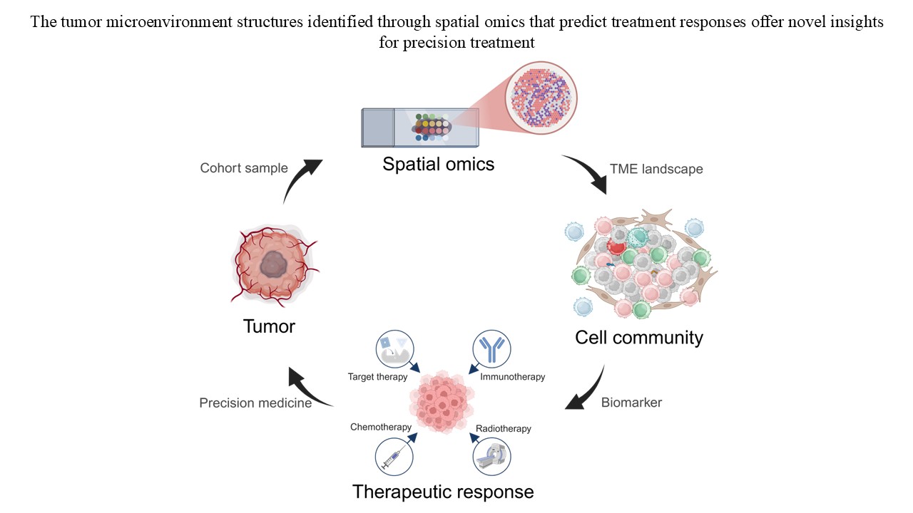
Despite advances in cancer therapies, treatment responses remain highly variable due to the complexity and heterogeneity of the tumor microenvironment. The tumor microenvironment comprises malignant, immune, stromal, and endothelial cells, along with extracellular matrix and soluble factors, all organized into spatially distinct communities that evolve dynamically throughout tumor progression and therapy. These spatial structures orchestrate tumor behavior, immune evasion, and drug resistance. Recent breakthroughs in spatial omics technologies, including spatial transcriptomics and spatial proteomics, have enabled high-resolution, multiplexed mapping of tissue architecture and molecular characteristics. These technologies provide valuable insights into how the spatial organization of cells and signaling networks within the tumor microenvironment influences therapeutic efficacy. Notably, specific structures, such as tertiary lymphoid structures, fibroblast-mediated stromal barriers, and vascular heterogeneity have been identified as spatial determinants of treatment response. By delineating cellular communities and their interactions, spatial omics technologies can reduce intratumoral complexity into clinically interpretable modules. This review summarizes the diversity of these spatial structures and their relationships with treatment outcomes in immunotherapy, chemotherapy, radiotherapy, and targeted therapy. In addition, it highlights present challenges in data integration, analytical standardization, and functional validation, and discusses future directions for incorporating spatial omics technologies into precision medicine.
- Bayik D, Lathia JD. Cancer stem cell-immune cell crosstalk in tumour progression. Nat Rev Cancer. 2021;21(8):526-536. doi: 10.1038/s41568-021-00366-w
- Fu T, Dai LJ, Wu SY, et al. Spatial architecture of the immune microenvironment orchestrates tumor immunity and therapeutic response. J Hematol Oncol. 2021;14(1):98. doi: 10.1186/s13045-021-01103-4
- Walsh LA, Quail DF. Decoding the tumor microenvironment with spatial technologies. Nat Immunol. 2023;24(12):1982-1993. doi: 10.1038/s41590-023-01678-9
- Jin MZ, Jin WL. The updated landscape of tumor microenvironment and drug repurposing. Signal Transduct Target Ther. 2020;5(1):166. doi: 10.1038/s41392-020-00280-x
- Lu MY, Sater HA, Mahmood F. Multiplex computational pathology for treatment response predication. Cancer Cell. 2021;39(8):1053-1055. doi: 10.1016/j.ccell.2021.07.014
- Elhanani O, Ben-Uri R, Keren L. Spatial profiling technologies illuminate the tumor microenvironment. Cancer Cell. 2023;41(3):404-420. doi: 10.1016/j.ccell.2023.01.010
- Dirkse A, Golebiewska A, Buder T, et al. Stem cell-associated heterogeneity in Glioblastoma results from intrinsic tumor plasticity shaped by the microenvironment. Nat Commun. 2019;10(1):1787. doi: 10.1038/s41467-019-09853-z
- Sheng W, Zhang C, Mohiuddin TM, et al. Multiplex immunofluorescence: A powerful tool in cancer immunotherapy. Int J Mol Sci. 2023;24(4):3086. doi: 10.3390/ijms24043086
- Giesen C, Wang HA, Schapiro D, et al. Highly multiplexed imaging of tumor tissues with subcellular resolution by mass cytometry. Nat Methods. 2014;11(4):417-422. doi: 10.1038/nmeth.2869
- Ståhl PL, Salmén F, Vickovic S, et al. Visualization and analysis of gene expression in tissue sections by spatial transcriptomics. Science. 2016;353(6294):78-82. doi: 10.1126/science.aaf2403
- Merritt CR, Ong GT, Church SE, et al. Multiplex digital spatial profiling of proteins and RNA in fixed tissue. Nat Biotechnol. 2020;38(5):586-599. doi: 10.1038/s41587-020-0472-9
- Lin JR, Fallahi-Sichani M, Chen JY, Sorger PK. Cyclic immunofluorescence (CycIF), a highly multiplexed method for single-cell imaging. Curr Protoc Chem Biol. 2016;8(4):251-264. doi: 10.1002/cpch.14
- Goltsev Y, Samusik N, Kennedy-Darling J, et al. Deep profiling of mouse splenic architecture with CODEX multiplexed imaging. Cell. 2018;174(4):968-981.e15. doi: 10.1016/j.cell.2018.07.010
- Radtke AJ, Kandov E, Lowekamp B, et al. IBEX: A versatile multiplex optical imaging approach for deep phenotyping and spatial analysis of cells in complex tissues. Proc Natl Acad Sci U S A. 2020;117(52):33455-33465. doi: 10.1073/pnas.2018488117
- Keren L, Bosse M, Thompson S, et al. MIBI-TOF: A multiplexed imaging platform relates cellular phenotypes and tissue structure. Sci Adv. 2019;5(10):eaax5851. doi: 10.1126/sciadv.aax5851
- Mund A, Coscia F, Kriston A, et al. Deep visual proteomics defines single-cell identity and heterogeneity. Nat Biotechnol. 2022;40(8):1231-1240. doi: 10.1038/s41587-022-01302-5
- Helmink BA, Reddy SM, Gao J, et al. B cells and tertiary lymphoid structures promote immunotherapy response. Nature. 2020;577(7791):549-555. doi: 10.1038/s41586-019-1922-8
- Petitprez F, de Reyniès A, Keung EZ, et al. B cells are associated with survival and immunotherapy response in sarcoma. Nature. 2020;577(7791):556-560. doi:10.1038/ s41586-019-1906-8
- Cabrita R, Lauss M, Sanna A, et al. Tertiary lymphoid structures improve immunotherapy and survival in melanoma. Nature. 2020;577(7791):561-565. doi: 10.1038/s41586-019-1914-8
- Williams HL, Frei AL, Koessler T, et al. The current landscape of spatial biomarkers for prediction of response to immune checkpoint inhibition. NPJ Precis Oncol. 2024;8(1):178. doi: 10.1038/s41698-024-00671-1
- Watson SS, Duc B, Kang Z, et al. Microenvironmental reorganization in brain tumors following radiotherapy and recurrence revealed by hyperplexed immunofluorescence imaging. Nat Commun. 2024;15(1):3226. doi: 10.1038/s41467-024-47185-9
- Liu W, Puri A, Fu D, et al. Dissecting the tumor microenvironment in response to immune checkpoint inhibitors via single-cell and spatial transcriptomics. Clin Exp Metastasis. 2024;41(4):313-332. doi: 10.1007/s10585-023-10246-2
- Dunn GP, Old LJ, Schreiber RD. The three Es of cancer immunoediting. Annu Rev Immunol. 2004;22:329-360. doi: 10.1146/annurev.immunol.22.012703.104803
- Koebel CM, Vermi W, Swann JB, et al. Adaptive immunity maintains occult cancer in an equilibrium state. Nature. 2007;450(7171):903-907. doi: 10.1038/nature06309
- Schreiber RD, Old LJ, Smyth MJ. Cancer immunoediting: Integrating immunity’s roles in cancer suppression and promotion. Science. 2011;331(6024):1565-1570. doi: 10.1126/science.1203486
- Shankaran V, Ikeda H, Bruce AT, et al. IFNgamma and lymphocytes prevent primary tumour development and shape tumour immunogenicity. Nature. 2001;410(6832):1107-1111. doi: 10.1038/35074122
- Galon J, Costes A, Sanchez-Cabo F, et al. Type, density, and location of immune cells within human colorectal tumors predict clinical outcome. Science. 2006;313(5795):1960- 1964. doi: 10.1126/science.1129139
- Mlecnik B, Tosolini M, Kirilovsky A, et al. Histopathologic-based prognostic factors of colorectal cancers are associated with the state of the local immune reaction. J Clin Oncol. 2011;29(6):610-618. doi: 10.1200/jco.2010.30.5425
- Hanahan D, Weinberg RA. Hallmarks of cancer: The next generation. Cell. 2011;144(5):646-674. doi: 10.1016/j.cell.2011.02.013
- Lu Q, Kou D, Lou S, et al. Nanoparticles in tumor microenvironment remodeling and cancer immunotherapy. J Hematol Oncol. 2024;17(1):16. doi: 10.1186/s13045-024-01535-8
- Zhang L, Zheng H, Jiang ST, et al. Worldwide research trends on tumor burden and immunotherapy: A bibliometric analysis. Int J Surg. 2024;110(3):1699-1710. doi: 10.1097/js9.0000000000001022
- Couzin-Frankel J. Breakthrough of the year 2013. Cancer immunotherapy. Science. 2013;342(6165):1432-1433. doi: 10.1126/science.342.6165.1432
- Maude SL, Frey N, Shaw PA, et al. Chimeric antigen receptor T cells for sustained remissions in leukemia. N Engl J Med. 2014;371(16):1507-1517. doi: 10.1056/NEJMoa1407222
- Geckin B, Konstantin Föhse F, Domínguez-Andrés J, Netea MG. Trained immunity: Implications for vaccination. Curr Opin Immunol. 2022;77:102190. doi: 10.1016/j.coi.2022.102190
- Quesada JR, Hersh EM, Manning J, et al. Treatment of hairy cell leukemia with recombinant alpha-interferon. Blood. 1986;68(2):493-497.
- Andtbacka RH, Kaufman HL, Collichio F, et al. Talimogene laherparepvec improves durable response rate in patients with advanced melanoma. J Clin Oncol. 2015;33(25):2780-2788. doi: 10.1200/jco.2014.58.3377
- Fridman WH, Pagès F, Sautès-Fridman C, Galon J. The immune contexture in human tumours: Impact on clinical outcome. Nat Rev Cancer. 2012;12(4):298-306. doi: 10.1038/nrc3245
- Sun K, Xu Y, Zhang L, et al. A phase 2 trial of enhancing immune checkpoint blockade by stereotactic radiation and in situ virus gene therapy in metastatic triple-negative breast cancer. Clin Cancer Res. 2022;28(20):4392-4401. doi: 10.1158/1078-0432.Ccr-22-0622
- Wang XQ, Danenberg E, Huang CS, et al. Spatial predictors of immunotherapy response in triple-negative breast cancer. Nature. 2023;621(7980):868-876. doi: 10.1038/s41586-023-06498-3
- Yehan Z, Sheng Q, Hong Y, et al. To develop a prognostic model for neoadjuvant immunochemotherapy efficacy in esophageal squamous cell carcinoma by analyzing the immune microenvironment. Front Immunol. 2024;15:1312380. doi: 10.3389/fimmu.2024.1312380
- Wu C, Zhang G, Wang L, et al. Spatial proteomic profiling elucidates immune determinants of neoadjuvant chemo-immunotherapy in esophageal squamous cell carcinoma. Oncogene. 2024;43(37):2751-2767. doi: 10.1038/s41388-024-03123-z
- Lim CJ, Nguyen PHD, Wasser M, et al. Immunological hallmarks for clinical response to BCG in bladder cancer. Front Immunol. 2020;11:615091. doi: 10.3389/fimmu.2020.615091
- Xiao M, Tu L, Zhou T, He Y, Li X, Zuo Q. Predictive model based on multiple immunofluorescence quantitative analysis for pathological complete response to neoadjuvant immunochemotherapy in lung squamous cell carcinoma. Front Oncol. 2024;14:1396439. doi: 10.3389/fonc.2024.1396439
- Parra ER, Zhang J, Duose DY, et al. Multi-omics analysis reveals immune features associated with immunotherapy benefit in patients with squamous cell lung cancer from phase III lung-MAP S1400I trial. Clin Cancer Res. 2024;30(8):1655-1668. doi: 10.1158/1078-0432.Ccr-23-0251
- Berrell N, Monkman J, Donovan M, et al. Spatial resolution of the head and neck cancer tumor microenvironment to identify tumor and stromal features associated with therapy response. Immunol Cell Biol. 2024;102(9):830-846. doi: 10.1111/imcb.12811
- Hayes JD, Dinkova-Kostova AT, Tew KD. Oxidative stress in cancer. Cancer Cell. 2020;38(2):167-197. doi: 10.1016/j.ccell.2020.06.001
- Larroquette M, Guegan JP, Besse B, et al. Spatial transcriptomics of macrophage infiltration in non-small cell lung cancer reveals determinants of sensitivity and resistance to anti-PD1/PD-L1 antibodies. J Immunother Cancer. 2022;10(5):e003890. doi: 10.1136/jitc-2021-003890
- Monkman J, Kim H, Mayer A, et al. Multi-omic and spatial dissection of immunotherapy response groups in non-small cell lung cancer. Immunology. 2023;169(4):487-502. doi: 10.1111/imm.13646
- Peranzoni E, Lemoine J, Vimeux L, et al. Macrophages impede CD8 T cells from reaching tumor cells and limit the efficacy of anti-PD-1 treatment. Proc Natl Acad Sci U S A. 2018;115(17):E4041-E4050. doi: 10.1073/pnas.1720948115
- Leader AM, Grout JA, Maier BB, et al. Single-cell analysis of human non-small cell lung cancer lesions refines tumor classification and patient stratification. Cancer Cell. 2021;39(12):1594-1609.e12. doi: 10.1016/j.ccell.2021.10.009
- Park S, Ock CY, Kim H, et al. Artificial intelligence-powered spatial analysis of tumor-infiltrating lymphocytes as complementary biomarker for immune checkpoint inhibition in non-small-cell lung cancer. J Clin Oncol. 2022;40(17):1916-1928. doi: 10.1200/jco.21.02010
- Fan G, Xie T, Tang L, Li L, Han X, Shi Y. The co-location of CD14+APOE+ cells and MMP7+ tumour cells contributed to worse immunotherapy response in non-small cell lung cancer. Clin Transl Med. 2024;14(9):e70009. doi: 10.1002/ctm2.70009
- Ma J, Deng Y, Zhang M, Zhang Q. Spatial tertiary lymphoid structures imply response to anti-PD-1 plus anlotinib in advanced non-small cell lung cancer. Immunology. 2024;173:536-551. doi: 10.1111/imm.13841
- Moldoveanu D, Ramsay L, Lajoie M, et al. Spatially mapping the immune landscape of melanoma using imaging mass cytometry. Sci Immunol. 2022;7(70):eabi5072. doi: 10.1126/sciimmunol.abi5072
- Dammeijer F, van Gulijk M, Mulder EE, et al. The PD-1/ PD-L1-checkpoint restrains T cell immunity in tumor-draining lymph nodes. Cancer Cell. 2020;38(5):685-700.e8. doi: 10.1016/j.ccell.2020.09.001
- Antoranz A, Van Herck Y, Bolognesi MM, et al. Mapping the immune landscape in metastatic melanoma reveals localized cell-cell interactions that predict immunotherapy response. Cancer Res. 2022;82(18):3275-3290. doi: 10.1158/0008-5472.Can-22-0363
- Gartrell RD, Marks DK, Hart TD, et al. Quantitative analysis of immune infiltrates in primary melanoma. Cancer Immunol Res. 2018;6(4):481-493. doi: 10.1158/2326-6066.Cir-17-0360
- Toki MI, Merritt CR, Wong PF, et al. High-plex predictive marker discovery for melanoma immunotherapy-treated patients using digital spatial profiling. Clin Cancer Res. 2019;25(18):5503-5512. doi: 10.1158/1078-0432.Ccr-19-0104
- Mi H, Ho WJ, Yarchoan M, Popel AS. Multi-scale spatial analysis of the tumor microenvironment reveals features of cabozantinib and nivolumab efficacy in hepatocellular carcinoma. Front Immunol. 2022;13:892250. doi: 10.3389/fimmu.2022.892250
- Huang CX, Lao XM, Wang XY, et al. Pericancerous cross-presentation to cytotoxic T lymphocytes impairs immunotherapeutic efficacy in hepatocellular carcinoma. Cancer Cell. 2024;42:2082-2097.e10. doi: 10.1016/j.ccell.2024.10.012
- Sheng J, Zhang J, Wang L, et al. Topological analysis of hepatocellular carcinoma tumour microenvironment based on imaging mass cytometry reveals cellular neighbourhood regulated reversely by macrophages with different ontogeny. Gut. 2022;71(6):1176-1191. doi: 10.1136/gutjnl-2021-324339
- Li Z, Pai R, Gupta S, et al. Presence of onco-fetal neighborhoods in hepatocellular carcinoma is associated with relapse and response to immunotherapy. Nat Cancer. 2024;5(1):167-186. doi: 10.1038/s43018-023-00672-2
- Sharma A, Seow JJW, Dutertre CA, et al. Onco-fetal reprogramming of endothelial cells drives immunosuppressive macrophages in hepatocellular carcinoma. Cell. 2020;183(2):377-394.e21. doi: 10.1016/j.cell.2020.08.040
- Sharma A, Blériot C, Currenti J, Ginhoux F. Oncofetal reprogramming in tumour development and progression. Nat Rev Cancer. 2022;22(10):593-602. doi: 10.1038/s41568-022-00497-8
- Wang H, Liang Y, Liu Z, et al. POSTN(+) cancer-associated fibroblasts determine the efficacy of immunotherapy in hepatocellular carcinoma. J Immunother Cancer. 2024;12(7):e008721. doi: 10.1136/jitc-2023-008721
- Liu Y, Xun Z, Ma K, et al. Identification of a tumour immune barrier in the HCC microenvironment that determines the efficacy of immunotherapy. J Hepatol. 2023;78(4):770-782. doi: 10.1016/j.jhep.2023.01.011
- Gil-Jimenez A, van Dijk N, Vos JL, et al. Spatial relationships in the urothelial and head and neck tumor microenvironment predict response to combination immune checkpoint inhibitors. Nat Commun. 2024;15(1):2538. doi: 10.1038/s41467-024-46450-1
- Reinstein ZZ, Zhang Y, Ospina OE, et al. Preexisting skin-resident CD8 and γδ T-cell circuits mediate immune response in Merkel cell carcinoma and predict immunotherapy efficacy. Cancer Discov. 2024;14(9):1631-1652. doi: 10.1158/2159-8290.Cd-23-0798
- Davidson G, Helleux A, Vano YA, et al. Mesenchymal-like tumor cells and myofibroblastic cancer-associated fibroblasts are associated with progression and immunotherapy response of clear cell renal cell carcinoma. Cancer Res. 2023;83(17):2952-2969. doi: 10.1158/0008-5472.Can-22-3034
- Gu X, Enane F, Tohme R, et al. PBRM1 loss in kidney cancer unbalances the proximal tubule master transcription factor hub to repress proximal tubule differentiation. Cell Rep. 2021;36(12):109747. doi: 10.1016/j.celrep.2021.109747
- Kieffer Y, Hocine HR, Gentric G, et al. Single-cell analysis reveals fibroblast clusters linked to immunotherapy resistance in cancer. Cancer Discov. 2020;10(9):1330-1351. doi: 10.1158/2159-8290.Cd-19-1384
- Tanaka M, Lum L, Hu K, et al. Tumor cell heterogeneity drives spatial organization of the intratumoral immune response. J Exp Med. 2025;222(6):e20242282. doi: 10.1084/jem.20242282
- Ma C, Yang C, Peng A, et al. Pan-cancer spatially resolved single-cell analysis reveals the crosstalk between cancer-associated fibroblasts and tumor microenvironment. Mol Cancer. 2023;22(1):170. doi: 10.1186/s12943-023-01876-x
- Tumeh PC, Harview CL, Yearley JH, et al. PD-1 blockade induces responses by inhibiting adaptive immune resistance. Nature. 2014;515(7528):568-571. doi: 10.1038/nature13954
- Gooden MJ, de Bock GH, Leffers N, Daemen T, Nijman HW. The prognostic influence of tumour-infiltrating lymphocytes in cancer: A systematic review with meta-analysis. Br J Cancer. 2011;105(1):93-103. doi: 10.1038/bjc.2011.189
- Yang Y, Attwood K, Bshara W, et al. High intratumoral CD8(+) T-cell infiltration is associated with improved survival in prostate cancer patients undergoing radical prostatectomy. Prostate. 2021;81(1):20-28. doi: 10.1002/pros.24068
- Gros A, Robbins PF, Yao X, et al. PD-1 identifies the patient-specific CD8⁺ tumor-reactive repertoire infiltrating human tumors. J Clin Invest. 2014;124(5):2246-2259. doi: 10.1172/jci73639
- Simon S, Labarriere N. PD-1 expression on tumor-specific T cells: Friend or foe for immunotherapy? Oncoimmunology. 2017;7(1):e1364828. doi: 10.1080/2162402x.2017.1364828
- Weigelin B, den Boer AT, Wagena E, et al. Cytotoxic T cells are able to efficiently eliminate cancer cells by additive cytotoxicity. Nat Commun. 2021;12(1):5217. doi: 10.1038/s41467-021-25282-3
- Gide TN, Silva IP, Quek C, et al. Close proximity of immune and tumor cells underlies response to anti-PD-1 based therapies in metastatic melanoma patients. Oncoimmunology. 2020;9(1):1659093. doi: 10.1080/2162402x.2019.1659093
- Bocchialini G, Lagrasta C, Madeddu D, et al. Spatial architecture of tumour-infiltrating lymphocytes as a prognostic parameter in resected non-small-cell lung cancer. Eur J Cardiothorac Surg. 2020;58(3):619-628. doi: 10.1093/ejcts/ezaa098
- Nirmal AJ, Maliga Z, Vallius T, et al. The spatial landscape of progression and immunoediting in primary melanoma at single-cell resolution. Cancer Discov. 2022;12(6):1518-1541. doi: 10.1158/2159-8290.Cd-21-1357
- Zhang S, Deshpande A, Verma BK, et al. Informing virtual clinical trials of hepatocellular carcinoma with spatial multi-omics analysis of a human neoadjuvant immunotherapy clinical trial. bioRxiv; 2023. [Preprint]. doi: 10.1101/2023.08.11.553000
- Aloisi F, Pujol-Borrell R. Lymphoid neogenesis in chronic inflammatory diseases. Nat Rev Immunol. 2006;6(3):205-217. doi: 10.1038/nri1786
- Drayton DL, Liao S, Mounzer RH, Ruddle NH. Lymphoid organ development: From ontogeny to neogenesis. Nat Immunol. 2006;7(4):344-353. doi: 10.1038/ni1330
- Sautès-Fridman C, Petitprez F, Calderaro J, Fridman WH. Tertiary lymphoid structures in the era of cancer immunotherapy. Nat Rev Cancer. 2019;19(6):307-325. doi: 10.1038/s41568-019-0144-6
- Gu-Trantien C, Loi S, Garaud S, et al. CD4⁺ follicular helper T cell infiltration predicts breast cancer survival. J Clin Invest. 2013;123(7):2873-92. doi: 10.1172/jci67428
- Goc J, Germain C, Vo-Bourgais TK, et al. Dendritic cells in tumor-associated tertiary lymphoid structures signal a Th1 cytotoxic immune contexture and license the positive prognostic value of infiltrating CD8+ T cells. Cancer Res. 2014;74(3):705-715. doi: 10.1158/0008-5472.Can-13-1342
- Hennequin A, Derangère V, Boidot R, et al. Tumor infiltration by Tbet+ effector T cells and CD20+ B cells is associated with survival in gastric cancer patients. Oncoimmunology. 2016;5(2):e1054598. doi: 10.1080/2162402x.2015.1054598
- Kroeger DR, Milne K, Nelson BH. Tumor-infiltrating plasma cells are associated with tertiary lymphoid structures, cytolytic T-cell responses, and superior prognosis in ovarian cancer. Clin Cancer Res. 2016;22(12):3005-3015. doi: 10.1158/1078-0432.Ccr-15-2762
- Di Caro G, Bergomas F, Grizzi F, et al. Occurrence of tertiary lymphoid tissue is associated with T-cell infiltration and predicts better prognosis in early-stage colorectal cancers. Clin Cancer Res. 2014;20(8):2147-2158. doi: 10.1158/1078-0432.Ccr-13-2590
- McMullen TP, Lai R, Dabbagh L, Wallace TM, de Gara CJ. Survival in rectal cancer is predicted by T cell infiltration of tumour-associated lymphoid nodules. Clin Exp Immunol. 2010;161(1):81-88. doi: 10.1111/j.1365-2249.2010.04147.x
- Baratin M, Simon L, Jorquera A, et al. T cell zone resident macrophages silently dispose of apoptotic cells in the lymph node. Immunity. 2017;47(2):349-362.e5. doi: 10.1016/j.immuni.2017.07.019
- Barone F, Gardner DH, Nayar S, Steinthal N, Buckley CD, Luther SA. Stromal fibroblasts in tertiary lymphoid structures: A novel target in chronic inflammation. Front Immunol. 2016;7:477. doi: 10.3389/fimmu.2016.00477
- Fridman WH, Meylan M, Pupier G, Calvez A, Hernandez I, Sautès-Fridman C. Tertiary lymphoid structures and B cells: An intratumoral immunity cycle. Immunity. 2023;56(10):2254-2269. doi: 10.1016/j.immuni.2023.08.009
- Schumacher TN, Thommen DS. Tertiary lymphoid structures in cancer. Science. 2022;375(6576):eabf9419. doi: doi:10.1126/science.abf9419
- Fridman WH, Meylan M, Petitprez F, Sun CM, Italiano A, Sautès-Fridman C. B cells and tertiary lymphoid structures as determinants of tumour immune contexture and clinical outcome. Nat Rev Clin Oncol. 2022;19(7):441-457. doi: 10.1038/s41571-022-00619-z
- Kang J, Lee JH, Cha H, et al. Systematic dissection of tumor-normal single-cell ecosystems across a thousand tumors of 30 cancer types. Nat Commun. 2024;15(1):4067. doi: 10.1038/s41467-024-48310-4
- Meylan M, Petitprez F, Becht E, et al. Tertiary lymphoid structures generate and propagate anti-tumor antibody-producing plasma cells in renal cell cancer. Immunity. 2022;55(3):527-541.e5. doi: 10.1016/j.immuni.2022.02.001
- Xu Z, Wang Q, Zhang Y, et al. Exploiting tertiary lymphoid structures gene signature to evaluate tumor microenvironment infiltration and immunotherapy response in colorectal cancer. Front Oncol. 2024;14:1383096. doi: 10.3389/fonc.2024.1383096
- Glentis A, Oertle P, Mariani P, et al. Cancer-associated fibroblasts induce metalloprotease-independent cancer cell invasion of the basement membrane. Nat Commun. 2017;8(1):924. doi: 10.1038/s41467-017-00985-8
- Sun X, Wu B, Chiang HC, et al. Tumour DDR1 promotes collagen fibre alignment to instigate immune exclusion. Nature. 2021;599(7886):673-678. doi: 10.1038/s41586-021-04057-2
- Xiao Z, Tan Y, Cai Y, et al. Nanodrug removes physical barrier to promote T-cell infiltration for enhanced cancer immunotherapy. J Control Release. 2023;356:360-372. doi: 10.1016/j.jconrel.2023.02.029
- You R, Liu YP, Xie YL, et al. Hyperfractionation compared with standard fractionation in intensity-modulated radiotherapy for patients with locally advanced recurrent nasopharyngeal carcinoma: A multicentre, randomised, open-label, phase 3 trial. Lancet. 2023;401(10380):917-927. doi: 10.1016/s0140-6736(23)00269-6
- Tang R, Yin J, Liu Y, Xue J. FLASH radiotherapy: A new milestone in the field of cancer radiotherapy. Cancer Lett. 2024;587:216651. doi: 10.1016/j.canlet.2024.216651
- Pratx G, Kapp DS. Ultra-high-dose-rate FLASH irradiation may spare hypoxic stem cell niches in normal tissues. Int J Radiat Oncol Biol Phys. 2019;105(1):190-192. doi: 10.1016/j.ijrobp.2019.05.030
- Yang Y, Tian W, Su L, et al. Tumor-infiltrating cytotoxic T cells and tumor-associated macrophages correlate with the outcomes of neoadjuvant chemoradiotherapy for locally advanced rectal cancer. Front Oncol. 2021;11:743540. doi: 10.3389/fonc.2021.743540
- Lan J, Sun L, Xu F, et al. M2 macrophage-derived exosomes promote cell migration and invasion in colon cancer. Cancer Res. 2019;79(1):146-158. doi: 10.1158/0008-5472.Can-18-0014
- Zhang S, Li N, Wang F, et al. Characterization of the tumor microenvironment and identification of spatially predictive biomarkers associated with beneficial neoadjuvant chemoradiotherapy in locally advanced rectal cancer. Pharmacol Res. 2023;197:106974. doi: 10.1016/j.phrs.2023.106974
- Tian W, Yang Y, Qin Q, et al. Vimentin and tumor-stroma ratio for neoadjuvant chemoradiotherapy response prediction in locally advanced rectal cancer. Cancer Sci. 2023;114(2):619-629. doi: 10.1111/cas.15610
- Kaufmann J, Haist M, Kur IM, et al. Tumor-stroma contact ratio - a novel predictive factor for tumor response to chemoradiotherapy in locally advanced oropharyngeal cancer. Transl Oncol. 2024;46:102019. doi: 10.1016/j.tranon.2024.102019
- Chaib S, López-Domínguez JA, Lalinde-Gutiérrez M, et al. The efficacy of chemotherapy is limited by intratumoral senescent cells expressing PD-L2. Nat Cancer. 2024;5(3):448-462. doi: 10.1038/s43018-023-00712-x
- Verdaguer H, Saurí T, Acosta DA, et al. ESMO scale for clinical actionability of molecular targets driving targeted treatment in patients with cholangiocarcinoma. Clin Cancer Res. 2022;28(8):1662-1671. doi: 10.1158/1078-0432.Ccr-21-2384
- Kulasinghe A, Monkman J, Shah ET, Matigian N, Adams MN, O’Byrne K. Spatial profiling identifies prognostic features of response to adjuvant therapy in triple negative breast cancer (TNBC). Front Oncol. 2021;11:798296. doi: 10.3389/fonc.2021.798296
- O’Rourke CJ, Salati M, Rae C, et al. Molecular portraits of patients with intrahepatic cholangiocarcinoma who diverge as rapid progressors or long survivors on chemotherapy. Gut. 2024;73(3):496-508. doi: 10.1136/gutjnl-2023-330748
- Cai S, Zhao M, Yang G, et al. Modified spatial architecture of regulatory T cells after neoadjuvant chemotherapy in non-small cell lung cancer patients. Int Immunopharmacol. 2024;137:112434. doi: 10.1016/j.intimp.2024.112434
- Yang G, Hu M, Cai S, et al. Optimizing the spatial immune landscape of CD103(+)CD8(+) tissue-resident memory T cells in non-small cell lung cancer by neoadjuvant chemotherapy. Cell Oncol (Dordr). 2024;47:1957-1971. doi: 10.1007/s13402-024-00980-4
- Huan X, Zou K, Zhang P, et al. Neoadjuvant chemotherapy is linked to an amended anti-tumorigenic microenvironment in gastric cancer. Int Immunopharmacol. 2024;127:111352. doi: 10.1016/j.intimp.2023.111352
- Zhao Y, Li D, Zhuang J, et al. Comprehensive multi-omics analysis of resectable locally advanced gastric cancer: Assessing response to neoadjuvant camrelizumab and chemotherapy in a single-center, open-label, single-arm phase II trial. Clin Transl Med. 2024;14(5):e1674. doi: 10.1002/ctm2.1674
- Bao X, Li Q, Chen D, et al. A multiomics analysis-assisted deep learning model identifies a macrophage-oriented module as a potential therapeutic target in colorectal cancer. Cell Rep Med. 2024;5(2):101399. doi: 10.1016/j.xcrm.2024.101399
- Shiau C, Cao J, Gregory MT, et al. Therapy-associated remodeling of pancreatic cancer revealed by single-cell spatial transcriptomics and optimal transport analysis. bioRxiv. 2023. [Preprint]. doi: 10.1101/2023.06.28.546848
- Hwang WL, Jagadeesh KA, Guo JA, et al. Single-nucleus and spatial transcriptome profiling of pancreatic cancer identifies multicellular dynamics associated with neoadjuvant treatment. Nat Genet. 2022;54(8):1178-1191. doi: 10.1038/s41588-022-01134-8
- Zeng Z, Zhao Y, Chen Q, et al. Hypoxic exosomal HIF- 1α-stabilizing circZNF91 promotes chemoresistance of normoxic pancreatic cancer cells via enhancing glycolysis. Oncogene. 2021;40(36):5505-5517. doi: 10.1038/s41388-021-01960-w
- Lin J, Xia L, Oyang L, et al. The POU2F1-ALDOA axis promotes the proliferation and chemoresistance of colon cancer cells by enhancing glycolysis and the pentose phosphate pathway activity. Oncogene. 2022;41(7):1024-1039. doi: 10.1038/s41388-021-02148-y
- Weinstein IB. Cancer. Addiction to oncogenes--the Achilles heal of cancer. Science. 2002;297(5578):63-64. doi: 10.1126/science.1073096
- Jin H, Wang L, Bernards R. Rational combinations of targeted cancer therapies: background, advances and challenges. Nat Rev Drug Discov. 2023;22(3):213-234. doi: 10.1038/s41573-022-00615-z
- Maemondo M, Inoue A, Kobayashi K, et al. Gefitinib or chemotherapy for non-small-cell lung cancer with mutated EGFR. N Engl J Med. 2010;362(25):2380-2388. doi: 10.1056/NEJMoa0909530
- McNamara KL, Caswell-Jin JL, Joshi R, et al. Spatial proteomic characterization of HER2-positive breast tumors through neoadjuvant therapy predicts response. Nat Cancer. 2021;2(4):400-413. doi: 10.1038/s43018-021-00190-z
- Coker EA, Stewart A, Ozer B, et al. Individualized prediction of drug response and rational combination therapy in NSCLC using artificial intelligence-enabled studies of acute phosphoproteomic changes. Mol Cancer Ther. 2022;21(6):1020-1029. doi: 10.1158/1535-7163.Mct-21-0442
- Zhou Tran Y, Minozada R, Cao X, et al. Immediate adaptation analysis implicates BCL6 as an EGFR-TKI combination therapy target in NSCLC. Mol Cell Proteomics. 2020;19(6):928-943. doi: 10.1074/mcp.RA120.002036
- De Angelis C, Nagi C, Hoyt CC, et al. Evaluation of the predictive role of tumor immune infiltrate in patients with HER2-positive breast cancer treated with neoadjuvant anti- HER2 therapy without chemotherapy. Clin Cancer Res. 2020;26(3):738-745. doi: 10.1158/1078-0432.Ccr-19-1402
- Valenza C, Guidi L, Battaiotto E, et al. Targeting HER2 heterogeneity in breast and gastrointestinal cancers. Trends Cancer. 2024;10(2):113-123. doi: 10.1016/j.trecan.2023.11.001
- Jiang L, Zhao X, Li Y, et al. The tumor immune microenvironment remodeling and response to HER2- targeted therapy in HER2-positive advanced gastric cancer. IUBMB Life. 2024;76(7):420-436. doi: 10.1002/iub.2804
- Zhang D, Ni Y, Wang Y, et al. Spatial heterogeneity of tumor microenvironment influences the prognosis of clear cell renal cell carcinoma. J Transl Med. 2023;21(1):489. doi: 10.1186/s12967-023-04336-8
- Zhang S, Yuan L, Danilova L, et al. Spatial transcriptomics analysis of neoadjuvant cabozantinib and nivolumab in advanced hepatocellular carcinoma identifies independent mechanisms of resistance and recurrence. Genome Med. 2023;15(1):72. doi: 10.1186/s13073-023-01218-y
- Arora R, Cao C, Kumar M, et al. Spatial transcriptomics reveals distinct and conserved tumor core and edge architectures that predict survival and targeted therapy response. Nat Commun. 2023;14(1):5029. doi: 10.1038/s41467-023-40271-4
- Ziemys A, Kim M, Menzies AM, et al. Integration of digital pathologic and transcriptomic analyses connects tumor-infiltrating lymphocyte spatial density with clinical response to BRAF inhibitors. Front Oncol. 2020;10:757. doi: 10.3389/fonc.2020.00757
- Siravegna G, Sartore-Bianchi A, Nagy RJ, et al. Plasma HER2 (ERBB2) copy number predicts response to HER2- targeted therapy in metastatic colorectal cancer. Clin Cancer Res. 2019;25(10):3046-3053. doi: 10.1158/1078-0432.Ccr-18-3389
- Marusyk A, Janiszewska M, Polyak K. Intratumor heterogeneity: The rosetta stone of therapy resistance. Cancer Cell. 2020;37(4):471-484. doi: 10.1016/j.ccell.2020.03.007
- Hou Y, Shen R, Chaudhary S, Gao F, Li Z. Correlation of expression of breast biomarkers in primary and metastatic breast carcinomas: A single-institution experience. Acta Cytol. 2016;60(5):481-489. doi: 10.1159/000449400
- Caswell-Jin JL, McNamara K, Reiter JG, et al. Clonal replacement and heterogeneity in breast tumors treated with neoadjuvant HER2-targeted therapy. Nat Commun. 2019;10(1):657. doi: 10.1038/s41467-019-08593-4
- Rye IH, Trinh A, Saetersdal AB, et al. Intratumor heterogeneity defines treatment-resistant HER2+ breast tumors. Mol Oncol. 2018;12(11):1838-1855. doi: 10.1002/1878-0261.12375
- Filho OM, Viale G, Stein S, et al. Impact of HER2 heterogeneity on treatment response of early-stage HER2- positive breast cancer: Phase II neoadjuvant clinical trial of T-DM1 combined with pertuzumab. Cancer Discov. 2021;11(10):2474-2487. doi: 10.1158/2159-8290.Cd-20-1557