

Research progress and application prospects of in vitro human skin models

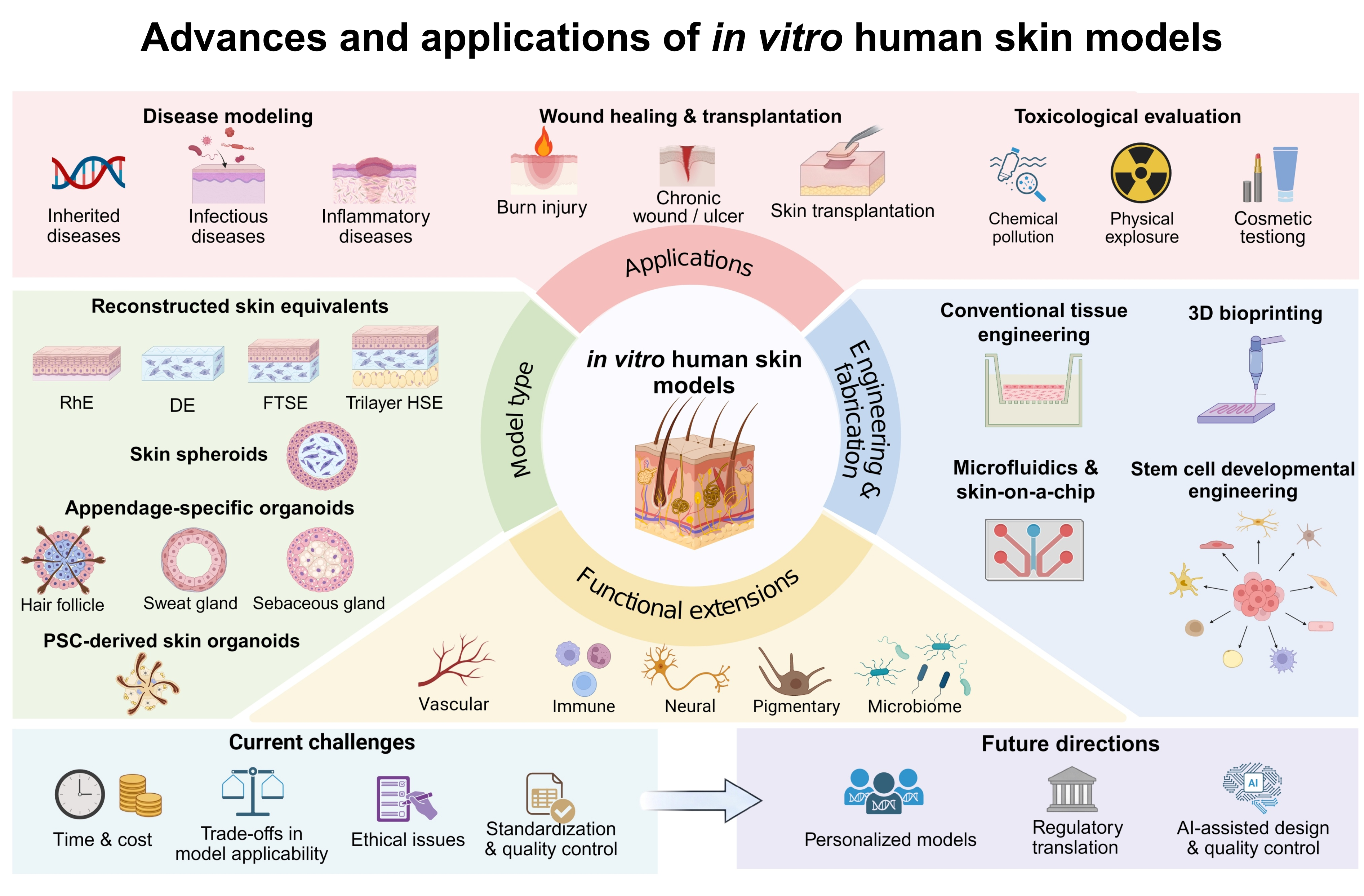
In vitro human skin models have evolved from reconstructed epidermal and full-thickness tissues to more diverse platforms that combine multicellular organization with increasingly precise control over tissue architecture and the culture microenvironment. This growing diversity increases the need for clear classification and fit-for-purpose evaluation. This review organizes current in vitro human skin models according to biological architecture, including reconstructed epidermal, dermal, full-thickness, and multilayered equivalents, spheroids, and skin organoids, and separately considers vascular, immune, neural, pigmentary, and microbial components as functional extensions. We further examine engineering strategies to construct, mature, and control these systems, including conventional tissue engineering, biomaterial design, three-dimensional bioprinting, microfluidic platforms, and stem-cell-based differentiation. For wound healing, toxicological testing, and disease modeling, we examine which functions each model reproduces, how reproducibly it performs, and how far it has progressed toward experimental, clinical, or regulatory validation. Future development should prioritize models that reproduce the biological functions required for a defined application while remaining experimentally controllable, reproducible, and appropriately validated for that context of use.
- Moniz T, Costa Lima SA, Reis S. Human skin models: From healthy to disease-mimetic systems; characteristics and applications. Br J Pharmacol. 2020;177(19):4314-4329. doi: 10.1111/bph.15184
- Moreci RS, Lechler T. Epidermal structure and differentiation. Curr Biol. 2020;30(4):R144-R149. doi: 10.1016/j.cub.2020.01.004
- Bento-Lopes L, Cabaco LC, Charneca J, Neto MV, Seabra MC, Barral DC. Melanin’s Journey from Melanocytes to Keratinocytes: Uncovering the Molecular Mechanisms of Melanin Transfer and Processing. Int J Mol Sci. 2023;24(14):11289. doi: 10.3390/ijms241411289
- Deckers J, Hammad H, Hoste E. Langerhans Cells: Sensing the Environment in Health and Disease. Front Immunol. 2018;9:93. doi: 10.3389/fimmu.2018.00093
- Galati A, Gallo RL, Cavagnero KJ. Multifaceted Roles of Dermal Fibroblasts in Skin Health and Disease. Dermatol Surg. 2025;51(9S):S5-S11. doi: 10.1097/DSS.0000000000004668
- Kabashima K, Honda T, Ginhoux F, Egawa G. The immunological anatomy of the skin. Nat Rev Immunol. 2019;19(1):19-30. doi: 10.1038/s41577-018-0084-5
- Fluhr JW, Moore DJ, Lane ME, Lachmann N, Rawlings AV. Epidermal barrier function in dry, flaky and sensitive skin: A narrative review. J Eur Acad Dermatol Venereol. 2024;38(5):812-820. doi: 10.1111/jdv.19745
- Charkoudian N. Skin blood flow in adult human thermoregulation: how it works, when it does not, and why. Mayo Clin Proc. 2003;78(5):603-612. doi: 10.4065/78.5.603
- Lee SY, Jeon S, Park KY, Hong JY. Human-Relevant In Vitro Skin Models: From Regulatory-Validated Platforms to Emerging Technologies for Translational Dermatology. Exp Dermatol. 2026;35(7):e70312. doi: 10.1111/exd.70312
- Zeng D, Li S, Du F, et al. Advances in engineered organoid models of skin for biomedical research. Burns Trauma. 2025;13:tkaf016. doi: 10.1093/burnst/tkaf016
- Jia YY, Atwood SX. Diversity of human skin three-dimensional organotypic cultures. Curr Opin Genet Dev. 2024;89:102275. doi: 10.1016/j.gde.2024.102275
- Hofmann E, Schwarz A, Fink J, Kamolz LP, Kotzbeck P. Modelling the Complexity of Human Skin In Vitro. Biomedicines. 2023;11(3):794. doi: 10.3390/biomedicines11030794
- Ali N, Hosseini M, Vainio S, Taieb A, Cario-Andre M, Rezvani HR. Skin equivalents: skin from reconstructions as models to study skin development and diseases. Br J Dermatol. 2015;173(2):391-403. doi: 10.1111/bjd.13886
- Wang D, Majid M, Chen J, Wu C, Chen Y, Ni F. Skin Organoids for Disease Modelling, Drug Screening and Regenerative Medicine: Recent Advances and Future Perspectives. Wound Repair Regen. 2026;34(1):e70117. doi: 10.1111/wrr.70117
- Quilez C, Bebiano LB, Jones E, et al. Targeting the Complexity of In Vitro Skin Models: A Review of Cutting-Edge Developments. J Invest Dermatol. 2024;144(12):2650-2670. doi: 10.1016/j.jid.2024.04.032
- Green H, Kehinde O, Thomas J. Growth of cultured human epidermal cells into multiple epithelia suitable for grafting. Proc Natl Acad Sci USA. 1979;76(11):5665-5668. doi: 10.1073/pnas.76.11.5665
- Bell E, Ivarsson B, Merrill C. Production of a tissue-like structure by contraction of collagen lattices by human fibroblasts of different proliferative potential in vitro. Proc Natl Acad Sci USA. 1979;76(3):1274-1278. doi: 10.1073/pnas.76.3.1274
- Prunieras M, Regnier M, Woodley D. Methods for cultivation of keratinocytes with an air-liquid interface. J Invest Dermatol. 1983;81(1 Suppl):28s–33s. doi: 10.1111/1523-1747.ep12540324
- Poumay Y, Dupont F, Marcoux S, Leclercq-Smekens M, Herin M, Coquette A. A simple reconstructed human epidermis: preparation of the culture model and utilization in in vitro studies. Arch Dermatol Res. 2004;296(5):203-211. doi: 10.1007/s00403-004-0507-y
- Ponec M, Weerheim A, Kempenaar J, et al. The formation of competent barrier lipids in reconstructed human epidermis requires the presence of vitamin C. J Invest Dermatol. 1997;109(3):348-355. doi: 10.1111/1523-1747.ep12336024
- Ponec M, Boelsma E, Weerheim A, Mulder A, Bouwstra J, Mommaas M. Lipid and ultrastructural characterization of reconstructed skin models. Int J Pharm. 2000;203(1-2):211-225. doi: 10.1016/s0378-5173(00)00459-2
- Thakoersing VS, van Smeden J, Mulder AA, Vreeken RJ, El Ghalbzouri A, Bouwstra JA. Increased presence of monounsaturated fatty acids in the stratum corneum of human skin equivalents. J Invest Dermatol. 2013;133(1):59-67. doi: 10.1038/jid.2012.262
- Mieremet A, Rietveld M, Absalah S, van Smeden J, Bouwstra JA, El Ghalbzouri A. Improved epidermal barrier formation in human skin models by chitosan modulated dermal matrices. PLoS ONE. 2017;12(3):e0174478. doi: 10.1371/journal.pone.0174478
- Bouwstra JA, Helder RWJ, El Ghalbzouri A. Human skin equivalents: Impaired barrier function in relation to the lipid and protein properties of the stratum corneum. Adv Drug Deliv Rev. 2021;175:113802. doi: 10.1016/j.addr.2021.05.012
- Netzlaff F, Kaca M, Bock U, et al. Permeability of the reconstructed human epidermis model Episkin in comparison to various human skin preparations. Eur J Pharm Biopharm. 2007;66(1):127-134. doi: 10.1016/j.ejpb.2006.08.012
- Organisation for Economic Co-operation and Development. Test No. 431: In Vitro Skin Corrosion: Reconstructed Human Epidermis (RHE) Test Method. Online. OECD Publishing. 2025. Accessed September 20, 2026. doi: 10.1787/9789264264618-en
- Organisation for Economic Co-operation and Development. Test No. 439: In Vitro Skin Irritation: Reconstructed Human Epidermis Test Method. Online. OECD Publishing. 2025. Accessed September 20, 2026. doi: 10.1787/9789264242845-en
- Organisation for Economic Co-operation and Development. Performance Standards for the Assessment of Proposed Similar or Modified In Vitro Reconstructed Human Epidermis (RHE) Test Methods for Skin Corrosion Testing as described in TG 431. Online. OECD Publishing. 2015. Accessed September 20, 2026. doi: 10.1787/b7c3d922-en
- Organisation for Economic Co-operation and Development. Performance Standards for the Assessment of Proposed Similar or Modified In Vitro Reconstructed Human Epidermis (RhE) Test Methods for Skin Irritation Testing as described in TG 439. Online. OECD Publishing. 2015. doi: 10.1787/745487b9-en
- Netzlaff F, Lehr CM, Wertz PW, Schaefer UF. The human epidermis models EpiSkin, SkinEthic and EpiDerm: an evaluation of morphology and their suitability for testing phototoxicity, irritancy, corrosivity, and substance transport. Eur J Pharm Biopharm. 2005;60(2):167-178. doi: 10.1016/j.ejpb.2005.03.004
- El Ghalbzouri A, Lamme E, Ponec M. Crucial role of fibroblasts in regulating epidermal morphogenesis. Cell Tissue Res. 2002;310(2):189-199. doi: 10.1007/s00441-002-0621-0
- Huet F, Severino-Freire M, Cheret J, et al. Reconstructed human epidermis for in vitro studies on atopic dermatitis: A review. J Dermatol Sci. 2018;89(3):213-218. doi: 10.1016/j.jdermsci.2017.11.015
- Lee DY, Lee JH, Yang JM, Lee ES, Park KH, Mun GH. A new dermal equivalent: the use of dermal fibroblast culture alone without exogenous materials. J Dermatol Sci. 2006;43(2):95-104. doi: 10.1016/j.jdermsci.2006.03.007
- El Ghalbzouri A, Commandeur S, Rietveld MH, Mulder AA, Willemze R. Replacement of animal-derived collagen matrix by human fibroblast-derived dermal matrix for human skin equivalent products. Biomaterials. 2009;30(1):71-78. doi: 10.1016/j.biomaterials.2008.09.002
- Haydont V, Neiveyans V, Perez P, et al. Fibroblasts from the Human Skin Dermo-Hypodermal Junction are Distinct from Dermal Papillary and Reticular Fibroblasts and from Mesenchymal Stem Cells and Exhibit a Specific Molecular Profile Related to Extracellular Matrix Organization and Modeling. Cells. 2020;9(2):368. doi: 10.3390/cells9020368
- Passariello R, Imparato G, Casale C, Urciuolo F, Netti PA. An immunocompetent bioengineered human dermal equivalent to recapitulate scar tissue formation in vitro. Acta Biomater. 2025;204:293-311. doi: 10.1016/j.actbio.2025.08.027
- Janson DG, Saintigny G, van Adrichem A, Mahe C, El Ghalbzouri A. Different gene expression patterns in human papillary and reticular fibroblasts. J Invest Dermatol. 2012;132(11):2565-2572. doi: 10.1038/jid.2012.192
- Janson D, Rietveld M, Mahe C, Saintigny G, El Ghalbzouri A. Differential effect of extracellular matrix derived from papillary and reticular fibroblasts on epidermal development in vitro. Eur J Dermatol. 2017;27(3):237-246. doi: 10.1684/ejd.2017.2984
- Louis A, Mergen C, Rietveld MH, et al. Fibroblast-derived matrix human skin equivalents reveal distinct roles for papillary and reticular fibroblasts in human skin barrier integrity and aging. Sci Rep. 2026;16(1):17668. doi: 10.1038/s41598-026-48648-3
- Janson D, Saintigny G, Mahe C, El Ghalbzouri A. Papillary fibroblasts differentiate into reticular fibroblasts after prolonged in vitro culture. Exp Dermatol. 2013;22(1):48-53. doi: 10.1111/exd.12069
- Philippeos C, Telerman SB, Oules B, et al. Spatial and Single-Cell Transcriptional Profiling Identifies Functionally Distinct Human Dermal Fibroblast Subpopulations. J Invest Dermatol. 2018;138(4):811-825. doi: 10.1016/j.jid.2018.01.016
- Tan SH, Chua DAC, Tang JRJ, Bonnard C, Leavesley D, Liang K. Design of hydrogel-based scaffolds for in vitro three-dimensional human skin model reconstruction. Acta Biomater. 2022;153:13-37. doi: 10.1016/j.actbio.2022.09.068
- Footner E, Firipis K, Liu E, et al. Layer-by-Layer Analysis of In Vitro Skin Models. ACS Biomater Sci Eng. 2023;9(11):5933-5952. doi: 10.1021/acsbiomaterials.3c00283
- Kimura S, Sekiya S, Yamashiro S, Kikuchi T, Haga M, Shimizu T. The heterogeneity of dermal mesenchymal cells reproduced in skin equivalents regulates barrier function and elasticity. EMBO Rep. 2026;27(9):2319-2344. doi: 10.1038/s44319-026-00757-w
- Marionnet C, Pierrard C, Vioux-Chagnoleau C, Sok J, Asselineau D, Bernerd F. Interactions between fibroblasts and keratinocytes in morphogenesis of dermal epidermal junction in a model of reconstructed skin. J Invest Dermatol. 2006;126(5):971-979. doi: 10.1038/sj.jid.5700230
- El Ghalbzouri A, Jonkman MF, Dijkman R, Ponec M. Basement membrane reconstruction in human skin equivalents is regulated by fibroblasts and/or exogenously activated keratinocytes. J Invest Dermatol. 2005;124(1):79-86. doi: 10.1111/j.0022-202X.2004.23549.x
- Huber B, Link A, Linke K, Gehrke SA, Winnefeld M, Kluger PJ. Integration of Mature Adipocytes to Build-Up a Functional Three-Layered Full-Skin Equivalent. Tissue Eng Part C Methods. 2016;22(8):756-764. doi: 10.1089/ten.TEC.2016.0141
- Workman VL, Giblin AV, Green NH, MacNeil S, Hearnden V. Development of a tissue-engineered skin model with epidermal, dermal and hypodermal components. In Vitro Model. 2023;2(6):297-306. doi: 10.1007/s44164-023-00058-9
- Jager J, Vahav I, Thon M, et al. Reconstructed Human Skin with Hypodermis Shows Essential Role of Adipose Tissue in Skin Metabolism. Tissue Eng Regen Med. 2024;21(3):499-511. doi: 10.1007/s13770-023-00621-1
- Avelino TM, Harb SV, Adamoski D, et al. Unveiling the impact of hypodermis on gene expression for advancing bioprinted full-thickness 3D skin models. Commun Biol. 2024;7(1):1437. doi: 10.1038/s42003-024-07106-4
- Kruglikov IL, Zhang Z, Scherer PE. Skin aging: Dermal adipocytes metabolically reprogram dermal fibroblasts. Bioessays. 2022;44(1):e2100207. doi: 10.1002/bies.202100207
- Feng X, Li GY, Ramier A, Eltony AM, Yun SH. In vivo stiffness measurement of epidermis, dermis, and hypodermis using broadband Rayleigh-wave optical coherence elastography. Acta Biomater. 2022;146:295-305. doi: 10.1016/j.actbio.2022.04.030
- Gibbs S, Murli S, De Boer G, Mulder A, Mommaas AM, Ponec M. Melanosome capping of keratinocytes in pigmented reconstructed epidermis--effect of ultraviolet radiation and 3-isobutyl-1-methyl-xanthine on melanogenesis. Pigment Cell Res. 2000;13(6):458-466. doi: 10.1034/j.1600-0749.2000.130608.x
- Rimal R, Muduli S, Desai P, et al. Vascularized 3D Human Skin Models in the Forefront of Dermatological Research. Adv Healthc Mater. 2024;13(9):e2303351. doi: 10.1002/adhm.202303351
- Pang Y, Liu B, Jiang J, et al. Recent advances in immunocompetent human skin-on-chip models: Construction strategies and biomedical applications. Bioact Mater. 2026;64:94-134. doi: 10.1016/j.bioactmat.2026.04.030
- Scheurer J, Sauer B, Focken J, et al. Histological and functional characterization of 3D human skin models mimicking the inflammatory skin diseases psoriasis and atopic dermatitis. Dis Model Mech. 2024;17(1):dmm050541. doi: 10.1242/dmm.050541
- Tripetchr T, Dubau M, Hedtrich S, Kleuser B. A hair-follicle reconstructed in vitro immunocompetent skin model for prediction of the sensitizing potential of chemicals. Arch Toxicol. 2025;99(10):4131-4144. doi: 10.1007/s00204-025-04130-z
- Hindle SA, Bachas Brook H, Chrysanthou A, Chambers ES, Caley MP, Connelly JT. Replicating Dynamic Immune Responses at Single-Cell Resolution within a Microfluidic Human Skin Equivalent. Adv Sci (Weinh). 2025;12(10):e2415717. doi: 10.1002/advs.202415717
- Attiogbe E, Larochelle S, Chaib Y, et al. An in vitro autologous, vascularized, and immunocompetent Tissue Engineered Skin model obtained by the self-assembled approach. Acta Biomater. 2023;168:361-371. doi: 10.1016/j.actbio.2023.06.045
- Griffoni C, Neidhart B, Yang K, et al. In vitro skin culture media influence the viability and inflammatory response of primary macrophages. Sci Rep. 2021;11(1):7070. doi: 10.1038/s41598-021-86486-7
- Vidal SEL, Tamamoto KA, Nguyen H, Abbott RD, Cairns DM, Kaplan DL. 3D biomaterial matrix to support long term, full thickness, immuno-competent human skin equivalents with nervous system components. Biomaterials. 2019;198:194-203. doi: 10.1016/j.biomaterials.2018.04.044
- Genin E, Guerber L, Aymard E, et al. Skin innervation and its in vitro modeling. Front Cell Neurosci. 2026;20:1904623. doi: 10.3389/fncel.2026.1904623
- Freer M, Darling N, Goncalves K, Mills KJ, Przyborski S. Development of a mammalian neurosensory full-thickness skin equivalent and its application to screen sensitizing stimuli. Bioeng Transl Med. 2023;8(3):e10484. doi: 10.1002/btm2.10484
- Malheiro A, Thon M, Lourenco AF, et al. A Humanized In Vitro Model of Innervated Skin for Transdermal Analgesic Testing. Macromol Biosci. 2023;23(1):e2200387. doi: 10.1002/mabi.202200387
- Pepin R, Ringuet J, Beaudet MJ, et al. Sensory neurons increase keratinocyte proliferation through CGRP release in a tissue engineered in vitro model of innervation in psoriasis. Acta Biomater. 2024;182:1-13. doi: 10.1016/j.actbio.2024.05.021
- Guo Z, Tong CK, Jackow J, et al. Engineering human skin model innervated with itch sensory neuron-like cells differentiated from induced pluripotent stem cells. Bioeng Transl Med. 2022;7(1):e10247. doi: 10.1002/btm2.10247
- Park J, Kim J, Cho S, Shin W. In vitro platforms for reconstructing skin-microbiome interactions. Trends Biotechnol. 2026;44(10):2926-2940. doi: 10.1016/j.tibtech.2026.02.006
- Kissner MS, Rosin A, Sprenger H, et al. Elucidating the Impact of Bacterial Composition and Diversity on Skin Homeostasis Using a Human 3D In Vitro Skin Model. Microorganisms. 2026;14(8):1722. doi: 10.3390/microorganisms14081722
- Vitry G, Angdisen J, Sawant MA, et al. Using a full thickness bioengineered human skin equivalent as a model for radiation biology research. Sci Rep. 2025;15(1):34702. doi: 10.1038/s41598-025-17153-4
- Maloney AL, Crawford T, Hurlbut J, et al. A multi-strain human skin microbiome model provides a testbed for disease modeling. Front Microbiomes. 2025;4:1473292. doi: 10.3389/frmbi.2025.1473292
- Nuwayhid R, Ngoc-Huyen N, Lippmann N, et al. Synergistic tissue destruction by Staphylococcus aureus and Staphylococcus epidermidis in a 3D human skin biofilm equivalent. Biofilm. 2026;11:100361. doi: 10.1016/j.bioflm.2026.100361
- Ngari C, Poulet V, Percoco G, Peno-Mazzarino L, Seyer D. Early skin colonization by Staphylococcus epidermidis and Staphylococcus aureus reveals environment-dependent synergistic effects. Front Microbiol. 2025;16:1677214. doi: 10.3389/fmicb.2025.1677214
- Mieremet A, Rietveld M, van Leijden B, et al. Advancing human skin models by integrating skin microbes for next-generation research. Sci Rep. 2026;16(1):13182. doi: 10.1038/s41598-026-44005-6
- Bi O, Caballero-Lima D, Sikkink S, et al. Do Melanocytes Have a Role in Controlling Epidermal Bacterial Colonisation and the Skin Microbiome? Exp Dermatol. 2025;34(3):e70071. doi: 10.1111/exd.70071
- Pupovac A, Senturk B, Griffoni C, Maniura-Weber K, Rottmar M, McArthur SL. Toward Immunocompetent 3D Skin Models. Adv Healthc Mater. 2018;7(12):e1701405. doi: 10.1002/adhm.201701405
- Ebner-Peking P, Krisch L, Wolf M, et al. Self-assembly of differentiated progenitor cells facilitates spheroid human skin organoid formation and planar skin regeneration. Theranostics. 2021;11(17):8430-8447. doi: 10.7150/thno.59661
- Chen Z, Kalhori D, Rakhshani F, et al. Hydrodynamically generated multilayer skin spheroids enable in vitro screening of biologically active ingredients and toxicity tests. Sci Adv. 2025;11(19):eadu1251. doi: 10.1126/sciadv.adu1251
- Xie J, Yang Q, Zhang Y, Zheng K, Geng H, Wu Y. The Bioengineering of Microspheric Skin Organoids and Their Application in Drug Screening. Adv Sci (Weinh). 2025;12(22):e2416863. doi: 10.1002/advs.202416863
- Kageyama T, Shimizu A, Anakama R, et al. Reprogramming of three-dimensional microenvironments for in vitro hair follicle induction. Sci Adv. 2022;8(42):eadd4603. doi: 10.1126/sciadv.add4603
- Pusceddu T, Ardondi L, Cavaleri MP, et al. From sebocytes to skin engineering human sebaceous organoids within a reconstructed dermal matrix. Front Toxicol. 2026;8:1874806. doi: 10.3389/ftox.2026.1874806
- Sun X, Xiang J, Chen R, et al. Sweat Gland Organoids Originating from Reprogrammed Epidermal Keratinocytes Functionally Recapitulated Damaged Skin. Adv Sci (Weinh). 2021;8(22):e2103079. doi: 10.1002/advs.202103079
- Lee J, Rabbani CC, Gao H, et al. Hair-bearing human skin generated entirely from pluripotent stem cells. Nature. 2020;582(7812):399-404. doi: 10.1038/s41586-020-2352-3
- Gopee NH, Winheim E, Olabi B, et al. A prenatal skin atlas reveals immune regulation of human skin morphogenesis. Nature. 2024;635(8039):679-689. doi: 10.1038/s41586-024-08002-x
- Lee J, van der Valk WH, Serdy SA, et al. Generation and characterization of hair-bearing skin organoids from human pluripotent stem cells. Nat Protoc. 2022;17(5):1266-1305. doi: 10.1038/s41596-022-00681-y
- Jung SY, You HJ, Kim MJ, Ko G, Lee S, Kang KS. Wnt-activating human skin organoid model of atopic dermatitis induced by Staphylococcus aureus and its protective effects by Cutibacterium acnes. iScience. 2022;25(10):105150. doi: 10.1016/j.isci.2022.105150
- Quilez C, Jeon EY, Pappalardo A, Pathak P, Abaci HE. Efficient Generation of Skin Organoids from Pluripotent Cells via Defined Extracellular Matrix Cues and Morphogen Gradients in a Spindle-Shaped Microfluidic Device. Adv Healthc Mater. 2024;13(20):e2400405. doi: 10.1002/adhm.202400405
- Lavigne EG, Cavagnino A, Steinschneider R, Breton L, Baraibar MA, Jager S. Oxidative damage prevention in human skin and sensory neurons by a salicylic acid derivative. Free Radic Biol Med. 2022;181:98-104. doi: 10.1016/j.freeradbiomed.2022.01.029
- Regnier M, Staquet MJ, Schmitt D, Schmidt R. Integration of Langerhans cells into a pigmented reconstructed human epidermis. J Invest Dermatol. 1997;109(4):510-512. doi: 10.1111/1523-1747.ep12336627
- Bechetoille N, Vachon H, Gaydon A, et al. A new organotypic model containing dermal-type macrophages. Exp Dermatol. 2011;20(12):1035-1037. doi: 10.1111/j.1600-0625.2011.01383.x
- Bell E, Ehrlich HP, Buttle DJ, Nakatsuji T. Living tissue formed in vitro and accepted as skin-equivalent tissue of full thickness. Science. 1981;211(4486):1052-1054. doi: 10.1126/science.7008197
- Ackermann K, Borgia SL, Korting HC, Mewes KR, Schafer-Korting M. The Phenion full-thickness skin model for percutaneous absorption testing. Skin Pharmacol Physiol. 2010;23(2):105-112. doi: 10.1159/000265681
- Kim BS, Gao G, Kim JY, Cho DW. 3D Cell Printing of Perfusable Vascularized Human Skin Equivalent Composed of Epidermis, Dermis, and Hypodermis for Better Structural Recapitulation of Native Skin. Adv Healthc Mater. 2019;8(7):e1801019. doi: 10.1002/adhm.201801019
- Gaviria Agudelo C, Karchalla L, Chandler Z, McNutt P. A Vascularized, Adipose-Containing Human Skin Equivalent Enables Long-Term Culture and Models Orthopoxvirus-Mediated Immune Suppression. J Funct Biomater. 2026;17(8):374. doi: 10.3390/jfb17080374
- Priya S, Siew K, Sawadkar P, et al. Bioengineering vascularised composite skin equivalents via Living Assembly through Templated Tissue Integration and Construct Engineering (LATTICE). Virtual Phys Prototyp. 2026;21(1):e2696679. doi: 10.1080/17452759.2026.2696679
- Higgins CA, Chen JC, Cerise JE, Jahoda CA, Christiano AM. Microenvironmental reprogramming by three-dimensional culture enables dermal papilla cells to induce de novo human hair-follicle growth. Proc Natl Acad Sci USA. 2013;110(49):19679-19688. doi: 10.1073/pnas.1309970110
- Martin BA, Viegas J, Dalmolin LF, et al. Development of a Sensory Neuron-Integrated Skin Spheroid Model for the Evaluation of Neuropeptide-Based Topical Delivery Systems. ACS Biomater Sci Eng. 2025;11(6):3503-3522. doi: 10.1021/acsbiomaterials.5c00141
- Klaka P, Grudl S, Banowski B, et al. A novel organotypic 3D sweat gland model with physiological functionality. PLoS ONE. 2017;12(8):e0182752. doi: 10.1371/journal.pone.0182752
- Oules B, Philippeos C, Segal J, et al. Contribution of GATA6 to homeostasis of the human upper pilosebaceous unit and acne pathogenesis. Nat Commun. 2020;11(1):5067. doi: 10.1038/s41467-020-18784-z
- Wei Z, Liu X, Ooka M, et al. Two-Dimensional Cellular and Three-Dimensional Bio-Printed Skin Models to Screen Topical-Use Compounds for Irritation Potential. Front Bioeng Biotechnol. 2020;8:109. doi: 10.3389/fbioe.2020.00109
- Organisation for Economic Co-operation and Development. Test No. 428: Skin Absorption: In Vitro Method. Online. OECD Publishing. 2004. Accessed September 20, 2026, doi: 10.1787/9789264071087-en
- Kocsis D, Kichou H, Dome K, et al. Structural and Functional Analysis of Excised Skins and Human Reconstructed Epidermis with Confocal Raman Spectroscopy and in Microfluidic Diffusion Chambers. Pharmaceutics. 2022;14(8):1689. doi: 10.3390/pharmaceutics14081689
- Dijkhoff IM, Petracca B, Prieux R, Valacchi G, Rothen-Rutishauser B, Eeman M. Cultivating a Three-dimensional Reconstructed Human Epidermis at a Large Scale. J Vis Exp. 2021;(171):e61802. doi: 10.3791/61802
- Organisation for Economic Co-operation and Development. Test No. 498: In Vitro Phototoxicity: Reconstructed Human Epidermis Phototoxicity Test Method. Online. OECD Publishing. 2026. Accessed September 20, 2026. doi: 10.1787/7b2f9ea0-en
- Flasza M, Kemp P, Shering D, et al. Development and manufacture of an investigational human living dermal equivalent (ICX-SKN). Regen Med. 2007;2(6):903-918. doi: 10.2217/17460751.2.6.903
- Reisinger K, Blatz V, Brinkmann J, et al. Validation of the 3D Skin Comet assay using full thickness skin models: Transferability and reproducibility. Mutat Res Genet Toxicol Environ Mutagen. 2018;827:27-41. doi: 10.1016/j.mrgentox.2018.01.003
- Lenzi E, Vertti-Quintero N, Husson J, et al. Spherical Skin Model: Stratified Co-Culture of Fibroblasts and Keratinocytes on Spherical Beads Toward Compound Screening. Adv Healthc Mater. 2026;15(13):e03250. doi: 10.1002/adhm.202503250
- Motter Catarino C, Cigaran Schuck D, Dechiario L, Karande P. Incorporation of hair follicles in 3D bioprinted models of human skin. Sci Adv. 2023;9(41):eadg0297. doi: 10.1126/sciadv.adg0297
- Sun J, Ahmed I, Brown J, Khosrotehrani K, Shafiee A. The empowering influence of air-liquid interface culture on skin organoid hair follicle development. Burns Trauma. 2025;13:tkae070. doi: 10.1093/burnst/tkae070
- Metallo CM, Ji L, de Pablo JJ, Palecek SP. Retinoic acid and bone morphogenetic protein signaling synergize to efficiently direct epithelial differentiation of human embryonic stem cells. Stem Cells. 2008;26(2):372-380. doi: 10.1634/stemcells.2007-0501
- Guenou H, Nissan X, Larcher F, et al. Human embryonic stem-cell derivatives for full reconstruction of the pluristratified epidermis: a preclinical study. Lancet. 2009;374(9703):1745-1753. doi: 10.1016/S0140-6736(09)61496-3
- Metallo CM, Azarin SM, Moses LE, Ji L, de Pablo JJ, Palecek SP. Human embryonic stem cell-derived keratinocytes exhibit an epidermal transcription program and undergo epithelial morphogenesis in engineered tissue constructs. Tissue Eng Part A. 2010;16(1):213-223. doi: 10.1089/ten.TEA.2009.0325
- Itoh M, Kiuru M, Cairo MS, Christiano AM. Generation of keratinocytes from normal and recessive dystrophic epidermolysis bullosa-induced pluripotent stem cells. Proc Natl Acad Sci USA. 2011;108(21):8797-8802. doi: 10.1073/pnas.1100332108
- Sah SK, Kanaujiya JK, Chen IP, Reichenberger EJ. Generation of Keratinocytes from Human Induced Pluripotent Stem Cells Under Defined Culture Conditions. Cell Reprogram. 2021;23(1):1-13. doi: 10.1089/cell.2020.0046
- Nissan X, Larribere L, Saidani M, et al. Functional melanocytes derived from human pluripotent stem cells engraft into pluristratified epidermis. Proc Natl Acad Sci USA. 2011;108(36):14861-14866. doi: 10.1073/pnas.1019070108
- Gledhill K, Guo Z, Umegaki-Arao N, Higgins CA, Itoh M, Christiano AM. Melanin Transfer in Human 3D Skin Equivalents Generated Exclusively from Induced Pluripotent Stem Cells. PLoS ONE. 2015;10(8):e0136713. doi: 10.1371/journal.pone.0136713
- Veraitch O, Mabuchi Y, Matsuzaki Y, et al. Induction of hair follicle dermal papilla cell properties in human induced pluripotent stem cell-derived multipotent LNGFR(+)THY-1(+) mesenchymal cells. Sci Rep. 2017;7:42777. doi: 10.1038/srep42777
- Kim Y, Park N, Rim YA, et al. Establishment of a complex skin structure via layered co-culture of keratinocytes and fibroblasts derived from induced pluripotent stem cells. Stem Cell Res Ther. 2018;9(1):217. doi: 10.1186/s13287-018-0958-2
- Gnedeva K, Vorotelyak E, Cimadamore F, et al. Derivation of hair-inducing cell from human pluripotent stem cells. PLoS ONE. 2015;10(1):e0116892. doi: 10.1371/journal.pone.0116892
- Itoh M, Umegaki-Arao N, Guo Z, Liu L, Higgins CA, Christiano AM. Generation of 3D skin equivalents fully reconstituted from human induced pluripotent stem cells (iPSCs). PLoS ONE. 2013;8(10):e77673. doi: 10.1371/journal.pone.0077673
- Trottier V, Marceau-Fortier G, Germain L, Vincent C, Fradette J. IFATS collection: Using human adipose-derived stem/stromal cells for the production of new skin substitutes. Stem Cells. 2008;26(10):2713-2723. doi: 10.1634/stemcells.2008-0031
- Sheikholeslam M, Wright MEE, Jeschke MG, Amini-Nik S. Biomaterials for Skin Substitutes. Adv Healthc Mater. 2018;7(5):1700897. doi: 10.1002/adhm.201700897
- Lotz C, Schmid FF, Oechsle E, Monaghan MG, Walles H, Groeber-Becker F. Cross-linked Collagen Hydrogel Matrix Resisting Contraction To Facilitate Full-Thickness Skin Equivalents. ACS Appl Mater Interfaces. 2017;9(24):20417-20425. doi: 10.1021/acsami.7b04017
- Li X, Wang C, Xiong Q, et al. A photocurable and thermocurable composite hydrogel and the application in a contraction resistant full-thickness skin model. Biofabrication. 2025;17(1):015043. doi: 10.1088/1758-5090/ad905e
- Girardeau-Hubert S, Lynch B, Zuttion F, et al. Impact of microstructure on cell behavior and tissue mechanics in collagen and dermal decellularized extra-cellular matrices. Acta Biomater. 2022;143:100-114. doi: 10.1016/j.actbio.2022.02.035
- Blackstone BN, Malara MM, Baumann ME, McFarland KL, Supp DM, Powell HM. Fractional CO(2) laser micropatterning of cell-seeded electrospun collagen scaffolds enables rete ridge formation in 3D engineered skin. Acta Biomater. 2020;102:287-297. doi: 10.1016/j.actbio.2019.11.051
- Connon CJ, Gouveia RM. Autogenous Biofabrication of Nativelike, Scaffold-Free Human Skin Equivalents Using a Smart, Enzyme-Degradable Tissue Templating Coating. ACS Appl Bio Mater. 2019;2(2):838-847. doi: 10.1021/acsabm.8b00685
- Thangavel P, Saravanakumar I, Sundaram MK, Rathinam B, Muthuvijayan V. Preparation and characterization of a jelly fig (Ficus awkeotsang Makino) polysaccharide-based bioactive 3D scaffold for improved vascularization and skin tissue engineering applications. Int J Biol Macromol. 2024;259(Pt 1):129199. doi: 10.1016/j.ijbiomac.2024.129199
- Hajipour FP, Feyzbakhsh A, Maleknia L, Ahanian I. Electrospun scaffold with bioactive polyurethane shell infused with propolis and starch-hyaluronic acid core: An advanced therapeutic platform for skin tissue engineering. Int J Biol Macromol. 2025;288:138745. doi: 10.1016/j.ijbiomac.2024.138745
- Ganguly K, Jin H, Dutta SD, Patel DK, Patil TV, Lim KT. Magnetic field-assisted aligned patterning in an alginate-silk fibroin/nanocellulose composite for guided wound healing. Carbohydr Polym. 2022;287:119321. doi: 10.1016/j.carbpol.2022.119321
- Xu J, Fang H, Zheng S, et al. A biological functional hybrid scaffold based on decellularized extracellular matrix/gelatin/chitosan with high biocompatibility and antibacterial activity for skin tissue engineering. Int J Biol Macromol. 2021;187:840-849. doi: 10.1016/j.ijbiomac.2021.07.162
- Zhang J, Wang Y, Liu Y, et al. Reinforced Granular Hydrogels Scaffolds with Tunable Physicochemical Properties for Advanced Skin Tissue Engineering. Adv Sci (Weinh). 2025;12(21):e2415634. doi: 10.1002/advs.202415634
- Fartasch M, Ponec M. Improved barrier structure formation in air-exposed human keratinocyte culture systems. J Invest Dermatol. 1994;102(3):366-374. doi: 10.1111/1523-1747.ep12371797
- Lee DY, Yang JM, Park KH. A dermal equivalent developed from fibroblast culture alone: effect of EGF and insulin. Wound Repair Regen. 2007;15(6):936-939. doi: 10.1111/j.1524-475X.2007.00310.x
- Wu WS, Chen LR, Chen KH. Platelet-Rich Plasma (PRP): Molecular Mechanisms, Actions and Clinical Applications in Human Body. Int J Mol Sci. 2025;26(21):10804. doi: 10.3390/ijms262110804
- Qiu M, He Y, Zhang H, Zheng Y, Shi X, Yang J. Platelet-Rich Plasma (PRP) Based on Simple and Efficient Integrated Preparation Precises Quantitatively for Skin Wound Repair. Int J Mol Sci. 2024;25(17):9340. doi: 10.3390/ijms25179340
- do Amaral R, Zayed NMA, Pascu EI, et al. Functionalising Collagen-Based Scaffolds With Platelet-Rich Plasma for Enhanced Skin Wound Healing Potential. Front Bioeng Biotechnol. 2019;7:371. doi: 10.3389/fbioe.2019.00371
- Zhang J, Wehrle E, Rubert M, Muller R. 3D Bioprinting of Human Tissues: Biofabrication, Bioinks, and Bioreactors. Int J Mol Sci. 2021;22(8):3971. doi: 10.3390/ijms22083971
- Weng T, Zhang W, Xia Y, et al. 3D bioprinting for skin tissue engineering: Current status and perspectives. J Tissue Eng. 2021;12:20417314211028574. doi: 10.1177/20417314211028574
- Murphy SV, Atala A. 3D bioprinting of tissues and organs. Nat Biotechnol. 2014;32(8):773-785. doi: 10.1038/nbt.2958
- Kim BS, Lee JS, Gao G, Cho DW. Direct 3D cell-printing of human skin with functional transwell system. Biofabrication. 2017;9(2):025034. doi: 10.1088/1758-5090/aa71c8
- Groll J, Burdick JA, Cho DW, et al. A definition of bioinks and their distinction from biomaterial inks. Biofabrication. 2018;11(1):013001. doi: 10.1088/1758-5090/aaec52
- Gungor-Ozkerim PS, Inci I, Zhang YS, Khademhosseini A, Dokmeci MR. Bioinks for 3D bioprinting: an overview. Biomater Sci. 2018;6(5):915-946. doi: 10.1039/c7bm00765e
- Tripathi S, Dash M, Chakraborty R, et al. Engineering considerations in the design of tissue specific bioink for 3D bioprinting applications. Biomater Sci. 2024;13(1):93-129. doi: 10.1039/d4bm01192a
- Wei L, Li Z, Li J, et al. An approach for mechanical property optimization of cell-laden alginate-gelatin composite bioink with bioactive glass nanoparticles. J Mater Sci Mater Med. 2020;31(11):103. doi: 10.1007/s10856-020-06440-3
- Fan Y, Fu C, Wang J, et al. Zinc-enhanced methacrylated collagen bioinks with dual crosslinking for high-resolution DLP bioprinting of personalized anti-aging skin therapy. Int J Biol Macromol. 2025;330(Pt 4):148163. doi: 10.1016/j.ijbiomac.2025.148163
- Fu C, Liu G, Fan Y, Wang J, Tian X, Xiao J. Pro-angiogenic dual-crosslinked collagen bioinks for precise cell-laden DLP 3D printing and rapid vascularized diabetic wound repair. Int J Biol Macromol. 2026;343(Pt 2):150408. doi: 10.1016/j.ijbiomac.2026.150408
- Levato R, Jungst T, Scheuring RG, Blunk T, Groll J, Malda J. From Shape to Function: The Next Step in Bioprinting. Adv Mater. 2020;32(12):e1906423. doi: 10.1002/adma.201906423
- Jin R, Cui Y, Chen H, et al. Three-dimensional bioprinting of a full-thickness functional skin model using acellular dermal matrix and gelatin methacrylamide bioink. Acta Biomater. 2021;131:248-261. doi: 10.1016/j.actbio.2021.07.012
- Li W, Li J, Pan C, Lee JS, Kim BS, Gao G. Light-based 3D bioprinting techniques for illuminating the advances of vascular tissue engineering. Mater Today Bio. 2024;29:101286. doi: 10.1016/j.mtbio.2024.101286
- Maggiotto F, Dalla Valle E, Fietta A, Visentin LM, Giomo M, Cimetta E. 3D bioprinting of a perfusable skin-on-chip model suitable for drug testing and wound healing studies. Mater Today Bio. 2025;33:101974. doi: 10.1016/j.mtbio.2025.101974
- Wang K, Lan X, Chen J, et al. One-Step Coaxial 3D Printing of Pre-Vascularized Skin Organoid Models with ADSC Microspheres for Enhanced Wound Healing. Adv Sci (Weinh). 2026;13(9):e17409. doi: 10.1002/advs.202517409
- Andl T, Zhou L, Zhang Y. The dermal papilla dilemma and potential breakthroughs in bioengineering hair follicles. Cell Tissue Res. 2023;391(2):221-233. doi: 10.1007/s00441-022-03730-w
- Abaci HE, Coffman A, Doucet Y, et al. Tissue engineering of human hair follicles using a biomimetic developmental approach. Nat Commun. 2018;9(1):5301. doi: 10.1038/s41467-018-07579-y
- Risueno I, Valencia L, Jorcano JL, Velasco D. Skin-on-a-chip models: General overview and future perspectives. APL Bioeng. 2021;5(3):030901. doi: 10.1063/5.0046376
- Zoio P, Oliva A. Skin-on-a-Chip Technology: Microengineering Physiologically Relevant In Vitro Skin Models. Pharmaceutics. 2022;14(3):682. doi: 10.3390/pharmaceutics14030682
- Cho SW, Malick H, Kim SJ, Grattoni A. Advances in Skin-on-a-Chip Technologies for Dermatological Disease Modeling. J Invest Dermatol. 2024;144(8):1707-1715. doi: 10.1016/j.jid.2024.01.031
- Biggs LC, Kim CS, Miroshnikova YA, Wickstrom SA. Mechanical Forces in the Skin: Roles in Tissue Architecture, Stability, and Function. J Invest Dermatol. 2020;140(2):284-290. doi: 10.1016/j.jid.2019.06.137
- Kaiser K, Bendixen SM, Sorensen JA, Brewer JR. From static to dynamic: The influence of mechanotransduction on skin equivalents analyzed by bioimaging and RNAseq. Mater Today Bio. 2024;25:101010. doi: 10.1016/j.mtbio.2024.101010
- Tokuyama E, Nagai Y, Takahashi K, Kimata Y, Naruse K. Mechanical Stretch on Human Skin Equivalents Increases the Epidermal Thickness and Develops the Basement Membrane. PLoS ONE. 2015;10(11):e0141989. doi: 10.1371/journal.pone.0141989
- Quan Q, Weng D, Li X, et al. Analysis of drug efficacy for inflammatory skin on an organ-chip system. Front Bioeng Biotechnol. 2022;10:939629. doi: 10.3389/fbioe.2022.939629
- Martinelli C, Bocconi A, Milone S, et al. A 3D millifluidic model of a dermal perivascular microenvironment on a chip. Lab Chip. 2025;25(3):423-439. doi: 10.1039/d4lc00898g
- Morrison AI, Sjoerds MJ, Vonk LA, Gibbs S, Koning JJ. In vitro immunity: an overview of immunocompetent organ-on-chip models. Front Immunol. 2024;15:1373186. doi: 10.3389/fimmu.2024.1373186
- Sutterby E, Thurgood P, Baratchi S, Khoshmanesh K, Pirogova E. Microfluidic Skin-on-a-Chip Models: Toward Biomimetic Artificial Skin. Small. 2020;16(39):e2002515. doi: 10.1002/smll.202002515
- Mori N, Morimoto Y, Takeuchi S. Skin integrated with perfusable vascular channels on a chip. Biomaterials. 2017;116:48-56. doi: 10.1016/j.biomaterials.2016.11.031
- Sun S, Jin L, Zheng Y, Zhu J. Modeling human HSV infection via a vascularized immune-competent skin-on-chip platform. Nat Commun. 2022;13(1):5481. doi: 10.1038/s41467-022-33114-1
- Zhang J, Zhang Q, Fang X, Guo G, Wang X. Advances in skin-on-a-chip technology: Functional innovations and translational challenges. TrAC Trends Anal Chem. 2026;194:118500. doi: 10.1016/j.trac.2025.118500
- Song HJ, Lim HY, Chun W, et al. Development of 3D skin-equivalent in a pump-less microfluidic chip. J Ind Eng Chem. 2018;60:355-359. doi: 10.1016/j.jiec.2017.11.022
- Wang YI, Shuler ML. UniChip enables long-term recirculating unidirectional perfusion with gravity-driven flow for microphysiological systems. Lab Chip. 2018;18(17):2563-2574. doi: 10.1039/c8lc00394g
- Ismayilzada N, Tarar C, Dabbagh SR, et al. Skin-on-a-chip technologies towards clinical translation and commercialization. Biofabrication. 2024;16(4):042001. doi: 10.1088/1758-5090/ad5f55
- Zhang J, Chen Z, Zhang Y, et al. Construction of a high fidelity epidermis-on-a-chip for scalable in vitro irritation evaluation. Lab Chip. 2021;21(19):3804-3818. doi: 10.1039/d1lc00099c
- Tawade P, Mastrangeli M. Integrated Electrochemical and Optical Biosensing in Organs-on-Chip. Chembiochem. 2024;25(3):e202300560. doi: 10.1002/cbic.202300560
- Wikswo JP, Curtis EL, Eagleton ZE, et al. Scaling and systems biology for integrating multiple organs-on-a-chip. Lab Chip. 2013;13(18):3496-3511. doi: 10.1039/c3lc50243k
- Lee HR, Sung JH. Multiorgan-on-a-chip for realization of gut-skin axis. Biotechnol Bioeng. 2022;119(9):2590-2601. doi: 10.1002/bit.28164
- Ko B, Son J, In Won J, et al. Gut microbe-skin axis on a chip for reproducing the inflammatory crosstalk. Lab Chip. 2025;25(11):2609-2619. doi: 10.1039/d4lc01010h
- Tavares RSN, Tao TP, Maschmeyer I, et al. Toxicity of topically applied drugs beyond skin irritation: Static skin model vs. Two organs-on-a-chip. Int J Pharm. 2020;589:119788. doi: 10.1016/j.ijpharm.2020.119788
- Lee JS, Kim J, Cui B, et al. Hybrid skin chips for toxicological evaluation of chemical drugs and cosmetic compounds. Lab Chip. 2022;22(2):343-353. doi: 10.1039/d1lc00550b
- Ronaldson-Bouchard K, Teles D, Yeager K, et al. A multi-organ chip with matured tissue niches linked by vascular flow. Nat Biomed Eng. 2022;6(4):351-371. doi: 10.1038/s41551-022-00882-6
- Tomlinson L, Ramsden D, Leite SB, et al. Considerations from an International Regulatory and Pharmaceutical Industry (IQ MPS Affiliate) Workshop on the Standardization of Complex In Vitro Models in Drug Development. Adv Biol (Weinh). 2024;8(8):e2300131. doi: 10.1002/adbi.202300131
- Man Y, Liu Y, Chen Q, et al. Organoids-On-a-Chip for Personalized Precision Medicine. Adv Healthc Mater. 2024;13(30):e2401843. doi: 10.1002/adhm.202401843
- Lee J, Bӧscke R, Tang PC, Hartman BH, Heller S, Koehler KR. Hair Follicle Development in Mouse Pluripotent Stem Cell-Derived Skin Organoids. Cell Rep. 2018;22(1):242-254. doi: 10.1016/j.celrep.2017.12.007
- Shafiee A, Sun J, Ahmed IA, et al. Development of Physiologically Relevant Skin Organoids from Human Induced Pluripotent Stem Cells. Small. 2024;20(16):e2304879. doi: 10.1002/smll.202304879
- Zaulyanov L, Kirsner RS. A review of a bi-layered living cell treatment (Apligraf) in the treatment of venous leg ulcers and diabetic foot ulcers. Clin Interv Aging. 2007;2(1):93-98. doi: 10.2147/ciia.2007.2.1.93
- US Food and Drug Administration. Premarket Approval (PMA): Apligraf (Graftskin), P950032. US Food and Drug Administration. 1998. Accessed September 20, 2026. https://www.accessdata.fda.gov/scripts/cdrh/cfdocs/cfpma/pma.cfm?id=P950032
- US Food and Drug Administration. Apligraf (Graftskin), PMA supplement P950032/S016. US Food and Drug Administration. 2000. Accessed September 20, 2026. https://www.accessdata.fda.gov/scripts/cdrh/cfdocs/cfpma/pma.cfm?id=P950032S016
- Falanga V, Sabolinski M. A bilayered living skin construct (APLIGRAF) accelerates complete closure of hard-to-heal venous ulcers. Wound Repair Regen. 1999;7(4):201-207. doi: 10.1046/j.1524-475x.1999.00201.x
- Gibson ALF, Holmes JHt, Shupp JW, et al. A phase 3, open-label, controlled, randomized, multicenter trial evaluating the efficacy and safety of StrataGraft(R) construct in patients with deep partial-thickness thermal burns. Burns. 2021;47(5):1024-1037. doi: 10.1016/j.burns.2021.04.021
- US Food and Drug Administration. STRATAGRAFT (allogeneic cultured keratinocytes and dermal fibroblasts in murine collagen-dsat), prescribing information. US Food and Drug Administration. 2024. Accessed October 17, 2026. https://www.fda.gov/media/150129/download
- US Food and Drug Administration. Purple Book: StrataGraft, BLA 125730. FDA Purple Book. 2021. Accessed September 20, 2026. https://purplebooksearch.fda.gov/index.cfm?event=productdetails&blaNo=125730
- Mallinckrodt plc. Annual Report for the fiscal year ended December 27, 2024 (Form 10-K). US Securities and Exchange Commission. 2025. Accessed September 20, 2026. https://www.sec.gov/Archives/edgar/data/1567892/000156789225000010/mnk-20241227.htm
- US Food and Drug Administration. Humanitarian Device Exemption (HDE): Epicel (cultured epidermal autografts), H990002. US Food and Drug Administration. 2007. Accessed September 20, 2026. https://www.accessdata.fda.gov/scripts/cdrh/cfdocs/cfhde/hde.cfm?id=H990002
- US Food and Drug Administration. Product classification: cultured epithelial autograft (Epicel), product code OCE. US Food and Drug Administration. Accessed September 20, 2026. https://www.accessdata.fda.gov/scripts/cdrh/cfdocs/cfPCD/classification.cfm?id=OCE
- Lo CH, Chong E, Akbarzadeh S, Brown WA, Cleland H. A systematic review: Current trends and take rates of cultured epithelial autografts in the treatment of patients with burn injuries. Wound Repair Regen. 2019;27(6):693-701. doi: 10.1111/wrr.12748
- US Food and Drug Administration. Premarket Approval (PMA): Dermagraft, P000036. US Food and Drug Administration. 2001. Accessed September 20, 2026. https://www.accessdata.fda.gov/scripts/cdrh/cfdocs/cfpma/pma.cfm?id=P000036
- Hart CE, Loewen-Rodriguez A, Lessem J. Dermagraft: Use in the Treatment of Chronic Wounds. Adv Wound Care (New Rochelle). 2012;1(3):138-141. doi: 10.1089/wound.2011.0282
- Marston WA, Hanft J, Norwood P, Pollak R, Dermagraft Diabetic Foot Ulcer Study G. The efficacy and safety of Dermagraft in improving the healing of chronic diabetic foot ulcers: results of a prospective randomized trial. Diabetes Care. 2003;26(6):1701-1705. doi: 10.2337/diacare.26.6.1701
- Organogenesis Holdings Inc. Quarterly Report for the quarter ended June 30, 2026 (Form 10-Q). US Securities and Exchange Commission. 2026. Accessed September 20, 2026. https://www.sec.gov/Archives/edgar/data/1661181/000119312526338049/orgo-20260630.htm
- Boyce ST, Lalley AL. Tissue engineering of skin and regenerative medicine for wound care. Burns Trauma. 2018;6:4. doi: 10.1186/s41038-017-0103-y
- Guo S, Dipietro LA. Factors affecting wound healing. J Dent Res. 2010;89(3):219-229. doi: 10.1177/0022034509359125
- Pastar I, Balukoff NC, Marjanovic J, Chen VY, Stone RC, Tomic-Canic M. Molecular Pathophysiology of Chronic Wounds: Current State and Future Directions. Cold Spring Harb Perspect Biol. 2023;15(4):a041243. doi: 10.1101/cshperspect.a041243
- Patiño MI, Restrepo LM, Becerra NY, van der Mei HC, van Kooten TG, Sharma PK. Nonviral Expression of LL-37 in a Human Skin Equivalent to Prevent Infection in Skin Wounds. Hum Gene Ther. 2021;32(19-20):1147-1157. doi: 10.1089/hum.2021.034
- Rahman MM, Arellano CL, Banakh I, et al. Bioengineered Skin from a Platelet-Derived Hydrogel Repairs Full Thickness Wounds in a Pre-Clinical Mouse Model. Int J Mol Sci. 2025;26(20):9988. doi: 10.3390/ijms26209988
- Miyazaki H, Tsunoi Y, Akagi T, Sato S, Akashi M, Saitoh D. A novel strategy to engineer pre-vascularized 3-dimensional skin substitutes to achieve efficient, functional engraftment. Sci Rep. 2019;9(1):7797. doi: 10.1038/s41598-019-44113-6
- Douglas A, Chen Y, Elloso M, Levschuk A, Jeschke MG. Bioprinting-By-Design of Hydrogel-Based Biomaterials for In Situ Skin Tissue Engineering. Gels. 2025;11(2):110. doi: 10.3390/gels11020110
- Albanna M, Binder KW, Murphy SV, et al. In Situ Bioprinting of Autologous Skin Cells Accelerates Wound Healing of Extensive Excisional Full-Thickness Wounds. Sci Rep. 2019;9(1):1856. doi: 10.1038/s41598-018-38366-w
- Cheng RY, Eylert G, Gariepy JM, et al. Handheld instrument for wound-conformal delivery of skin precursor sheets improves healing in full-thickness burns. Biofabrication. 2020;12(2):025002. doi: 10.1088/1758-5090/ab6413
- European Parliament and Council of the European Union. Regulation (EC) No 1223/2009 of the European Parliament and of the Council of 30 November 2009 on cosmetic products. EUR-Lex. 2009. Accessed September 20, 2026. https://eur-lex.europa.eu/eli/reg/2009/1223/oj
- Spielmann H, Hoffmann S, Liebsch M, et al. The ECVAM international validation study on in vitro tests for acute skin irritation: report on the validity of the EPISKIN and EpiDerm assays and on the Skin Integrity Function Test. Altern Lab Anim. 2007;35(6):559-601. doi: 10.1177/026119290703500614
- Organisation for Economic Co-operation and Development. Guidance Document for the Conduct of Skin Absorption Studies. Online. OECD Publishing. 2004. Accessed September 20, 2026. doi: 10.1787/9789264078796-en
- Schafer-Korting M, Bock U, Gamer A, et al. Reconstructed human epidermis for skin absorption testing: results of the German prevalidation study. Altern Lab Anim. 2006;34(3):283-294. doi: 10.1177/026119290603400312
- Catarino CM, do Nascimento Pedrosa T, Pennacchi PC, et al. Skin corrosion test: a comparison between reconstructed human epidermis and full thickness skin models. Eur J Pharm Biopharm. 2018;125:51-57. doi: 10.1016/j.ejpb.2018.01.002
- Pfuhler S, Pirow R, Downs TR, et al. Validation of the 3D reconstructed human skin Comet assay, an animal-free alternative for following-up positive results from standard in vitro genotoxicity assays. Mutagenesis. 2021;36(1):19-35. doi: 10.1093/mutage/geaa009
- Pfuhler S, Downs TR, Hewitt NJ, et al. Validation of the 3D reconstructed human skin micronucleus (RSMN) assay: an animal-free alternative for following-up positive results from standard in vitro genotoxicity assays. Mutagenesis. 2021;36(1):1-17. doi: 10.1093/mutage/geaa035
- Organisation for Economic Co-operation and Development. Test No. 442D: In vitro skin sensitisation: Assays addressing the adverse outcome pathway key event on keratinocyte activation. OECD Publishing. 2026. Accessed September 20, 2026. doi: 10.1787/9789264229822-en
- Cottrez F, Boitel E, Ourlin JC, et al. SENS-IS, a 3D reconstituted epidermis based model for quantifying chemical sensitization potency: Reproducibility and predictivity results from an inter-laboratory study. Toxicol In Vitro. 2016;32:248-260. doi: 10.1016/j.tiv.2016.01.007
- Organisation for Economic Co-operation and Development. Work plan for the OECD Test Guidelines Programme (TGP). Online. OECD Publishing. 2026. Accessed September 20, 2026. https://www.oecd.org/content/dam/oecd/en/topics/policy-sub-issues/testing-of-chemicals/work-plan-test-guidelines.pdf
- Salminen AT, Davis KJ, Felton RP, et al. Parallel evaluation of alternative skin barrier models and excised human skin for dermal absorption studies in vitro. Toxicol In Vitro. 2023;91:105630. doi: 10.1016/j.tiv.2023.105630
- Hewitt NJ, Edwards RJ, Fritsche E, et al. Use of human in vitro skin models for accurate and ethical risk assessment: metabolic considerations. Toxicol Sci. 2013;133(2):209-217. doi: 10.1093/toxsci/kft080
- Gotz C, Pfeiffer R, Tigges J, et al. Xenobiotic metabolism capacities of human skin in comparison with a 3D epidermis model and keratinocyte-based cell culture as in vitro alternatives for chemical testing: activating enzymes (Phase I). Exp Dermatol. 2012;21(5):358-363. doi: 10.1111/j.1600-0625.2012.01486.x
- Fitoussi R, Faure MO, Beauchef G, Achard S. Human skin responses to environmental pollutants: A review of current scientific models. Environ Pollut. 2022;306:119316. doi: 10.1016/j.envpol.2022.119316
- Dickman RA, Aga DS. A review of recent studies on toxicity, sequestration, and degradation of per- and polyfluoroalkyl substances (PFAS). J Hazard Mater. 2022;436:129120. doi: 10.1016/j.jhazmat.2022.129120
- Wang Z, Yuan G, Sun M, et al. Decorative cosmetics and skin care products contribute significantly to short-chain perfluoroalkyl carboxylates exposure. J Hazard Mater. 2025;495:138846. doi: 10.1016/j.jhazmat.2025.138846
- Han JS, Jang S, Son HY, et al. Subacute dermal toxicity of perfluoroalkyl carboxylic acids: comparison with different carbon-chain lengths in human skin equivalents and systemic effects of perfluoroheptanoic acid in Sprague Dawley rats. Arch Toxicol. 2020;94(2):523-539. doi: 10.1007/s00204-019-02634-z
- Magnani ND, Muresan XM, Belmonte G, et al. Skin Damage Mechanisms Related to Airborne Particulate Matter Exposure. Toxicol Sci. 2016;149(1):227-236. doi: 10.1093/toxsci/kfv230
- Simpson A, DiColandrea T, Przyborski S. Assessing the impact of airborne particulate pollution on human skin utilizing a novel human skin equivalent containing MUTZ-3-derived Langerhans cells. Bioeng Transl Med. 2025;10(2):e10738. doi: 10.1002/btm2.10738
- Liao Z, Nie J, Sun P. The impact of particulate matter (PM2.5) on skin barrier revealed by transcriptome analysis: Focusing on cholesterol metabolism. Toxicol Rep. 2020;7:1-9. doi: 10.1016/j.toxrep.2019.11.014
- Song GB, Nam J, Ji S, et al. Deciphering the links: Fragmented polystyrene as a driver of skin inflammation. J Hazard Mater. 2024;480:135815. doi: 10.1016/j.jhazmat.2024.135815
- Cavallero S, Neves Granito R, Stockholm D, Azzolin P, Martin MT, Fortunel NO. Exposure of Human Skin Organoids to Low Genotoxic Stress Can Promote Epithelial-to-Mesenchymal Transition in Regenerating Keratinocyte Precursor Cells. Cells. 2020;9(8):1912. doi: 10.3390/cells9081912
- Briganti S, Camera E, Ciriza J, et al. Modeling aging in a culture dish: towards the development of more sophisticated in vitro models of human skin aging. Ageing Res Rev. 2026;118:103109. doi: 10.1016/j.arr.2026.103109
- Yao Y, Zhang Z, Zhang J, et al. In vitro modelling of extracellular matrix changes during skin aging: from static 2D to 3D dynamic microphysiological systems. Microsyst Nanoeng. 2026;12:70. doi: 10.1038/s41378-026-01170-y
- Shakel Z, Costa Lima SA, Reis S. Strategies to make human skin models based on cellular senescence for ageing research. Ageing Res Rev. 2024;100:102430. doi: 10.1016/j.arr.2024.102430
- Wu X, Zhang Y, Xu Y, Cao X, Zhao Y. Biomimetic gender-specific human skin model based on gonads/epidermis-on-a-chip. Bioact Mater. 2025;53:240-252. doi: 10.1016/j.bioactmat.2025.07.014
- Grice EA, Segre JA. The skin microbiome. Nat Rev Microbiol. 2011;9(4):244-253. doi: 10.1038/nrmicro2537
- Pinto D, Ciardiello T, Franzoni M, Pasini F, Giuliani G, Rinaldi F. Effect of commonly used cosmetic preservatives on skin resident microflora dynamics. Sci Rep. 2021;11(1):8695. doi: 10.1038/s41598-021-88072-3
- Masri S, Fauzi MB, Rajab NF, Lee WH, Zainal Abidin DA, Siew EL. In vitro 3D skin culture and its sustainability in toxicology: a narrative review. Artif Cells Nanomed Biotechnol. 2024;52(1):476-499. doi: 10.1080/21691401.2024.2407617
- Ingber DE. Human organs-on-chips for disease modelling, drug development and personalized medicine. Nat Rev Genet. 2022;23(8):467-491. doi: 10.1038/s41576-022-00466-9
- US Food and Drug Administration. Roadmap to Reducing Animal Testing in Preclinical Safety Studies. Online. U.S. Food and Drug Administration. Updated April 10, 2025. Accessed September 20, 2026. https://www.fda.gov/media/186092/download
- Stanton DN, Ganguli-Indra G, Indra AK, Karande P. Bioengineered Efficacy Models of Skin Disease: Advances in the Last 10 Years. Pharmaceutics. 2022;14(2):319. doi: 10.3390/pharmaceutics14020319
- Rielland M, Bernerd F, Camman M, Tan X, Seyler N. Development of pathological skin models: from conventional techniques to 3D bioprinting. Front Bioeng Biotechnol. 2025;13:1538774. doi: 10.3389/fbioe.2025.1538774
- Kamsteeg M, Bergers M, de Boer R, et al. Type 2 helper T-cell cytokines induce morphologic and molecular characteristics of atopic dermatitis in human skin equivalent. Am J Pathol. 2011;178(5):2091-2099. doi: 10.1016/j.ajpath.2011.01.037
- Pouliot-Berube C, Zaniolo K, Guerin SL, Pouliot R. Tissue-engineered human psoriatic skin supplemented with cytokines as an in vitro model to study plaque psoriasis. Regen Med. 2016;11(6):545-557. doi: 10.2217/rme-2016-0037
- Nestle FO, Kaplan DH, Barker J. Psoriasis. N Engl J Med. 2009;361(5):496-509. doi: 10.1056/NEJMra0804595
- Brunner PM, Guttman-Yassky E, Leung DY. The immunology of atopic dermatitis and its reversibility with broad-spectrum and targeted therapies. J Allergy Clin Immunol. 2017;139(4S):S65-S76. doi: 10.1016/j.jaci.2017.01.011
- Moon S, Kim DH, Shin JU. In Vitro Models Mimicking Immune Response in the Skin. Yonsei Med J. 2021;62(11):969-980. doi: 10.3349/ymj.2021.62.11.969
- van den Bogaard EH, Tjabringa GS, Joosten I, et al. Crosstalk between keratinocytes and T cells in a 3D microenvironment: a model to study inflammatory skin diseases. J Invest Dermatol. 2014;134(3):719-727. doi: 10.1038/jid.2013.417
- Suhrkamp I, Fonfara M, Magdalena M, et al. Th2-Polarised CD4(+) T Cells Enhance Staphylococcus aureus Growth in a 3D Skin Model. Clin Exp Allergy. 2025;55(5):443-445. doi: 10.1111/cea.70019
- Vahav I, Thon M, Koning JJ, Gibbs S, Thiel A, Loyal L. CD4(+) and CD8(+) T cells contribute to psoriasis phenotype when integrated into human skin equivalents for drug testing. Front Immunol. 2026;17:1790531. doi: 10.3389/fimmu.2026.1790531
- Ouwehand K, Spiekstra SW, Waaijman T, Scheper RJ, de Gruijl TD, Gibbs S. Technical advance: Langerhans cells derived from a human cell line in a full-thickness skin equivalent undergo allergen-induced maturation and migration. J Leukoc Biol. 2011;90(5):1027-1033. doi: 10.1189/jlb.0610374
- Al B, Traidl S, Holzscheck N, et al. Single-cell RNA sequencing reveals 2D cytokine assay can model atopic dermatitis more accurately than immune-competent 3D setup. Exp Dermatol. 2024;33(5):e15077. doi: 10.1111/exd.15077
- Liang PP, Huang XZ, Yi JL, et al. A Trichophyton Rubrum Infection Model Based on the Reconstructed Human Epidermis - Episkin(R). Chin Med J (Engl). 2016;129(1):54-58. doi: 10.4103/0366-6999.172573
- Haisma EM, Rietveld MH, de Breij A, van Dissel JT, El Ghalbzouri A, Nibbering PH. Inflammatory and antimicrobial responses to methicillin-resistant Staphylococcus aureus in an in vitro wound infection model. PLoS ONE. 2013;8(12):e82800. doi: 10.1371/journal.pone.0082800
- Meloni M, de Rooij B, Janssen FW, Rescigno F, Lombardi B. Targeted Antibacterial Endolysin to Treat Infected Wounds on 3D Full-Thickness Skin Model: XZ.700 Efficacy. Pharmaceutics. 2024;16(12):1539. doi: 10.3390/pharmaceutics16121539
- Ma J, Liu J, Gao D, et al. Establishment of Human Pluripotent Stem Cell-Derived Skin Organoids Enabled Pathophysiological Model of SARS-CoV-2 Infection. Adv Sci (Weinh). 2022;9(7):e2104192. doi: 10.1002/advs.202104192
- Li J, Ma J, Cao R, et al. A skin organoid-based infection platform identifies an inhibitor specific for HFMD. Nat Commun. 2025;16(1):2513. doi: 10.1038/s41467-025-57610-2
- Liang L, Zhou J, Leng L. Skin organoid diversity and applications in biomedical research: A comprehensive review. Cell Biomater. 2026;2(6):100492. doi: 10.1016/j.celbio.2026.100492
- Wang Z, Liu J, Wechsberg O, et al. Variants in KLF4 affecting residue Asp441 cause an autosomal dominant syndromic ichthyosis. Br J Dermatol. 2025;193(1):136-146. doi: 10.1093/bjd/ljaf062
- Nasrallah A, Rezvani HR, Kobaisi F, et al. Generation and characterization of CRISPR-Cas9-mediated XPC gene knockout in human skin cells. Sci Rep. 2024;14(1):30879. doi: 10.1038/s41598-024-81675-6
- Garriga-Cerda L, Pappalardo A, Lee CY, Kysar J, Myers K, Abaci HE. IPSC-derived organoid-sourced skin cells enable functional 3D skin modeling of recessive dystrophic epidermolysis bullosa. J Tissue Eng. 2025;16:20417314251397594. doi: 10.1177/20417314251397594
- Apaydin DC, Sadhnani G, Carlaw T, et al. Lipid nanoparticle-based non-viral in situ gene editing of congenital ichthyosis-causing mutations in human skin models. Cell Stem Cell. 2026;33(2):233-252.e12. doi: 10.1016/j.stem.2026.01.001
- Liu D, Cao D, Han R. Recent advances in therapeutic gene-editing technologies. Mol Ther. 2025;33(6):2619-2644. doi: 10.1016/j.ymthe.2025.03.026
- Palamenghi M, De Luca M, De Rosa L. The steep uphill path leading to ex vivo gene therapy for genodermatoses. Am J Physiol Cell Physiol. 2022;323(3):C896-C906. doi: 10.1152/ajpcell.00117.2022
- Hirsch T, Rothoeft T, Teig N, et al. Regeneration of the entire human epidermis using transgenic stem cells. Nature. 2017;551(7680):327-332. doi: 10.1038/nature24487
- Mavilio F, Pellegrini G, Ferrari S, et al. Correction of junctional epidermolysis bullosa by transplantation of genetically modified epidermal stem cells. Nat Med. 2006;12(12):1397-1402. doi: 10.1038/nm1504
- Eichstadt S, Barriga M, Ponakala A, et al. Phase 1/2a clinical trial of gene-corrected autologous cell therapy for recessive dystrophic epidermolysis bullosa. JCI Insight. 2019;4(19):e130554. doi: 10.1172/jci.insight.130554
- Izmiryan A, Ganier C, Bovolenta M, Schmitt A, Mavilio F, Hovnanian A. Ex Vivo COL7A1 Correction for Recessive Dystrophic Epidermolysis Bullosa Using CRISPR/Cas9 and Homology-Directed Repair. Mol Ther Nucleic Acids. 2018;12:554-567. doi: 10.1016/j.omtn.2018.06.008
- Anzalone AV, Koblan LW, Liu DR. Genome editing with CRISPR-Cas nucleases, base editors, transposases and prime editors. Nat Biotechnol. 2020;38(7):824-844. doi: 10.1038/s41587-020-0561-9
- Hong SA, Kim SE, Lee AY, et al. Therapeutic base editing and prime editing of COL7A1 mutations in recessive dystrophic epidermolysis bullosa. Mol Ther. 2022;30(8):2664-2679. doi: 10.1016/j.ymthe.2022.06.005
- Fink S, Fischer M, Spange S, et al. Cold Atmospheric Plasma Exerts Antimicrobial Effects in a 3D Skin Model of Cutaneous Candidiasis. Antibiotics (Basel). 2023;12(5):933. doi: 10.3390/antibiotics12050933
- Fink S, Burmester A, Hipler UC, Neumeister C, Gotz MR, Wiegand C. Efficacy of antifungal agents against fungal spores: An in vitro study using microplate laser nephelometry and an artificially infected 3D skin model. Microbiologyopen. 2022;11(1):e1257. doi: 10.1002/mbo3.1257
- Rioux G, Simard M, Morin S, Lorthois I, Guerin SL, Pouliot R. Development of a 3D psoriatic skin model optimized for infiltration of IL-17A producing T cells: Focus on the crosstalk between T cells and psoriatic keratinocytes. Acta Biomater. 2021;136:210-222. doi: 10.1016/j.actbio.2021.09.018
- Kim BS, Ahn M, Cho WW, Gao G, Jang J, Cho DW. Engineering of diseased human skin equivalent using 3D cell printing for representing pathophysiological hallmarks of type 2 diabetes in vitro. Biomaterials. 2021;272:120776. doi: 10.1016/j.biomaterials.2021.120776
- Li P, Pachis ST, Xu G, et al. Mpox virus infection and drug treatment modelled in human skin organoids. Nat Microbiol. 2023;8(11):2067-2079. doi: 10.1038/s41564-023-01489-6
- Yue L, Liang Y, Zhong P, et al. Human pluripotent stem cell-derived skin organoids enabled pathophysiological model of Mycobacterium tuberculosis infection. Nat Commun. 2025;16(1):10831. doi: 10.1038/s41467-025-65848-z
- Zhai H, Jin X, Moghaddam AO, et al. 3D organotypic skin models recapitulate autoantibody-driven pemphigus pathomechanisms and targeted therapeutic response. Sci Adv. 2026;12(2):eadz5003. doi: 10.1126/sciadv.adz5003
- Zhang Q, Sito L, Mao M, He J, Zhang YS, Zhao X. Current advances in skin-on-a-chip models for drug testing. Microphysiol Syst. 2018;2:4. doi: 10.21037/mps.2018.08.01
- Pamies D, Ekert J, Zurich MG, et al. Recommendations on fit-for-purpose criteria to establish quality management for microphysiological systems and for monitoring their reproducibility. Stem Cell Rep. 2024;19(5):604-617. doi: 10.1016/j.stemcr.2024.03.009
- Varkey M, Visscher DO, van Zuijlen PPM, Atala A, Yoo JJ. Skin bioprinting: the future of burn wound reconstruction? Burns Trauma. 2019;7:4. doi: 10.1186/s41038-019-0142-7
- Lee S, Rim YA, Kim J, et al. Guidelines for Manufacturing and Application of Organoids: Skin. Int J Stem Cells. 2024;17(2):182-193. doi: 10.15283/ijsc24045
- Lovell-Badge R, Anthony E, Barker RA, et al. ISSCR Guidelines for Stem Cell Research and Clinical Translation: The 2021 update. Stem Cell Rep. 2021;16(6):1398–1408. doi: 10.1016/j.stemcr.2021.05.012
- de Jongh D, Massey EK, the VANGUARD consortium, Bunnik EM. Organoids: a systematic review of ethical issues. Stem Cell Res Ther. 2022;13(1):337. doi: 10.1186/s13287-022-02950-9
- Zhu F, Nie G. Cell reprogramming: methods, mechanisms and applications. Cell Regen. 2025;14(1):12. doi: 10.1186/s13619-025-00229-x
- Mostina M, Sun J, Sim SL, et al. Coordinated Development of Immune Cell Populations in Vascularized Skin Organoids from Human Induced Pluripotent Stem Cells. Adv Healthc Mater. 2025;14(31):e02108. doi: 10.1002/adhm.202502108
- Guo JL, Januszyk M, Longaker MT. Machine Learning in Tissue Engineering. Tissue Eng Part A. 2023;29(1-2):2-19. doi: 10.1089/ten.TEA.2022.0128
- Maramraju S, Kowalczewski A, Kaza A, et al. AI-organoid integrated systems for biomedical studies and applications. Bioeng Transl Med. 2024;9(2):e10641. doi: 10.1002/btm2.10641
- Zamojski D, Gogler A, Scieglinska D, Marczyk M. EpidermaQuant: Unsupervised Detection and Quantification of Epidermal Differentiation Markers on H-DAB-Stained Images of Reconstructed Human Epidermis. Diagnostics (Basel). 2024;14(17):1904. doi: 10.3390/diagnostics14171904
- Sanchez MM, Orneles DN, Park BH, Morgan JT. Automated epidermal thickness quantification of in vitro human skin equivalents using optical coherence tomography. Biotechniques. 2022;72(5):194-200. doi: 10.2144/btn-2021-0123