

Mechano-encoding hydrogel microspheres for extracellular and pericellular matrix remodeling in musculoskeletal organoids

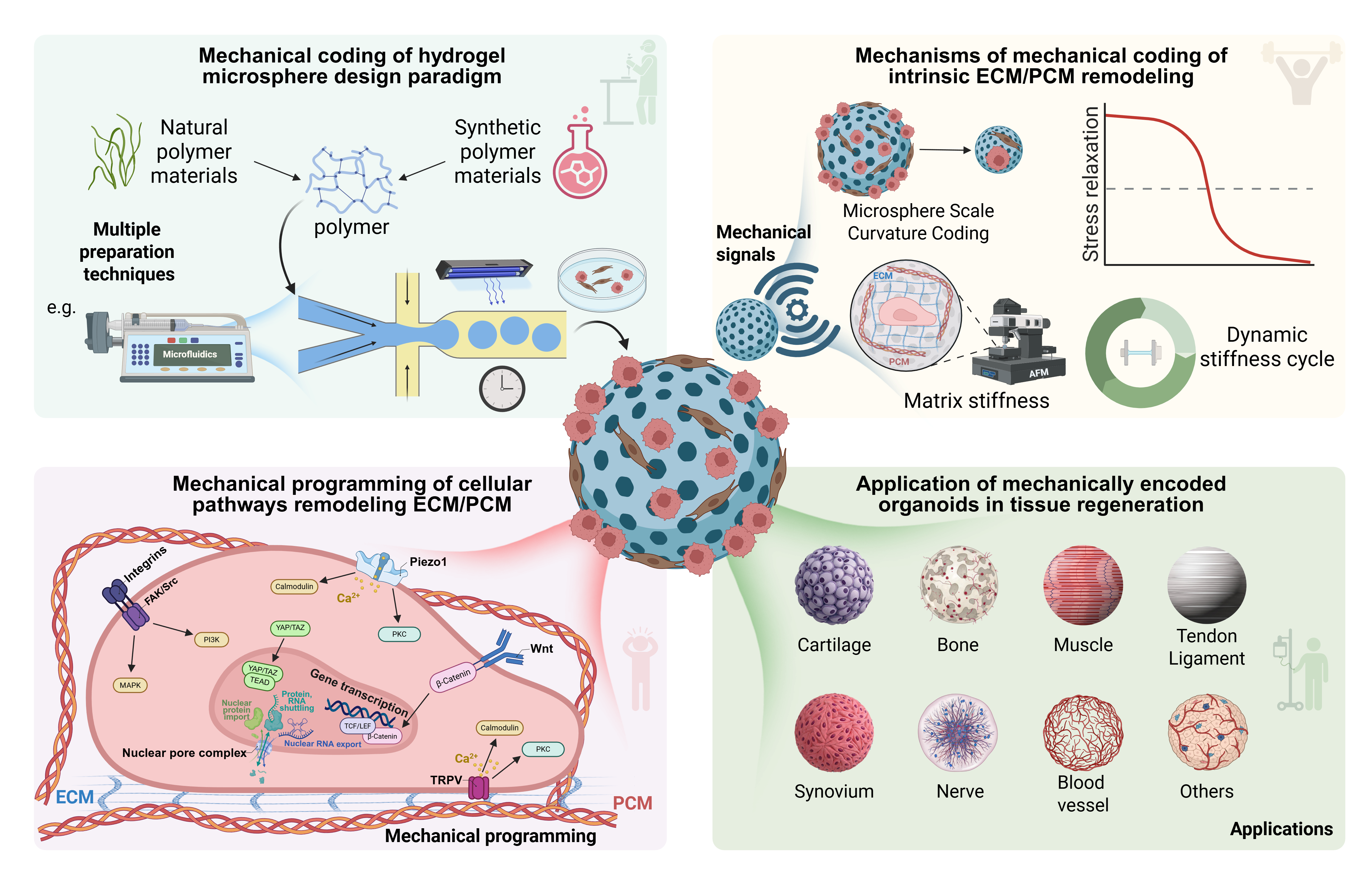
Organoids—three-dimensional, self-organizing living micro-tissue entities—are fundamentally reshaping the outcome of modern biomedical research through their remarkable capacity to recapitulate the topographical complexity and physiological fidelity of native tissues. Additionally, organoids are able to recapitulate the genetic and epigenetic signature of individual human beings, which is a crucial stepping stone for all personalized and regenerative medicine approaches. However, despite their ability to express selected lineage-specific markers, existing organoid culture platforms exhibit fundamental deficiencies in the spatiotemporal precision of extracellular matrix (ECM) and pericellular matrix (PCM) organization—a vexating limitation rooted in the absence of sophisticated spatiotemporal mechano-coding. Consequently, organoids frequently remain arrested in immature phenotypic states, severely constraining their translational efficacy in regenerative medicine. Addressing this critical bottleneck, hydrogel microsphere-mediated engineering strategies have emerged as a transformative paradigm. This review centers on innovative approaches with a focus on osteomuscular organoid models, elucidating their rational biomaterial design principles, unique mechano-coding advantages, and the core mechanisms by which they orchestrate the secretion and microstructural remodeling of endogenous ECM/PCM components. We conclude by prospectively exploring the potential of these microsphere-mediated systems to enhance their physiological accuracy and clinical translatability of in vitro musculoskeletal models.
- Zhang J, Wu Y, Shen T, et al. Organoid research: new concepts and new technologies. Burn Trauma 2026;14: tkag015. doi: 10.1093/burnst/tkag015
- Zeng Q, Xie D, Wang D, et al. Bioengineered materials-driven construction of musculoskeletal organoids in aging research: Strategies, applications, and future perspectives. OR. 2025;1(4):025450033. doi: 10.36922/OR025450033
- Yin Y, Zhou W, Zhu J, et al. Generation of self-organized neuromusculoskeletal tri-tissue organoids from human pluripotent stem cells. Cell Stem Cell. 2025;32(1):157-171.e8. doi: 10.1016/j.stem.2024.11.005
- Mostert D, van der Putten C, Sahlgren CM, Kurniawan NA, Bouten CVC. Bioengineering structural anisotropy in living tissues. Nat Rev Bioeng. 2025;3(9):727–741. doi: 10.1038/s44222-025-00338-x
- Gao Y, Zhang Z, Yao Y, et al. Engineering of tissue in microphysiological systems demonstrated by modelling skeletal muscle. Regen Biomater. 2025;12:rbaf059. doi: 10.1093/rb/rbaf059
- Shan B-H, Wu F-G. Hydrogel-Based Growth Factor Delivery Platforms: Strategies and Recent Advances. Adv Mater. 2024;36(5):e2210707. doi: 10.1002/adma.202210707
- Caballero Aguilar LM, Silva SM, Moulton SE. Growth factor delivery: Defining the next generation platforms for tissue engineering. J Control Release. 2019;306:40–58. doi: 10.1016/j.jconrel.2019.05.028
- Wang X, Lei Y, Jiang K, et al. Mito-battery: Micro-nanohydrogel microspheres for targeted regulation of cellular mitochondrial respiratory chain. Nano Today. 2023;49:101820. doi: 10.1016/j.nantod.2023.101820
- Daly AC, Riley L, Segura T, Burdick JA. Hydrogel microparticles for biomedical applications. Nat Rev Mater. 2020;5(1):20–43. doi: 10.1038/s41578-019-0148-6
- Zhang D, Liu W, Feng L, et al. Innovative Advances in Droplet Microfluidics. Research. 2025;8:0856. doi: 10.34133/research.0856
- Xiao P, Yuan H, Lei Y, et al. Lubrication Barriers Hydrogel Microspheres Improve Dynamic Tissue Repair via the Blockade of Inflammatory Communication. Adv Funct Mater. 2026;36(6):e15734. doi: 10.1002/adfm.202515734
- Zhang L, Luo Z, Chen H, Wu X, Zhao Y. Glycyrrhizic Acid Hydrogel Microparticles Encapsulated with Mesenchymal Stem Cell Exosomes for Wound Healing. Research. 2024;7:0496. doi: 10.34133/research.0496
- Liu H, Chen Z, Lin Q, et al. A multicellular self-organized probiotic platform for oral delivery enhances intestinal colonization. Nat Commun. 2025;16(1):7060. doi: 10.1038/s41467-025-62349-x
- Lei Y, Santos HA, Cui W. Advances and challenges in hydrogel microspheres for biomedical applications. Biomater Transl. 2024;5(3):203–204. doi: 10.12336/biomatertransl.2024.03.001
- Wang X, He J, Hong Y, et al. Spermidine-encapsulated chondroitin sulfate methacryloyl hydrogel delivery system for remodeling bone homeostasis in rheumatoid arthritis. Mater Today Bio. 2025;35:102531. doi: 10.1016/j.mtbio.2025.102531
- Chen Z, Lv Z, Zhang Z, et al. Advanced microfluidic devices for fabricating multi-structural hydrogel microsphere. Exploration. 2021;1(3):20210036. doi: 10.1002/EXP.20210036
- Hu X, Zhang MX, Xu FW, et al. Preparation Methods of Hydrogel Microspheres and Recent Advances in Their Application for Treating Diabetic Wounds. Int J Nanomed. 2025;20:10645–10666. doi: 10.2147/IJN.S527650
- Reihs EI, Stoegner A, Vasconez Martínez MG, et al. An Animal-Free Patient-Derived Tissue-Mimetic Biochip Model of the Human Synovial Membrane for Human- Relevant Osteoarthritis Research. Adv Healthc Mater. 2025;14(23):e2404799. doi: 10.1002/adhm.202404799
- Yuan N, Shao K, Huang S, Chen C. Chitosan, alginate, hyaluronic acid and other novel multifunctional hydrogel dressings for wound healing: A review. Int J Biol Macromol. 2023;240:124321. doi: 10.1016/j.ijbiomac.2023.124321
- Chi H, Qiu Y, Ye X, Shi J, Li Z. Preparation strategy of hydrogel microsphere and its application in skin repair. Front Bioeng Biotechnol. 2023;11:1239183. doi: 10.3389/fbioe.2023.1239183
- Chelu M, Calderon Moreno JM, Musuc AM, Popa M. Natural Regenerative Hydrogels for Wound Healing. Gels. 2024;10(9):547. doi: 10.3390/gels10090547
- Zhang Y, Chen H, Li J. Recent advances on gelatin methacrylate hydrogels with controlled microstructures for tissue engineering. Int J Biol Macromol. 2022;221:91–107. doi: 10.1016/j.ijbiomac.2022.08.171
- Chen S, Li J, Zheng L, Huang J, Wang M. Biomimicking trilayer scaffolds with controlled estradiol release for uterine tissue regeneration. Exploration. 2024;4(5):20230141. doi: 10.1002/EXP.20230141
- Chen Y, Yang ZR, Cheng Z, et al. Injectable hydrogel microspheres promoting inflammation modulation and nucleus pulposus-like differentiation for intervertebral disc regeneration. J Control Release. 2025;380:599–614. doi: 10.1016/j.jconrel.2025.02.016
- Ghobadi F, Saadatmand M, Simorgh S, Brouki Milan P. Microfluidic 3D cell culture: potential application of collagen hydrogels with an optimal dose of bioactive glasses. Sci Rep. 2025;15(1):569. doi: 10.1038/s41598-024-84346-8
- Wang Y, Lv H, Ren S, et al. Biological Functions of Macromolecular Protein Hydrogels in Constructing Osteogenic Microenvironment. ACS Biomater Sci Eng. Sep 9 2024;10(9):5513–5536. doi: 10.1021/acsbiomaterials.4c00910
- Hu X, Tang J, Yu H, Yang H, Lu X, Zheng D. Preparation of fish collagen and vancomycin microspheres based on microfluidic technology and its application in osteomyelitis. Front Bioeng Biotechnol. 2023;11:1249706. doi: 10.3389/fbioe.2023.1249706
- Suliman M, Alissa M, Alghamdi A. Collagen-based hydrogel encapsulated with SDF-1α microspheres accelerate diabetic wound healing in rats. Tissue Cell. 2025;95:102877. doi: 10.1016/j.tice.2025.102877
- Hakim Khalili M, Zhang R, Wilson S, Goel S, Impey SA, Aria AI. Additive Manufacturing and Physicomechanical Characteristics of PEGDA Hydrogels: Recent Advances and Perspective for Tissue Engineering. Polymers. 2023;15(10):2341. doi: 10.3390/polym15102341
- Yan Y, Vladisavljević GT, Lin Z, Yang H, Zhang X, Yuan W. PEGDA hydrogel microspheres with encapsulated salt for versatile control of protein crystallization. J Colloid Interface Sci. 2024;660:574–584. doi: 10.1016/j.jcis.2024.01.125
- Chen S, Shahar T, Cohen S. Thermo-controlled microfluidic generation of monodisperse alginate microspheres based on external gelation. RSC Adv. 2024;14(44):32021–32028. doi: 10.1039/d4ra07049f
- Zhang R, Lei L, Song Q, Li X. Calcium ion cross-linking alginate/dexamethasone sodium phosphate hybrid hydrogel for extended drug release. Colloids Surf B Biointerfaces. 2019;175:569–575. doi: 10.1016/j.colsurfb.2018.11.083
- Qin BQ, Wu SZ, Nie R, et al. SDF-1α/BMP-12 loaded biphasic sustained-release SIS hydrogel/SA microspheres composite for tendon regeneration. Biomaterials. 2025;320:123246. doi: 10.1016/j.biomaterials.2025.123246
- Gowda AHJ, Bu Y, Kudina O, et al. Design of tunable gelatin-dopamine based bioadhesives. Int J Biol Macromol. 2020;164:1384–1391. doi: 10.1016/j.ijbiomac.2020.07.195
- Wu K, Yan Z, Wu Z, et al. Recent Advances in the Preparation, Antibacterial Mechanisms, and Applications of Chitosan. J Funct Biomater. 2024;15(11):318. doi: 10.3390/jfb15110318
- Upadhyay A, Alimohammadi F, Tehrani R. Engineering Porosity-Tuned Chitosan Beads: Balancing Porosity, Kinetics, and Mechanical Integrity. ACS Omega. 2024;9(31):33857–33867. doi: 10.1021/acsomega.4c03583
- Li Y, Li X, Zhu L, Liu T, Huang L. Chitosan-based biomaterials for bone tissue engineering. Int J Biol Macromol. 2025;304(Pt 2):140923. doi: 10.1016/j.ijbiomac.2025.140923
- Xin C, Jia P, Zhao Y, et al. Antioxidant effects of Gastrodia elata polysaccharide-based hydrogels loaded with puerarin/ gelatin microspheres for D-galactose-induced aging-skin wound healing. Int J Biol Macromol. 2025;296:139809. doi: 10.1016/j.ijbiomac.2025.139809
- Du J, Liu Q, Diao X, Ma W, Liu G. Development of plantaricin RX-8 loaded pectin/4-carboxyphenylboric acid/carboxymethyl chitosan hydrogel microbead: A potential targeted oral delivery system. Int J Biol Macromol. 2025;292:139134. doi: 10.1016/j.ijbiomac.2024.139134
- Conzatti G, Nadal C, Berthelot J, et al. Chitosan-PNIPAM Thermogel Associated with Hydrogel Microspheres as a Smart Formulation for MSC Injection. ACS Appl Bio Mater. 2024;7(5):3033–3040. doi: 10.1021/acsabm.4c00071
- Ressler A. Chitosan-Based Biomaterials for Bone Tissue Engineering Applications: A Short Review. Polymers. 2022;14(16):3430. doi: 10.3390/polym14163430
- Lim BY, Azmi F, Ng SF. LL37 Microspheres Loaded on Activated Carbon-chitosan Hydrogel: Anti-bacterial and Anti-toxin Wound Dressing for Chronic Wound Infections. AAPS PharmSciTech. 2024;25(5):110. doi: 10.1208/s12249-024-02826-6
- Priya R, Yun Nam S, Cho W-S, Maruthupandy M. Graphene Reinforced Chitosan Nanocomposites for Drug Delivery. In: Swain SK, ed. Carbon Based Nanomaterials for Drug Delivery. Singapore: Springer; 2025:443–468. doi: 10.1007/978-981-97-8086-0_15
- Ramezani Farani M, Zare I, Mirshafiei M, et al. Graphene oxide-engineered chitosan nanoparticles: Synthesis, properties, and antibacterial activity for tissue engineering and regenerative medicine. Chem Eng J. 2025;509:160852. doi: 10.1016/j.cej.2025.160852
- Luo J, Huang H, Jiang J, Zheng W, Chen P, Bai H. Clearance of Intracellular Pathogens with Hyaluronic Acid Nanomicelles Responsive to H(2)S and pH. Molecules. 2024;29(24):5971. doi: 10.3390/molecules29245971
- Heydari M, Hosseinzadeh Colagar A, Sabour D, Khorasani HR. Evidence of Urtica dioica Agglutinin’s Antiproliferative and Anti-migratory Potentials on the Hyaluronic Acid- Overexpressing Prostate Cancer Cells. Planta Med. 2024;90(10):774–784. doi: 10.1055/a-2324-2250
- Wang Y-J, Jiang W-J, Zhao H-J, et al. A Bioorthogonal and Programmable Bacterial Delivery System for Spatiotemporally Targeted Therapy of Solid Tumors. Exploration. 2025;5(6):20240396. doi: 10.1002/EXP.20240396
- Liu J, Chen S, Zhang Z, et al. The oxidized hyaluronic acid hydrogels containing paeoniflorin microspheres regulates the polarization of M1/M2 macrophages to promote wound healing. Int J Biol Macromol. 2024;282(Pt 5):137107. doi: 10.1016/j.ijbiomac.2024.137107
- Yao Y, Wei G, Ding J, Cui W. [Injectable hydrogel microspheres experimental research for the treatment of osteoarthritis]. Zhongguo Xiu Fu Chong Jian Wai Ke Za Zhi. 2023;37(8):918–928. doi: 10.7507/1002-1892.202302105
- Lin C, Ying C, Xu Y, et al. Synergizing adaptive immunity and regenerative signals to enhance osteochondral defects repair. Bioact Mater. 2025;46:242–258. doi: 10.1016/j.bioactmat.2024.12.011
- Hou J, Lin Y, Zhu C, et al. Zwitterion-Lubricated Hydrogel Microspheres Encapsulated with Metformin Ameliorate Age- Associated Osteoarthritis. Adv Sci. 2024;11(30):e2402477. doi: 10.1002/advs.202402477
- Pritchard EM, Kaplan DL. Silk fibroin biomaterials for controlled release drug delivery. Expert Opin Drug Deliv. 2011;8(6):797–811. doi: 10.1517/17425247.2011.568936
- Ma K, Yang L, Li W, et al. Mussel-inspired multi-bioactive microsphere scaffolds for bone defect photothermal therapy. Mater Today Bio. 2024;29:101363. doi: 10.1016/j.mtbio.2024.101363
- Kaewpirom S, Piboonnithikasem S, Sroisroemsap P, Uttoom S, Boonsang S. Tailoring silk fibroin hydrophilicity and physicochemical properties using sugar alcohols for medical device coatings. Sci Rep. 14 2024;14(1):13781. doi: 10.1038/s41598-024-64450-5
- Tu H, Zhu M, Duan B, Zhang L. Recent Progress in High- Strength and Robust Regenerated Cellulose Materials. Adv Mater. 2021;33(28):e2000682. doi: 10.1002/adma.202000682
- Chen Y, Liu X, Zhou R, et al. Porous sodium alginate/ cellulose nanofiber composite hydrogel microspheres for heavy metal removal in wastewater. Int J Biol Macromol. 2024;278(Pt 3):135000. doi: 10.1016/j.ijbiomac.2024.135000
- Batta-Mpouma J, Kandhola G, Kaur J, et al. Cellulose nanocrystal-based hydrogel microspheres prepared via electrohydrodynamic processes for controlled release of bioactive compounds. Carbohydr Polym. 2025;356:123355. doi: 10.1016/j.carbpol.2025.123355
- Xu Z, Tian W, Wen C, et al. Cellulose-Based Cryogel Microspheres with Nanoporous and Controllable Wrinkled Morphologies for Rapid Hemostasis. Nano Lett. 2022;22(15):6350–6358. doi: 10.1021/acs.nanolett.2c02144
- Wang R, Fujie T, Itaya H, Wada N, Takahashi K. Force- Induced Alignment of Nanofibrillated Bacterial Cellulose for the Enhancement of Cellulose Composite Macrofibers. Int J Mol Sci. 2023;25(1):69. doi: 10.3390/ijms25010069
- Su M, Ruan L, Dong X, et al. Current state of knowledge on intelligent-response biological and other macromolecular hydrogels in biomedical engineering: A review. Int J Biol Macromol. 2023;227:472–492. doi: 10.1016/j.ijbiomac.2022.12.148
- Jiang F, Xu XW, Chen FQ, et al. Extraction, Modification and Biomedical Application of Agarose Hydrogels: A Review. Mar Drugs. 2023;21(5):299. doi: 10.3390/md21050299
- Huang Y, Peng S, Chen Y, Chu B. Agarose Hydrogels for Bone Tissue Engineering, from Injectables to Bioprinting. Gels. 2025;11(4): 255. doi: 10.3390/gels11040255
- Zhu S, Shou X, Kuang G, et al. Stimuli-responsive hydrogel microspheres encapsulated with tumor-cell-derived microparticles for malignant ascites treatment. Acta Biomater. 2025;192:328–339. doi: 10.1016/j.actbio.2024.11.034
- Ivanova I, Slavkova M, Popova T, et al. Agar Graft Modification with Acrylic and Methacrylic Acid for the Preparation of pH-Sensitive Nanogels for 5-Fluorouracil Delivery. Gels. 2024;10(3):165. doi: 10.3390/gels10030165
- Pang Q, Chen Z, Li X, et al. Cytokine-Activated Mesenchymal-Stem-Cell-Derived Extracellular Matrix Facilitates Cartilage Repair by Enhancing Chondrocyte Homeostasis and Chondrogenesis of Recruited Stem Cells. Research. 2025;8:0700. doi: 10.34133/research.0700
- Golebiowska AA, Intravaia JT, Sathe VM, Kumbar SG, Nukavarapu SP. Decellularized extracellular matrix biomaterials for regenerative therapies: Advances, challenges and clinical prospects. Bioact Mater. 2024;32:98–123. doi: 10.1016/j.bioactmat.2023.09.017
- Pang Q, Li X, Tang B, et al. Cartilage-anchoring ECM-functionalized microgels targeting miMOMP to suppress senescence and restore cartilage in osteoarthritis. Chem Eng J. 2025;525:170093. doi: 10.1016/j.cej.2025.170093
- Chen Y, Chen L-F, Wang Y, et al. Engineered dECM-based microsystem promotes cartilage regeneration in osteoarthritis by synergistically enhancing chondrogenesis of BMSCs and anti-inflammatory effect. Compos Part B Eng. 2025;290:111974. doi: 10.1016/j.compositesb.2024.111974
- Luo SC, Li MT, Wang YC, et al. Microfluidic GelMA/Bone- Derived Extracellular Matrix Microgels for Enhanced Stem Cell-Based Bone Regeneration. ACS Appl Bio Mater. 2026;9(2):906–920. doi: 10.1021/acsabm.5c01821
- Chen X, Zhang J, Lin T, et al. Bioactive Decellularized Extracellular Matrix Platform Integrating Multifunctional Nanozymes and Cell-Laden Microgels for Acute Liver Failure Treatment. ACS Nano. 2025;19(7):6890–6910. doi: 10.1021/acsnano.4c13709
- Im P, Shin H, Kim J. Tilapia-Derived Granular Hydrogel as a 3D Scaffold Promoting Rapid Wound Healing. Biomacromolecules. 2024;25(2):1153–1161. doi: 10.1021/acs.biomac.3c01137
- Dreifke MB, Ebraheim NA, Jayasuriya AC. Investigation of potential injectable polymeric biomaterials for bone regeneration. J Biomed Mater Res A. 2013;101A(8):2436–2447. doi: 10.1002/jbm.a.34521
- Santerre JP, Woodhouse K, Laroche G, Labow RS. Understanding the biodegradation of polyurethanes: from classical implants to tissue engineering materials. Biomaterials. 2005;26(35):7457–7470. doi: 10.1016/j.biomaterials.2005.05.079
- Mainardi A, Börsch A, Occhetta P, et al. An Organ-on-Chip Platform for Strain-Controlled, Tissue-Specific Compression of Cartilage and Mineralized Osteochondral Interface to Study Mechanical Overloading in Osteoarthritis. Adv Healthc Mater. 14(23):e2501588. doi: 10.1002/adhm.202501588
- Zhao X, Ma H, Han H, et al. Precision medicine strategies for spinal degenerative diseases: Injectable biomaterials with in situ repair and regeneration. Mater Today Bio. 2022;16:100336. doi: 10.1016/j.mtbio.2022.100336
- Shirbhate U, Bajaj P. Injectable and Self-Invigorating Hydrogel Applications in Dentistry and Periodontal Regeneration: A Literature Review. Cureus. 2022;14(9):e29248. doi: 10.7759/cureus.29248
- Bédouet L, Beilvert A, Servais E, Moine L. Degradable Hydrogel Microspheres for Drug Delivery: In Vitro Performance and Influence of E-Beam Sterilization. AAPS PharmSciTech. 2025;26(5):162. doi: 10.1208/s12249-025-03144-1
- Suk JS, Xu Q, Kim N, Hanes J, Ensign LM. PEGylation as a strategy for improving nanoparticle-based drug and gene delivery. Adv Drug Deliv Rev. 2016;99(Pt A):28–51. doi: 10.1016/j.addr.2015.09.012
- Li X, Xu Q, Johnson M, et al. A chondroitin sulfate based injectable hydrogel for delivery of stem cells in cartilage regeneration. Biomater Sci. 2021;9(11):4139–4148. doi: 10.1039/D1BM00482D
- Liang X, Zhong HJ, Ding H, et al. Polyvinyl Alcohol (PVA)- Based Hydrogels: Recent Progress in Fabrication, Properties, and Multifunctional Applications. Polymers. 2024;16(19): 2755. doi: 10.3390/polym16192755
- Allen MJ, Schoonmaker JE, Bauer TW, Williams PF, Higham PA, Yuan HA. Preclinical evaluation of a poly (vinyl alcohol) hydrogel implant as a replacement for the nucleus pulposus. Spine. 2004;29(5):515–523. doi: 10.1097/01.BRS.0000113871.67305.38
- Tsioptsias C, Fardis D, Ntampou X, Tsivintzelis I, Panayiotou C. Thermal Behavior of Poly(vinyl alcohol) in the Form of Physically Crosslinked Film. Polymers. 2023;15(8):1843. doi: 10.3390/polym15081843
- Rahman Khan MM, Rumon MMH. Synthesis of PVA-Based Hydrogels for Biomedical Applications: Recent Trends and Advances. Gels. 2025;11(2):88. doi: 10.3390/gels11020088
- Długa A, Bajer D, Kaczmarek H. Photochemical and Thermal Stability of Bionanocellulose/Poly(Vinyl Alcohol) Blends. Polymers. 2022;14(20):4364. doi: 10.3390/polym14204364
- Țăin Anastasiu AE, Bîrcă AC, Naulea AMI, Niculescu AG, Grumezescu AM, Croitoru GA. Alginate/PVA Hydrogel Incorporating HA-Liposomes and Aronia-Derived Silver Nanoparticles for Advanced Wound Management. Int J Mol Sci. 2025;26(18):9203. doi: 10.3390/ijms26189203
- Liu W, Lei L, Ma F, et al. A Dioscorea opposita Polysaccharide- Calcium Carbonate Microsphere-Doped Hydrogel for Accelerated Diabetic Wound Healing via Synergistic Glucose-Responsive Hypoglycemic and Anti-Inflammatory Effects. ACS Biomater Sci Eng. 2025;11(1):415–428. doi: 10.1021/acsbiomaterials.4c02090
- Karam JP, Muscari C, Sindji L, et al. Pharmacologically active microcarriers associated with thermosensitive hydrogel as a growth factor releasing biomimetic 3D scaffold for cardiac tissue-engineering. J Control Release. 2014;192:82–94. doi: 10.1016/j.jconrel.2014.06.052
- Ma S, Feng X, Liu F, Wang B, Zhang H, Niu X. The pro-inflammatory response of macrophages regulated by acid degradation products of poly(lactide-co-glycolide) nanoparticles. Eng Life Sci. 2021;21(10):709–720. doi: 10.1002/elsc.202100040
- Liu H, Slamovich EB, Webster TJ. Less harmful acidic degradation of poly(lacticco-glycolic acid) bone tissue engineering scaffolds through titania nanoparticle addition. Int J Nanomed. 2006;1(4):541–545. doi: 10.2147/nano.2006.1.4.541
- El Khatib M, Russo V, Prencipe G, et al. Amniotic Epithelial Stem Cells Counteract Acidic Degradation By-Products of Electrospun PLGA Scaffold by Improving Their Immunomodulatory Profile In Vitro. Cells. 2021;10(11):3221. doi: 10.3390/cells10113221
- Yu Z, Ma S, Wu M, et al. Self-assembling hydrogel loaded with 5-FU PLGA microspheres as a novel vitreous substitute for proliferative vitreoretinopathy. J Biomed Mater Res A. 2020;108(12):2435–2446. doi: 10.1002/jbm.a.36995
- Segneanu AE, Bejenaru LE, Bejenaru C, et al. Advancements in Hydrogels: A Comprehensive Review of Natural and Synthetic Innovations for Biomedical Applications. Polymers. 2025;17(15): 2026. doi: 10.3390/polym17152026
- Choi HS, Seo SA, Khang G, Rhee JM, Lee HB. Preparation and characterization of fentanyl-loaded PLGA microspheres: in vitro release profiles. Int J Pharm. 2002;234(1-2):195–203. doi: 10.1016/s0378-5173(01)00968-1
- Hauser CAE, Deng R, Mishra A, et al. Natural tri- to hexapeptides self-assemble in water to amyloid β-type fiber aggregates by unexpected α-helical intermediate structures. Proc Natl Acad Sci USA. 2011;108(4):1361–1366. doi: 10.1073/pnas.1014796108
- Mishra A, Loo Y, Deng R, et al. Ultrasmall natural peptides self-assemble to strong temperature-resistant helical fibers in scaffolds suitable for tissue engineering. Nano Today. 2011;6(3):232–239. doi: 10.1016/j.nantod.2011.05.001
- Susapto HH, Alhattab D, Abdelrahman S, et al. Ultrashort Peptide Bioinks Support Automated Printing of Large-Scale Constructs Assuring Long-Term Survival of Printed Tissue Constructs. Nano Lett. 2021;21(7):2719–2729. doi: 10.1021/acs.nanolett.0c04426
- Loo Y, Goktas M, Tekinay AB, Guler MO, Hauser CAE, Mitraki A. Self-Assembled Proteins and Peptides as Scaffolds for Tissue Regeneration. Adv Healthc Mater. 2015;4(16):2557–2586. doi: 10.1002/adhm.201500402
- Loo Y, Lakshmanan A, Ni M, Toh LL, Wang S, Hauser CAE. Peptide Bioink: Self-Assembling Nanofibrous Scaffolds for Three-Dimensional Organotypic Cultures. Nano Lett. 2015;15(10):6919–6925. doi: 10.1021/acs.nanolett.5b02859
- Ramirez-Calderon G, Susapto HH, Hauser CAE. Delivery of Endothelial Cell-Laden Microgel Elicits Angiogenesis in Self-Assembling Ultrashort Peptide Hydrogels In Vitro. ACS Appl Mater Interfaces. 2021;13(25):29281–29292. doi: 10.1021/acsami.1c03787
- Liu J, Du C, Chen J, et al. Hydrogel Microspheres Empowering Organ-on-a-Chip Systems: Innovations and Applications. Small. 2025;21(44):e2504563. doi: 10.1002/smll.202504563
- Tumarkin E, Kumacheva E. Microfluidic generation of microgels from synthetic and natural polymers. Chem Soc Rev. 2009;38(8):2161–2168. doi: 10.1039/b809915b
- Cheng S, Yang L, Dong M, et al. Metabolism-Regulating Microspheres: Design Principles, Therapeutic Applications Across Multisystem Diseases, and Future Perspectives. Research. 2026;9:1106. doi: 10.34133/research.1106
- Cheng S, Luo X, Yang L, et al. Design and applications of immunomodulatory microspheres: From material properties to multisystem disease therapies. Mater Today. 2026;92:925-951. doi: 10.1016/j.mattod.2025.12.035
- Xia X, Liu Y, Lu Y, et al. Retuning Mitochondrial Apoptosis/Mitophagy Balance via SIRT3-Energized and Microenvironment-Modulated Hydrogel Microspheres to Impede Osteoarthritis. Adv Health Mater. 2023;12(32):2302475. doi: 10.1002/adhm.202302475
- Lei Y, Wang Y, Shen J, et al. Injectable hydrogel microspheres with self-renewable hydration layers alleviate osteoarthritis. Sci Adv. 2022;8(5):eabl6449. doi: 10.1126/sciadv.abl6449
- Xiao P, Han X, Huang Y, et al. Reprogramming macrophages via immune cell mobilized hydrogel microspheres for osteoarthritis treatments. Bioact Mater. 2024;32:242–259. doi: 10.1016/j.bioactmat.2023.09.010
- Liu J, Liu J, Liu S, et al. Cascade targeting selenium nanoparticles-loaded hydrogel microspheres for multifaceted antioxidant defense in osteoarthritis. Biomaterials. 2025;318:123195. doi: 10.1016/j.biomaterials.2025.123195
- Mahou R, Vlahos AE, Shulman A, Sefton MV. Interpenetrating Alginate-Collagen Polymer Network Microspheres for Modular Tissue Engineering. ACS Biomater Sci Eng. 2018;4(11):3704–3712. doi: 10.1021/acsbiomaterials.7b00356
- Li X, Li L, Wang D, et al. Fabrication of polymeric microspheres for biomedical applications. Mater Horiz. 2024;11(12):2820–2855. doi: 10.1039/D3MH01641B
- Chen T, Cai Z, Zhao X, et al. Dynamic monitoring soft tissue healing via visualized Gd-crosslinked double network MRI microspheres. J Nanobiotechnol. 2024;22(1):289. doi: 10.1186/s12951-024-02549-7
- Zhang X, Zhu R, Wang X, et al. Core-Shell Microspheres Prepared Using Coaxial Electrostatic Spray for Local Chemotherapy of Solid Tumors. Pharmaceutics. 2023;16(1):45. doi: 10.3390/pharmaceutics16010045
- Wu Y, Liao IC, Kennedy SJ, et al. Electrosprayed core-shell microspheres for protein delivery. Chem Commun. 2010;46(26):4743–4745. doi: 10.1039/c0cc00535e
- Yang Y, Sha L, Zhao H, Guo Z, Wu M, Lu P. Recent advances in cellulose microgels: Preparations and functionalized applications. Adv Colloid Interface Sci. 2023;311:102815. doi: 10.1016/j.cis.2022.102815
- Zhu L, Li M, Liu X, Jin Y. Drug-Loaded PLGA Electrospraying Porous Microspheres for the Local Therapy of Primary Lung Cancer via Pulmonary Delivery. ACS Omega. 2017;2(5):2273–2279. doi: 10.1021/acsomega.7b00456
- Xie J, Ng WJ, Lee LY, Wang CH. Encapsulation of protein drugs in biodegradable microparticles by co-axial electrospray. J Colloid Interface Sci. 2008;317(2):469–476. doi: 10.1016/j.jcis.2007.09.082
- Helgeson ME, Chapin SC, Doyle PS. Hydrogel microparticles from lithographic processes: novel materials for fundamental and applied colloid science. Curr Opin Colloid Interface Sci. 2011;16(2):106–117. doi: 10.1016/j.cocis.2011.01.005
- Stornello DE, Kim J, Chen Z, Heaton K, Qazi TH. Controlling Microparticle Aspect Ratio via Photolithography for Injectable Granular Hydrogel Formation and Cell Delivery. ACS Biomater Sci Eng. 2025;11(2):1242–1252. doi: 10.1021/acsbiomaterials.4c02102
- Lee SA, Chung SE, Park W, Lee SH, Kwon S. Three-dimensional fabrication of heterogeneous microstructures using soft membrane deformation and optofluidic maskless lithography. Lab Chip. 2009;9(12):1670–1675. doi: 10.1039/b819999j
- Nichol JW, Koshy ST, Bae H, Hwang CM, Yamanlar S, Khademhosseini A. Cell-laden microengineered gelatin methacrylate hydrogels. Biomaterials. 2010;31(21):5536– 5544. doi: 10.1016/j.biomaterials.2010.03.064
- Palange AL, Mascolo DD, Ferreira M, et al. Boosting the Potential of Chemotherapy in Advanced Breast Cancer Lung Metastasis via Micro-Combinatorial Hydrogel Particles. Adv Sci. 2023;10(10):2205223. doi: 10.1002/advs.202205223
- Murphy SV, Atala A. 3D bioprinting of tissues and organs. Nat Biotechnol. 2014;32(8):773–785. doi: 10.1038/nbt.2958
- Zhu W, Ma X, Gou M, Mei D, Zhang K, Chen S. 3D printing of functional biomaterials for tissue engineering. Curr Opin Biotechnol. 2016;40:103–112. doi: 10.1016/j.copbio.2016.03.014
- Kotani T, Hananouchi T, Sakai S. Enhancing visible light-induced 3D bioprinting: alternating extruded support materials for bioink gelation. Biomed Mater. 2025;20(3):035005. doi: 10.1088/1748-605X/adc0d6
- Grottkau BE, Hui Z, Pang Y. Articular Cartilage Regeneration through Bioassembling Spherical Micro-Cartilage Building Blocks. Cells. 2022;11(20):3244. doi: 10.3390/cells11203244
- Wu CA, Zhu Y, Venkatesh A, Stark CJ, Lee SH, Woo YJ. Optimization of Freeform Reversible Embedding of Suspended Hydrogel Microspheres for Substantially Improved Three-Dimensional Bioprinting Capabilities. Tissue Eng Part C Methods. 2023;29(3):85–94. doi: 10.1089/ten.tec.2022.0214
- De Lorenzi F, Hansen N, Theek B, et al. Engineering Mesoscopic 3D Tumor Models with a Self-Organizing Vascularized Matrix. Adv Mater. 2024;36(5):e2303196. doi: 10.1002/adma.202303196
- Du C, Huang W, Lei Y. The application and prospects of 3D printable microgel in biomedical science and engineering. Int J Bioprint. 2023;9(5):753. doi: 10.18063/ijb.753
- Zhao H, Chen Y, Shao L, et al. Airflow-Assisted 3D Bioprinting of Human Heterogeneous Microspheroidal Organoids with Microfluidic Nozzle. Small. 2018;14(39):e1802630. doi: 10.1002/smll.201802630
- Zhang S, Li G, Man J, et al. Fabrication of Microspheres from High-Viscosity Bioink Using a Novel Microfluidic-Based 3D Bioprinting Nozzle. Micromachines. 2020;11(7):681. doi: 10.3390/mi11070681
- Cao Y, Tan J, Zhao H, et al. Bead-jet printing enabled sparse mesenchymal stem cell patterning augments skeletal muscle and hair follicle regeneration. Nat Commun. 2022;13(1):7463. doi: 10.1038/s41467-022-35183-8
- Clua-Ferré L, De Chiara F, Rodríguez-Comas J, et al. Collagen-Tannic Acid Spheroids for β-Cell Encapsulation Fabricated Using a 3D Bioprinter. Adv Mater Technol. 2022;7(7):2101696. doi: 10.1002/admt.202101696
- Gao Q, Liu Z, Lin Z, et al. 3D Bioprinting of Vessel-like Structures with Multilevel Fluidic Channels. ACS Biomater Sci Eng. 2017;3(3):399–408. doi: 10.1021/acsbiomaterials.6b00643
- Rocha Luiz ME, Carreira M, Nadine S, et al. Contactless 3D acoustic assembly of liquid capsules for bottom-up tissue engineering. Biomaterials. 2026;325:123555. doi: 10.1016/j.biomaterials.2025.123555
- Freitas S, Merkle HP, Gander B. Microencapsulation by solvent extraction/evaporation: reviewing the state of the art of microsphere preparation process technology. J Control Release. 2005;102(2):313–332. doi: 10.1016/j.jconrel.2004.10.015
- van der Kooij RS, Steendam R, Frijlink HW, Hinrichs WLJ. An overview of the production methods for core-shell microspheres for parenteral controlled drug delivery. Eur J Pharm Biopharm. 2022;170:24–42. doi: 10.1016/j.ejpb.2021.11.007
- Ren Z, Wang Y, Wu H, Cong H, Yu B, Shen Y. Preparation and application of hemostatic microspheres containing biological macromolecules and others. Int J Biol Macromol. 2024;257(Pt 1):128299. doi: 10.1016/j.ijbiomac.2023.128299
- Contessi Negrini N, Lipreri MV, Tanzi MC, Farè S. In vitro cell delivery by gelatin microspheres prepared in water-in-oil emulsion. J Mater Sci Mater Med. 2020;31(3):26. doi: 10.1007/s10856-020-6363-2
- Pradhan S, Clary JM, Seliktar D, Lipke EA. A three-dimensional spheroidal cancer model based on PEG-fibrinogen hydrogel microspheres. Biomaterials. 2017;115:141–154. doi: 10.1016/j.biomaterials.2016.10.052
- Muir VG, Qazi TH, Shan J, Groll J, Burdick JA. Influence of Microgel Fabrication Technique on Granular Hydrogel Properties. ACS Biomater Sci Eng. 2021;7(9):4269–4281. doi: 10.1021/acsbiomaterials.0c01612
- Lin CH, Srioudom JR, Sun W, et al. The use of hydrogel microspheres as cell and drug delivery carriers for bone, cartilage, and soft tissue regeneration. Biomater Transl. 2024;5(3):236–256. doi: 10.12336/biomatertransl.2024.03.003
- Puiggalí-Jou A, Asadikorayem M, Maniura-Weber K, Zenobi-Wong M. Growth factor–loaded sulfated microislands in granular hydrogels promote hMSCs migration and chondrogenic differentiation. Acta Biomater. 2023;166:69–84. doi: 10.1016/j.actbio.2023.03.045
- Jin S, Wang Y, Wu X, et al. Young Exosome Bio- Nanoparticles Restore Aging-Impaired Tendon Stem/ Progenitor Cell Function and Reparative Capacity. Adv Mater. 2023;35(18):e2211602. doi: 10.1002/adma.202211602
- Huang G, Shen H, Xu K, et al. Single-Cell Microgel Encapsulation Improves the Therapeutic Efficacy of Mesenchymal Stem Cells in Treating Intervertebral Disc Degeneration via Inhibiting Pyroptosis. Research. 2024;7:0311. doi: 10.34133/research.0311
- Baker BM, Chen CS. Deconstructing the third dimension: how 3D culture microenvironments alter cellular cues. J Cell Sci. 2012;125(Pt 13):3015–3024. doi: 10.1242/jcs.079509
- Raghavan S, Shen CJ, Desai RA, Sniadecki NJ, Nelson CM, Chen CS. Decoupling diffusional from dimensional control of signaling in 3D culture reveals a role for myosin in tubulogenesis. J Cell Sci. 2010;123(17):2877–2883. doi: 10.1242/jcs.055079
- Özkale B, Lou J, Özelçi E, et al. Actuated 3D microgels for single cell mechanobiology. Lab Chip. 2022;22(10):1962–1970. doi: 10.1039/d2lc00203e
- Schindler M, Siriwardena D, Kohler TN, et al. Agarose microgel culture delineates lumenogenesis in naive and primed human pluripotent stem cells. Stem Cell Rep. 2021;16(5):1347–1362. doi: 10.1016/j.stemcr.2021.04.009
- Munger C, Kohler TN, Slatery E, et al. Microgel culture and spatial identity mapping elucidate the signalling requirements for primate epiblast and amnion formation. Development. 2022;149(20):dev200263. doi: 10.1242/dev.200263
- Lavickova B, Kronabitter H, Cervera-Negueruela M, et al. Integrated Microfluidic Platform for High-Throughput Generation of Intestinal Organoids in Hydrogel Droplets. Adv Sci. 2026;13(13):e16507. doi: 10.1002/advs.202516507
- Zheng F, Tian R, Lu H, et al. Droplet Microfluidics Powered Hydrogel Microparticles for Stem Cell-Mediated Biomedical Applications. Small. 2024;20(42):e2401400. doi: 10.1002/smll.202401400
- Yang X, Wang L, Chen X, Wei N, Sun X, Xiao J. Digital light processing 3D bioprinting of type II collagen-based engineered cartilage organoids for enhanced chondrogenic regeneration. Chem Eng J. 2025;520:165969. doi: doi: 10.1016/j.cej.2025.165969
- Tevis KM, Colson YL, Grinstaff MW. Embedded Spheroids as Models of the Cancer Microenvironment. Adv Biosyst. 2017;1(10): 1700083. doi: 10.1002/adbi.201700083
- Fang G, Lu H, Rodriguez de la Fuente L, et al. Mammary Tumor Organoid Culture in Non-Adhesive Alginate for Luminal Mechanics and High-Throughput Drug Screening. Adv Sci. 2021;8(21):e2102418. doi: 10.1002/advs.202102418
- Sun Q, Tan SH, Chen Q, et al. Microfluidic Formation of Coculture Tumor Spheroids with Stromal Cells As a Novel 3D Tumor Model for Drug Testing. ACS Biomater Sci Eng. 2018;4(12):4425–4433. doi: 10.1021/acsbiomaterials.8b00904
- Lv C, Li S, Sang M, Cui T, Xie J. Construction of Microsphere Culture System for Human Mesenchymal Stem Cell Aggregates. Int J Mol Sci. 2025;26(13):6435. doi: 10.3390/ijms26136435
- Zhou C, Ji J, Fu M, et al. Hydrogel Microsphere-Based Alveolar Models for Toxicity Assessment and Pathogen Infection Studies. Bioengineering. 2025;13(1):17. doi: 10.3390/bioengineering13010017
- Kohler TN, De Jonghe J, Ellermann AL, et al. Plakoglobin is a mechanoresponsive regulator of naive pluripotency. Nat Commun. 2023;14(1):4022. doi: 10.1038/s41467-023-39515-0
- Huey DJ, Hu JC, Athanasiou KA. Unlike bone, cartilage regeneration remains elusive. Science. 2012;338(6109):917–921. doi: 10.1126/science.1222454
- Ahmed MS, Yun S, Kim HY, Ko S, Islam M, Nam KW. Hydrogels and Microgels: Driving Revolutionary Innovations in Targeted Drug Delivery, Strengthening Infection Management, and Advancing Tissue Repair and Regeneration. Gels. 2025;11(3):179. doi: 10.3390/gels11030179
- Ortega Quesada BA, Chauvin C, Martin E, Melvin A. Droplet microfluidics integrated with machine learning reveals how adipose-derived stem cells modulate endocrine response and tumor heterogeneity in ER(+) breast cancer. Lab Chip. 2025;25(15):3817–3830. doi: 10.1039/d5lc00320b
- Ochoa A, Gastélum G, Rocha J, Olguin LF. High-throughput bacterial co-encapsulation in microfluidic gel beads for discovery of antibiotic-producing strains. Analyst. 2023;148(22):5762–5774. doi: 10.1039/D3AN01101A
- Mulas C, Hodgson AC, Kohler TN, et al. Microfluidic platform for 3D cell culture with live imaging and clone retrieval. Lab Chip. 2020;20(14):2580–2591. doi: 10.1039/d0lc00165a
- Lei Y, Zhang Y, Zhou L, et al. Organoids and organs-on-chips for accelerating R&D and clinical translation in Orthopaedics: Emerging opportunities and regulatory pathways. J Orthop Translat. 2026;56:101021. doi: 10.1016/j.jot.2025.10.013
- Zhu Y, Cai L, Chen H, Zhao Y. Developing organs-on-chips for biomedicine. Sci Bull. 2022;67(11):1108–1111. doi: 10.1016/j.scib.2022.04.012
- You L, Chen Y, Zhang Z, et al. The FLOW Framework: a panvascular-on-a-chip platform to model systemic disease and guide panvascular interventional device suitcordance. Sci Bull. 2026;71(3):654–670. doi: 10.1016/j.scib.2025.12.051
- Pierfelice TV, D’Amico E, Petrini M, et al. A Systematic Review on Organ-on-a-Chip in PDMS or Hydrogel in Dentistry: An Update of the Literature. Gels. 2024;10(2):102. doi: 10.3390/gels10020102
- Geng Q, Xu Y, Hu Y, et al. Progress in the Application of Organoids-On-A-Chip in Diseases. Organogenesis. 2024;20(1):2386727. doi: 10.1080/15476278.2024.2386727
- Nejati B, Shahhosseini R, Hajiabbasi M, et al. Cancer-on-chip: a breakthrough organ-on-a-chip technology in cancer cell modeling. Med Biol Eng Comput. 2025;63(2):321–337. doi: 10.1007/s11517-024-03199-5
- Gong X, Yang C, Peng J, et al. Vascularized organoid-on-a-chip for centimeter-scale organoid cultivation. Bio-Des Manuf. 2025;8(3):410–422. doi: 10.1631/bdm.2400424
- Qian K, Wang Y, Li R, et al. Application and Prospects of Organoid-on-a-Chip in Research on the Intestinal Mucosal Barrier. Burn Trauma 2026;tkag029. doi: 10.1093/burnst/tkag029
- Cho Y, You J, Lee JH. Natural Polymer-Based Hydrogel Platforms for Organoid and Microphysiological Systems: Mechanistic Insights and Translational Perspectives. Polymers . 2025;17(15):2109. doi: 10.3390/polym17152109
- Qiao L, Chang S, Zou L, Zhang F, Cui C, Huang N. Multiorgan-on-a-chip: an advanced platform for disease modeling, drug toxicity assessment, and therapeutic screening. Bio-Des Manuf. 2025;8(6):1035–1062. doi: 10.1631/bdm.2500076
- Yang M, Shi Y, Wang F, et al. Hydrogel Microspheres as Versatile Platforms for Biomedical Research: Design, Properties, and Applications. MedComm. 2025;6(10):e70423. doi: 10.1002/mco2.70423
- Caires HR, Castillo-Fernández Ó, Sima N, et al. Advancing Organ-on-Chip Models With a Sacrificial Granular Hydrogel Strategy for Enhanced Permeability and Biomimicry. Small Methods. 2025;9(12):e00652. doi: 10.1002/smtd.202500652
- Pye JS, Zhang Y, Lin X, Yousefi M, Wen C, Li JJ. Joint-on-a- Chip: The Future of Preclinical Modelling of Joint Diseases. Adv Healthc Mater. 2025:e04487. doi: 10.1002/adhm.202504487
- Conceição F, Meneses J, Lebre F, et al. Sex-stratified osteochondral organ-on-chip model reveals sex-specific responses to inflammatory stimulation. Mater Today Bio. 2025;32:101728. doi: 10.1016/j.mtbio.2025.101728
- Lei L, Wen Z, Cao M, et al. The emerging role of Piezo1 in the musculoskeletal system and disease. Theranostics. 2024;14(10):3963–3983. doi: 10.7150/thno.96959
- Li J, Yang Y, Yi Z, et al. Microdroplet-Engineered Skeletal Muscle Organoids from Primary Tissue Recapitulate Parental Physiology with High Reproducibility. Research. 2025;8:0699. doi: 10.34133/research.0699
- Pueyo Moliner A, Ito K, Zaucke F, Kelly DJ, de Ruijter M, Malda J. Restoring articular cartilage: insights from structure, composition and development. Nat Rev Rheumatol. 2025;21(5):291–308. doi: 10.1038/s41584-025-01236-7
- Wilusz RE, Sanchez-Adams J, Guilak F. The structure and function of the pericellular matrix of articular cartilage. Matrix Biol. 2014;39:25–32. doi: 10.1016/j.matbio.2014.08.009
- Zhang W, Liu Y, Zhang H. Extracellular matrix: an important regulator of cell functions and skeletal muscle development. Cell Biosci. 2021;11(1):65. doi: 10.1186/s13578-021-00579-4
- Wohlgemuth RP, Brashear SE, Smith LR. Alignment, cross linking, and beyond: a collagen architect’s guide to the skeletal muscle extracellular matrix. Am J Physiol Cell Physiol. 2023;325(4):C1017–C1030. doi: 10.1152/ajpcell.00287.2023
- Kjaer M. Role of extracellular matrix in adaptation of tendon and skeletal muscle to mechanical loading. Physiol Rev. 2004;84(2):649–698.
- Conway R, O’Neill L, Gallagher P, et al. Ustekinumab for refractory giant cell arteritis: A prospective 52-week trial. Semin Arthritis Rheum. 2018;48(3):523–528. doi: 10.1016/j.semarthrit.2018.04.004
- Gilbert SJ, Bonnet CS, Blain EJ. Mechanical Cues: Bidirectional Reciprocity in the Extracellular Matrix Drives Mechano-Signalling in Articular Cartilage. Int J Mol Sci. 2021;22(24):13595. doi: 10.3390/ijms222413595
- Vincent TL, McClurg O, Troeberg L. The Extracellular Matrix of Articular Cartilage Controls the Bioavailability of Pericellular Matrix-Bound Growth Factors to Drive Tissue Homeostasis and Repair. Int J Mol Sci. 2022;23(11):6003. doi: 10.3390/ijms23116003
- Wilusz RE, Defrate LE, Guilak F. A biomechanical role for perlecan in the pericellular matrix of articular cartilage. Matrix Biol. 2012;31(6):320–327. doi: 10.1016/j.matbio.2012.05.002
- Chen L, Yang J, Cai Z, et al. Mitochondrial-Oriented Injectable Hydrogel Microspheres Maintain Homeostasis of Chondrocyte Metabolism to Promote Subcellular Therapy in Osteoarthritis. Research. 2024;7:0306. doi: 10.34133/research.0306
- Quan H, He Y, Xiao P, et al. Scaffold-Mediated Microenvironmental Modulation Targeting Osteoclasts for ONFH Niche Reprogramming. Research. 2025;8:1027. doi: 10.34133/research.1027
- Lampi MC, Reinhart-King CA. Targeting extracellular matrix stiffness to attenuate disease: From molecular mechanisms to clinical trials. Sci Transl Med. 2018;10(422):eaao0475. doi: 10.1126/scitranslmed.aao0475
- Liu E, Zhu D, Gonzalez Diaz E, Tong X, Yang F. Gradient Hydrogels for Optimizing Niche Cues to Enhance Cell- Based Cartilage Regeneration. Tissue Eng Part A. 2021;27(13- 14):929–939. doi: 10.1089/ten.tea.2020.0158
- Galarraga JH, Locke RC, Witherel CE, et al. Fabrication of MSC-laden composites of hyaluronic acid hydrogels reinforced with MEW scaffolds for cartilage repair. Biofabrication. 2022;14(1):014106. doi: 10.1088/1758-5090/ac3acb
- Asadikorayem M, Brunel LG, Weber P, Heilshorn SC, Zenobi-Wong M. Porosity dominates over microgel stiffness for promoting chondrogenesis in zwitterionic granular hydrogels. Biomater Sci. 2024;12(21):5504–5520. doi: 10.1039/D4BM00233D
- Walker M, Pringle EW, Ciccone G, et al. Mind the Viscous Modulus: The Mechanotransductive Response to the Viscous Nature of Isoelastic Matrices Regulates Stem Cell Chondrogenesis. Adv Healthc Mater. 2024;13(9):e2302571. doi: 10.1002/adhm.202302571
- Ai C, Liu L, Wong K, Tan XH, Goh JCH. The effect of chondroitin sulfate concentration and matrix stiffness on chondrogenic differentiation of mesenchymal stem cells. Biomater Sci. 2023;11(13):4557–4573. doi: 10.1039/D2BM01980A
- Roncada T, Bonithon R, Blunn G, Roldo M. Soft substrates direct stem cell differentiation into the chondrogenic lineage without the use of growth factors. J Tissue Eng. 2022;13:20417314221122121. doi: 10.1177/20417314221122121
- Žigon-Branc S, Markovic M, Van Hoorick J, et al. Impact of Hydrogel Stiffness on Differentiation of Human Adipose- Derived Stem Cell Microspheroids. Tissue Eng Part A. 2019;25(19-20):1369–1380. doi: 10.1089/ten.tea.2018.0237
- Bachmann B, Spitz S, Schädl B, et al. Stiffness Matters: Fine-Tuned Hydrogel Elasticity Alters Chondrogenic Redifferentiation. Front Bioeng Biotechnol. 2020;8:373. doi: 10.3389/fbioe.2020.00373
- Lee J, Jeon O, Koh J, et al. Micromechanical property mismatch between pericellular and extracellular matrices regulates stem cell articular and hypertrophic chondrogenesis. Matter. 2023;6(2):475–492. doi: 10.1016/j.matt.2022.11.008
- Chen Z, Lv Z, Zhuang Y, et al. Mechanical Signal-Tailored Hydrogel Microspheres Recruit and Train Stem Cells for Precise Differentiation. Adv Mater. 2023;35(40):2300180. doi: 10.1002/adma.202300180
- Chaudhuri O, Gu L, Klumpers D, et al. Hydrogels with tunable stress relaxation regulate stem cell fate and activity. Nat Mater. 2016;15(3):326–334. doi: 10.1038/nmat4489
- Chaudhuri O, Gu L, Darnell M, et al. Substrate stress relaxation regulates cell spreading. Nat Commun. 2015;6(1):6364. doi: 10.1038/ncomms7365
- Shi N, Wang J, Tang S, et al. Matrix Nonlinear Viscoelasticity Regulates Skeletal Myogenesis through MRTF Nuclear Localization and Nuclear Mechanotransduction. Small. 2024;20(9):e2305218. doi: 10.1002/smll.202305218
- Davidson MD, Prendergast ME, Ban E, et al. Programmable and contractile materials through cell encapsulation in fibrous hydrogel assemblies. Sci Adv. 2021;7(46):eabi8157. doi: 10.1126/sciadv.abi8157
- Bertsch P, Sacco P. The role of non-linear viscoelastic hydrogel mechanics in cell culture and transduction. Mater Today Bio. 2025;34:102188. doi: 10.1016/j.mtbio.2025.102188
- Nam S, Stowers R, Lou J, Xia Y, Chaudhuri O. Varying PEG density to control stress relaxation in alginate-PEG hydrogels for 3D cell culture studies. Biomaterials. 2019;200:15–24. doi: 10.1016/j.biomaterials.2019.02.004
- Bauer A, Gu L, Kwee B, et al. Hydrogel substrate stress-relaxation regulates the spreading and proliferation of mouse myoblasts. Acta Biomater. 2017;62:82–90. doi: 10.1016/j.actbio.2017.08.041
- Liu Y, Li L, Li X, et al. Viscoelastic hydrogels regulate adipose-derived mesenchymal stem cells for nucleus pulposus regeneration. Acta Biomater. 2024;180:244–261. doi: 10.1016/j.actbio.2024.04.017
- Zhao D, Li Y, Xiang L, et al. Cell Shock Absorption via Stress Relaxation Hydrogel Microspheres for Alleviating Endoplasmic Reticulum Stress in Chondrocytes. Research. 2025;8:0777. doi: 10.34133/research.0777
- Roth JG, Huang MS, Navarro RS, Akram JT, LeSavage BL, Heilshorn SC. Tunable hydrogel viscoelasticity modulates human neural maturation. Sci Adv. 2023;9(42):eadh8313. doi: 10.1126/sciadv.adh8313
- Li H, Zhao T, Yuan Z, et al. Cartilage lacuna-biomimetic hydrogel microspheres endowed with integrated biological signal boost endogenous articular cartilage regeneration. Bioact Mater. 2024;41:61–82. doi: 10.1016/j.bioactmat.2024.06.037
- Baptista D, Teixeira L, van Blitterswijk C, Giselbrecht S, Truckenmüller R. Overlooked? Underestimated? Effects of Substrate Curvature on Cell Behavior. Trends Biotechnol. 2019;37(8):838–854. doi: 10.1016/j.tibtech.2019.01.006
- Schamberger B, Ziege R, Anselme K, et al. Curvature in Biological Systems: Its Quantification, Emergence, and Implications across the Scales. Adv Mater. 2023;35(13):e2206110. doi: 10.1002/adma.202206110
- Lee SJ, Yang S. Substrate Curvature Restricts Spreading and Induces Differentiation of Human Mesenchymal Stem Cells. Biotechnol J. 2017;12(9). doi: 10.1002/biot.201700360
- Ding S-L, Zhao X-Y, Xiong W, et al. Cartilage Lacuna-Inspired Microcarriers Drive Hyaline Neocartilage Regeneration. Adv Mater. 2023;35(30):2212114. doi: 10.1002/adma.202212114
- Anderson DE, Johnstone B. Dynamic Mechanical Compression of Chondrocytes for Tissue Engineering: A Critical Review. Front Bioeng Biotechnol. 2017;5:76. doi: 10.3389/fbioe.2017.00076
- Vainieri ML, Wahl D, Alini M, van Osch GJVM, Grad S. Mechanically stimulated osteochondral organ culture for evaluation of biomaterials in cartilage repair studies. Acta Biomater. 2018;81:256–266. doi: 10.1016/j.actbio.2018.09.058
- Farcasanu MV, Ruiz TdLH, Brito FMJdS, et al. Dynamic Compression Improves Chondrogenesis in the Tissue Engineered Model of Cartilage. Biotechnol Bioeng. 2025;122(9):2574–2591. doi: 10.1002/bit.29026
- Davisson T, Kunig S, Chen A, Sah R, Ratcliffe A. Static and dynamic compression modulate matrix metabolism in tissue engineered cartilage. J Orthop Res. 2002;20(4):842–848.
- Mesallati T, Buckley CT, Nagel T, Kelly DJ. Scaffold architecture determines chondrocyte response to externally applied dynamic compression. Biomech Model Mechanobiol. 2013;12(5):889–899. doi: 10.1007/s10237-012-0451-2
- Zhao W, Cai G, Qian J, Geng J, Yao X, Zhang Y. Protein conformational transition microenvironment in silk fibroin hydrogels: proliferation and chondrogenesis of encapsulated stem cells. Regen Biomater. 2025;12:rbaf102. doi: 10.1093/rb/rbaf102
- Schneider MC, Lalitha Sridhar S, Vernerey FJ, Bryant SJ. Spatiotemporal neocartilage growth in matrix-metalloproteinase-sensitive poly(ethylene glycol) hydrogels under dynamic compressive loading: an experimental and computational approach. J Mater Chem B. 2020;8(14):2775– 2791. doi: 10.1039/C9TB02963J
- Xie W, Wei X, Kang H, et al. Static and Dynamic: Evolving Biomaterial Mechanical Properties to Control Cellular Mechanotransduction. Adv Sci. 2023;10(9):e2204594. doi: 10.1002/advs.202204594
- Alanko J, Mai A, Jacquemet G, et al. Integrin endosomal signalling suppresses anoikis. Nat Cell Biol. 2015;17(11):1412– 1421. doi: 10.1038/ncb3250
- Alanko J, Ivaska J. Endosomes: Emerging Platforms for Integrin-Mediated FAK Signalling. Trends Cell Biol. 2016;26(6):391–398. doi: 10.1016/j.tcb.2016.02.001
- Rainero E, Norman JC. Endosomal integrin signals for survival. Nat Cell Biol. 2015;17(11):1373–1375. doi: 10.1038/ncb3261
- Zhao Y, Dong X, Li Y, et al. Integrated Cross-Scale Manipulation and Modulable Encapsulation of Cell- Laden Hydrogel for Constructing Tissue-Mimicking Microstructures. Research. 2024;7:0414. doi: 10.34133/research.0414
- Shi S, Zhang W, Yu Y, et al. Dynamic matrix engineering promotes nascent protein deposition to drive cell migration and expedite Re-epithelization in chronic wound. Bioact Mater. 2026;56:455–467. doi: 10.1016/j.bioactmat.2025.10.020
- Dai S, Huang B, Wang P, et al. Exosome-anchored scholzite conversion coating on titanium with long-term stability and immunomodulatory ability to enhance osteogenesis. Chem Eng J. 2025;503:158142. doi: 10.1016/j.cej.2024.158142
- Li Q, He X-L, Chen S-J, et al. Excessive collagen type VII mediates pleural fibrosis via increasing extracellular matrix stiffness. J Clin Invest. 2025;135(24) doi: 10.1172/JCI188822
- Koo JH, Guan K-L. Interplay between YAP/TAZ and Metabolism. Cell Metab. 2018;28(2):196–206. doi: 10.1016/j.cmet.2018.07.010
- Totaro A, Panciera T, Piccolo S. YAP/TAZ upstream signals and downstream responses. Nat Cell Biol. 2018;20(8):888– 899. doi: 10.1038/s41556-018-0142-z
- Li X, Liu S, Han S, et al. Dynamic Stiffening Hydrogel with Instructive Stiffening Timing Modulates Stem Cell Fate In Vitro and Enhances Bone Remodeling In Vivo. Adv Healthc Mater. 2023;12(29):e2300326. doi: 10.1002/adhm.202300326
- İyisan N, Hausdörfer O, Wang C, et al. Mechanoactivation of Single Stem Cells in Microgels Using a 3D-Printed Stimulation Device. Small Methods. 2024;8(12):e2400272. doi: 10.1002/smtd.202400272
- Jiang Y, Zhang H, Wang J, Liu Y, Luo T, Hua H. Targeting extracellular matrix stiffness and mechanotransducers to improve cancer therapy. J Hematol Oncol. 2022;15(1):34. doi: 10.1186/s13045-022-01252-0
- Qin L, He T, Chen S, et al. Roles of mechanosensitive channel Piezo1/2 proteins in skeleton and other tissues. Bone Res. 2021;9(1):44. doi: 10.1038/s41413-021-00168-8
- Xiao B. Mechanisms of mechanotransduction and physiological roles of PIEZO channels. Nat Rev Mol Cell Biol. 2024;25(11):886–903. doi: 10.1038/s41580-024-00773-5
- Wang L, You X, Lotinun S, Zhang L, Wu N, Zou W. Mechanical sensing protein PIEZO1 regulates bone homeostasis via osteoblast-osteoclast crosstalk. Nat Commun. 2020;11(1):282. doi: 10.1038/s41467-019-14146-6
- Wu Y, Xu X, Liu F, et al. Three-Dimensional Matrix Stiffness Activates the Piezo1-AMPK-Autophagy Axis to Regulate the Cellular Osteogenic Differentiation. ACS Biomater Sci Eng. 2023;9(8):4735–4746. doi: 10.1021/acsbiomaterials.3c00419
- Chen W, Yi Z, Wang X, et al. Composite hydrogel-microsphere delivery system promotes early nerve-mediated bone regeneration and late-stage mechanotransduction-driven bone remodeling via sequential release of NGF and Yoda1. Bioact Mater. 2026;57:531–550. doi: 10.1016/j.bioactmat.2025.10.040
- Du J, Zu Y, Li J, et al. Extracellular matrix stiffness dictates Wnt expression through integrin pathway. Scientific Reports. 2016;6:20395. doi: 10.1038/srep20395
- Xue C, Chu Q, Shi Q, Zeng Y, Lu J, Li L. Wnt signaling pathways in biology and disease: mechanisms and therapeutic advances. Signal Transduct Target Ther. 2025;10(1):106. doi: 10.1038/s41392-025-02142-w
- Zhou Q, Ren X, Oberoi MK, et al. β-Catenin Limits Osteogenesis on Regenerative Materials in a Stiffness-Dependent Manner. Adv Healthc Mater. 2021;10(23):e2101467. doi: 10.1002/adhm.202101467
- Zhou Q, Lyu S, Bertrand AA, et al. Stiffness of Nanoparticulate Mineralized Collagen Scaffolds Triggers Osteogenesis via Mechanotransduction and Canonical Wnt Signaling. Macromol Biosci. 2021;21(3):e2000370. doi: 10.1002/mabi.202000370
- Deng A, Zhang H, Hu Y, et al. Microsphere Strategy to Generate Conformal Bone Organoid Units with Osteoimmunomodulation and Sustainable Oxygen Supply for Bone Regeneration. Adv Sci. 2025;12(32):e01437. doi: 10.1002/advs.202501437
- Humphrey JD, Dufresne ER, Schwartz MA. Mechanotransduction and extracellular matrix homeostasis. Nat Rev Mol Cell Biol. 2014;15(12):802–812. doi: 10.1038/nrm3896
- Saraswathibhatla A, Indana D, Chaudhuri O. Cell-extracellular matrix mechanotransduction in 3D. Nat Rev Mol Cell Biol. 2023;24(7):495–516. doi: 10.1038/s41580-023-00583-1
- Ji C, McCulloch CA. TRPV4 integrates matrix mechanosensing with Ca2+ signaling to regulate extracellular matrix remodeling. FEBS J. 2021;288(20):5867–5887. doi: 10.1111/febs.15665
- Yang Y, Xu Z, He S, et al. Developmental dynamics mimicking inversely engineered pericellular matrix for articular cartilage regeneration. Biomaterials. 2025;317:123066. doi: 10.1016/j.biomaterials.2024.123066
- Zhou R, Fu W, Vasylyev D, Waxman SG, Liu C-J. Ion channels in osteoarthritis: emerging roles and potential targets. Nat Rev Rheumatol. 2024;20(9):545–564. doi: 10.1038/s41584-024-01146-0
- Lin DH, Hoelz A. The Structure of the Nuclear Pore Complex (An Update). Annu Rev Biochem. 2019;88(1):725–783. doi: 10.1146/annurev-biochem-062917-011901
- Bley CJ, Nie S, Mobbs GW, et al. Architecture of the cytoplasmic face of the nuclear pore. Science. 2022;376(6598):eabm9129. doi: 10.1126/science.abm9129
- Li Z, Tang J, Zhou L, et al. MicroSphere 3D Structures Delay Tissue Senescence through Mechanotransduction. ACS Nano. 2025;19(2):2695–2714. doi: 10.1021/acsnano.4c14874
- Yu Z-H, Ji Y-C, Li K, et al. Stiffness of the extracellular matrix affects apoptosis of nucleus pulposus cells by regulating the cytoskeleton and activating the TRPV2 channel protein. Cell Signal. 2021;84:110005. doi: 10.1016/j.cellsig.2021.110005
- Zhu M, Zhang H, Zhou Q, et al. Dynamic GelMA/DNA Dual-Network Hydrogels Promote Woven Bone Organoid Formation and Enhance Bone Regeneration. Adv Mater. 2025;37(24):e2501254. doi: 10.1002/adma.202501254
- Zheng R, Zhang N, Mao S, et al. Fast bone regeneration using injectable fully biomimetic organoids with biomineralized and organic microenvironments. Biomater Sci. 2025;13(2):486–495. doi: 10.1039/D4BM01181C
- Gao Y, Li Q, Du Z, et al. HAMA-SBMA hydrogel with anti-inflammatory properties delivers cartilage organoids, boosting cartilage regeneration. J Nanobiotechnol. 2025;23(1):401. doi: 10.1186/s12951-025-03475-y
- Chen Z, Bo Q, Wang C, Xu Y, Fei X, Chen R. Single BMSC-derived cartilage organoids for gradient heterogeneous osteochondral regeneration by leveraging native vascular microenvironment. J Nanobiotechnol. 2025;23(1):325. doi: 10.1186/s12951-025-03403-0
- Luo P, Cheng Y, Luo Y, et al. Hydrogel Enhanced Organoid Multidirectional Differentiation via Yap/Tead4 Mechanotransduction for Accelerated Tissue Regeneration. ACS Appl Mater Interfaces. 2025;17(26):37601–37616. doi: 10.1021/acsami.5c06161
- Auletta B, Chiolerio P, Cecconi G, et al. Tissue-engineered neuromuscular organoids. Commun Biol. 2025;8(1):1074. doi: 10.1038/s42003-025-08484-z
- Kleuskens MWA, Crispim JF, van Doeselaar M, van Donkelaar CC, Janssen RPA, Ito K. Neo-cartilage formation using human nondegenerate versus osteoarthritic chondrocyte-derived cartilage organoids in a viscoelastic hydrogel. J Orthop Res. 2023;41(9):1902–1915. doi: 10.1002/jor.25540
- Menssen DMA, Feenstra JCA, Janssen RPA, Abinzano F, Ito K. Cartilage Organoids from Articular Chondroprogenitor Cells and Their Potential to Produce Neo-Hyaline Cartilage. Cartilage. 2025:19476035241313179. doi: 10.1177/19476035241313179
- Shen CY, Zhou QR, Wu X, et al. Accelerating cartilage regeneration with DNA-SF hydrogel sustained release system-based cartilage organoids. Mil Med Res. 2025;12(1):39. doi: 10.1186/s40779-025-00625-z
- Shen C, Wang J, Li G, et al. Boosting cartilage repair with silk fibroin-DNA hydrogel-based cartilage organoid precursor. Bioact Mater. 2024;35:429–444. doi: 10.1016/j.bioactmat.2024.02.016
- Sun Y, You Y, Wu Q, Hu R, Dai K. Senescence-targeted MicroRNA/Organoid composite hydrogel repair cartilage defect and prevention joint degeneration via improved chondrocyte homeostasis. Bioact Mater. 2024;39:427–442. doi: 10.1016/j.bioactmat.2024.05.036
- Wang J, Chen X, Li R, et al. Standardization and consensus in the development and application of bone organoids. Theranostics. 2025;15(2):682–706. doi: 10.7150/thno.105840
- Wang H, Li J, Feng H, et al. Engineering injectable composite scaffolds for enhanced bone healing: Integration of stem cells, hydrogels, and microspheres. Chem Eng J. 2025;507:160593. doi: 10.1016/j.cej.2025.160593
- Zhang L, Zhang Y, Wan Z, et al. Piezo1 mediated scaffold-free rapid generation of self-mineralized bone organoids via activating Wnt signaling. Mater Today Bio. 2026;36:102620. doi: 10.1016/j.mtbio.2025.102620
- Ding H, Chen D, Tan X, et al. Enhanced Bone Repair using Callus Organoids Derived from Urine-Derived Stem Cells with Silk Fibroin. Adv Healthc Mater. 2025;14(23):e2501852. doi: 10.1002/adhm.202501852
- Yang Z, Wang B, Liu W, et al. In situ self-assembled organoid for osteochondral tissue regeneration with dual functional units. Bioact Mater. 2023;27:200–215. doi: 10.1016/j.bioactmat.2023.04.002
- Shahriyari M, Rinn M, Hofemeier AD, Babych A, Zimmermann WH, Tiburcy M. Protocol to develop force-generating human skeletal muscle organoids. STAR Protoc. 2024;5(1):102794. doi: 10.1016/j.xpro.2023.102794
- Price FD, Matyas MN, Gehrke AR, et al. Organoid culture promotes dedifferentiation of mouse myoblasts into stem cells capable of complete muscle regeneration. Nat Biotechnol. 2025;43(6):889–903. doi: 10.1038/s41587-024-02344-7
- Chang TL, Borelli AN, Cutler AA, Olwin BB, Anseth KS. Myofibers cultured in viscoelastic hydrogels reveal the effects of integrin-binding and mechanosensing on muscle satellite cells. Acta Biomater. 2025;192:48–60. doi: 10.1016/j.actbio.2024.11.044
- Das S, Hilman MC, Yang F, Mourkioti F, Yang W, Cullen DK. Motor neurons and endothelial cells additively promote development and fusion of human iPSC-derived skeletal myocytes. Skelet Muscle. 2024;14(1):5. doi: 10.1186/s13395-024-00336-4
- Serna C, 3rd, Sandepudi K, Keate RL, et al. Incorporation of decellularized-ECM in graphene-based scaffolds enhances axonal outgrowth and branching in neuro-muscular co-cultures. Sci Prog. 2024;107(3):368504241281469. doi: 10.1177/00368504241281469
- Wang R, Wang F, Lu S, et al. Adipose-derived stem cell/ FGF19-loaded microfluidic hydrogel microspheres for synergistic restoration of critical ischemic limb. Bioact Mater. 2023;27:394–408. doi: 10.1016/j.bioactmat.2023.04.006
- Fang T, Zhang H, Xie Y, et al. Centimeter-Scale Self- Assembling Tendon Organoids Drive Tissue Regeneration. Adv Sci. 2025;12(43):e09453. doi: 10.1002/advs.202509453
- Graça AL, Kroner-Weigl N, Reyes Alcaraz V, Müller- Deubert S, Rudert M, Docheva D. Demonstration of Self- Assembled Cell Sheet Culture and Manual Generation of a 3D Tendon/Ligament-Like Organoid by using Human Dermal Fibroblasts. J Vis Exp. 2024;208. doi: 10.3791/66047
- Ding Z, Cai Y, Sun H, et al. Janus hydrogel microrobots with bioactive ions for the regeneration of tendon-bone interface. Nat Commun. 2025;16(1):2189. doi: 10.1038/s41467-025-57499-x
- Ma L, Zhang Z, Mu Y, et al. Generation of engineered human spinal cord organoid in channel-patterned collagen and transplantation for spinal cord regeneration. Biomaterials. 2026;327:123753. doi: 10.1016/j.biomaterials.2025.123753
- Nishijima T, Okuyama K, Shibata S, et al. Novel artificial nerve transplantation of human iPSC-derived neurite bundles enhanced nerve regeneration after peripheral nerve injury. Inflamm Regen. 2024;44(1):6. doi: 10.1186/s41232-024-00319-4
- Wang Z, Liu R, Liu Y, et al. Human Placenta Decellularized Extracellular Matrix Hydrogel Promotes the Generation of Human Spinal Cord Organoids with Dorsoventral Organization from Human Induced Pluripotent Stem Cells. ACS Biomater Sci Eng. 2024;10(5):3218–3231. doi: 10.1021/acsbiomaterials.4c00067
- Wang L, Gan J, Xu Z, et al. Laminin-Modified Porous GelMA Microspheres Sustain a Pro-Neurogenic Niche for Neural Stem Cells Transplantation in Spinal Cord Injury. Adv Funct Mater. 2025;35(50):e10129. doi: 10.1002/adfm.202510129
- Zhou R, Brislinger D, Fuchs J, et al. Vascularised organoids: Recent advances and applications in cancer research. Clin Transl Med. 2025;15(3):e70258. doi: 10.1002/ctm2.70258
- Sun D, Zhang K, Zheng F, et al. Matrix Viscoelasticity Controls Differentiation of Human Blood Vessel Organoids into Arterioles and Promotes Neovascularization in Myocardial Infarction. Adv Mater. 2025;37(5):e2410802. doi: 10.1002/adma.202410802
- Meijer EM, Koch SE, van Dijk CGM, et al. 3D Human iPSC Blood Vessel Organoids as a Source of Flow-Adaptive Vascular Cells for Creating a Human-Relevant 3D-Scaffold Based Macrovessel Model. Adv Biol. 2023;7(1):e2200137. doi: 10.1002/adbi.202200137
- Lou X, Wang F, Lv X, et al. Sequential angiogenic-osteogenic coupling via a spatiotemporally graded hydrogel enables vascularized bone organoids for critical-sized calvarial defect reconstruction. Compos Part B Eng. 2025;302:112553. doi: 10.1016/j.compositesb.2025.112553
- Jin X, Huang L, Wang X, et al. Synovial organoids: From fundamental construction to groundbreaking applications in arthritic disorders. J Orthop Translat. 2025;54:26–36. doi: 10.1016/j.jot.2025.07.004
- Wang X, He J, Zhang Q, He J, Wang Q. Constructing a 3D co-culture in vitro synovial tissue model for rheumatoid arthritis research. Mater Today Bio. 2025;31:101492. doi: 10.1016/j.mtbio.2025.101492
- Wang F, Liu J, Wen J, Sun Y, He M. Synovial Organoids and Cell Culture Models: An Ideal Platform for Exploring Traditional Chinese Medicine in Rheumatoid Arthritis. ACS Omega. 2025;10(33):36904–36916. doi: 10.1021/acsomega.5c06035
- Jiao Y, Lu S, Zhang J, Zhen J. Applications in osteochondral organoids for osteoarthritis research: from pathomimetic modeling to tissue engineering repair. Front Bioeng Biotechnol. 2025;13:1629608. doi: 10.3389/fbioe.2025.1629608
- Gao Q, Zhang X, Makarcyzk MJ, et al. Macrophage phenotypes modulate neoangiogenesis and fibroblast profiles in synovial-like organoid cultures. Osteoarthr Cartil. 2025;33(5):590–600. doi: 10.1016/j.joca.2025.02.777
- Khan S, Cho H, Hasty KA, Brown T, Bhogoju S, Subramanian A. Synoviocyte-chondrocyte triculture model for early-stage PTOA: fibronectin fragment-induced catabolic effects in vitro and in vivo. Front Bioeng Biotechnol. 2025;13:1683333. doi: 10.3389/fbioe.2025.1683333