

Appropriateness and outcomes of complementary radiological studies for pulmonary embolism diagnosis in routine clinical practice

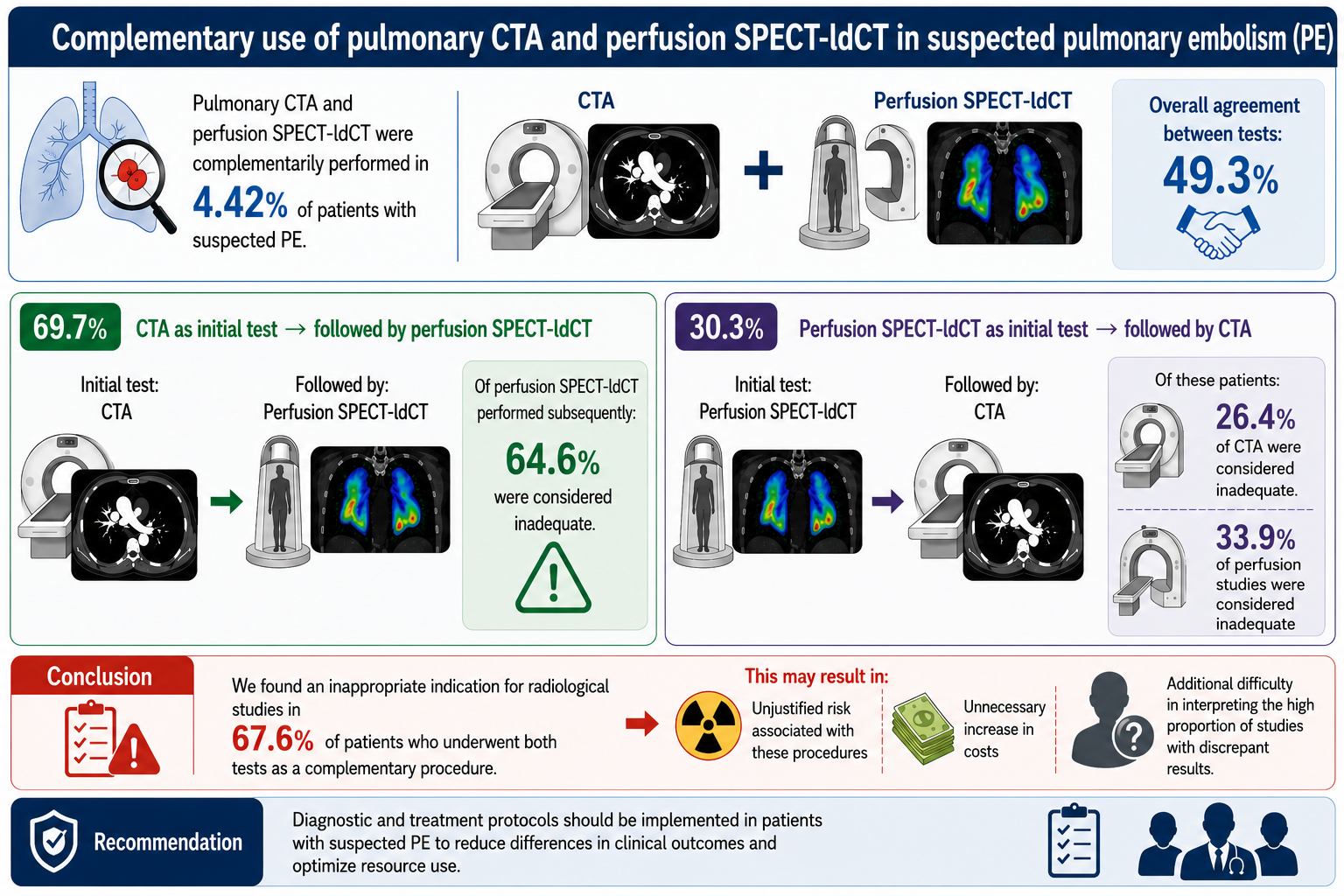
Background: The use of complementary radiological studies for suspected pulmonary embolism (PE) during the same clinical episode is uncommon and is usually due to a suboptimal or indeterminate initial study. However, the prevalence and decision-making in this clinical scenario are uncertain. Aim: The objective of this study was to determine the appropriateness of complementary studies (computed tomography angiography [CTA] and perfusion single-photon emission computed tomography/low-dose computed tomography [SPECT/ldCT]) in patients with suspected acute PE in real-world clinical practice. Methods: We analyzed all patients who underwent both tests for suspected PE during the same clinical process over a 10-year period. Results: Pulmonary CTA and perfusion SPECT/ldCT were performed as complementary studies in 4.42% of patients with suspected PE. In 69.7% of these patients, CTA was the initial diagnostic test and was subsequently followed by perfusion SPECT/ldCT, of which 64.6% were considered to have been inappropriately indicated. In 30.3% of patients, an initial lung perfusion SPECT/ldCT was followed by CTA; of these, 26.4% of CTA and 33.9% of perfusion studies were considered inappropriate. The overall agreement between the results of both tests was 49.3%. Conclusion: At least one imaging test was considered inappropriately indicated in 67.6% of patients who underwent both tests. This may result in unjustified risk associated with these procedures, unnecessary increase in costs, and additional difficulty in interpreting the high proportion of studies with discrepant results. Diagnostic and treatment protocols should be implemented in patients with suspected PE to reduce differences in clinical outcomes and optimize resource use. Relevance for patients: Inappropriate indications for radiological studies may cause unnecessary harm to certain patients, especially with regard to contrast-induced nephropathy.
- Raskob GE, Angchaisuksiri P, Blanco AN, et al. Thrombosis: a major contributor to the global disease burden. J Thromb Haemost. 2014;12(10):1580-1590. doi: 10.1111/jth.12698
- Wendelboe AM, Raskob GE. Global Burden of Thrombosis. Circ Res. 2016;118(9):1340-1347. doi: 10.1161/circresaha.115.306841
- Lehnert P, Lange T, Møller C, Olsen P, Carlsen J. Acute Pulmonary Embolism in a National Danish Cohort: Increasing Incidence and Decreasing Mortality. Thromb Haemost. 2018;118(03):539-546. doi: 10.1160/th17-08-0531
- Cohen AT, Agnelli G, Anderson FA, et al. Venous thromboembolism (VTE) in Europe. Thromb Haemost. 2007;98(10):756-764. doi: 10.1160/th07-03-0212
- Barco S, Woersching AL, Spyropoulos AC, Piovella F, Mahan CE. European Union-28: An annualised cost-of-illness model for venous thromboembolism. Thromb Haemost. 2016;115(04):800-808. doi: 10.1160/th15-08-0670
- Wells PS, Anderson D, Rodger M, et al. Derivation of a Simple Clinical Model to Categorize Patients Probability of Pulmonary Embolism: Increasing the Models Utility with the SimpliRED D-dimer. Thromb Haemost. 2000;83(03):416- 420. doi: 10.1055/s-0037-1613830
- Klok FA, Mos ICM, Nijkeuter M, et al. Simplification of the Revised Geneva Score for Assessing Clinical Probability of Pulmonary Embolism. Arch Intern Med. 2008;168(19):2131. doi: 10.1001/archinte.168.19.2131
- Konstantinides SV, Meyer G, Becattini C, et al. 2019 ESC Guidelines for the diagnosis and management of acute pulmonary embolism developed in collaboration with the European Respiratory Society (ERS). Eur Heart J. 2019;41(4):543-603. doi: 10.1093/eurheartj/ehz405
- Bae K, Jeon KN, Cho SB, et al. Improved Opacification of a Suboptimally Enhanced Pulmonary Artery in Chest CT: Experience Using a Dual-Layer Detector Spectral CT. Am J Roentgenol. 2018;210(4):734-741. doi: 10.2214/ajr.17.18537
- Zuin M, Bikdeli B, Ballard-Hernandez J, et al. International Clinical Practice Guideline Recommendations for Acute Pulmonary Embolism. J Am Coll Cardiol. 2024;84(16):1561- 1577. doi: 10.1016/j.jacc.2024.07.044
- Prentice D, Wipke-Tevis DD. Adherence to Best Practice Advice for Diagnosis of Pulmonary Embolism. Clin Nurse Spec. 2022;36(1):52-61. doi: 10.1097/nur.0000000000000642
- Ibáñez-Bravo S, Banzo I, Quirce R, et al. Ventilation/ Perfusion SPECT lung scintigraphy and computed tomography pulmonary angiography in patients with clinical suspicion of pulmonary embolism. Rev Española De Med Nucl E ImagenMol. 2016;35(4):215-220. doi: 10.1016/j.remn.2015.12.008
- Lu Y, Macapinlac HA. Perfusion SPECT/CT to diagnose pulmonary embolism during COVID-19 pandemic. Eur J Nucl Med Mol Imaging. 2020;47(9):2064-2065. doi: 10.1007/s00259-020-04851-6
- Torbicki A, Perrier A, Konstantinides S, et al. Guidelines on the diagnosis and management of acute pulmonary embolism. Eur Heart J. 2008;29(18):2276-2315. doi: 10.1093/eurheartj/ehn310
- Perrier A, Roy PM, Sanchez O, et al. Multidetector-Row Computed Tomography in Suspected Pulmonary Embolism. N Engl J Med. 2005;352(17):1760-1768. doi: 10.1056/nejmoa042905
- Stein PD, Fowler SE, Goodman LR, et al. Multidetector Computed Tomography for Acute Pulmonary Embolism. N Engl J Med. 2006;354(22):2317-2327. doi: 10.1056/nejmoa052367
- Writing Group for the Christopher Study Investigators*. Effectiveness of Managing Suspected Pulmonary Embolism Using an Algorithm Combining Clinical Probability, D-Dimer Testing, and Computed Tomography. JAMA. 2006;295(2):172–179. doi: 10.1001/jama.295.2.172
- Curtis BR, Cox M, Poplawski M, Lyshchik A. Low yield of ventilation and perfusion imaging for the evaluation of pulmonary embolism after indeterminate CT pulmonary angiography. Emerg Radiol. 2017;24(5):525-530. doi: 10.1007/s10140-017-1503-9
- Bates DDB, Tkacz JN, LeBedis CA, Holalkere N. Suboptimal CT pulmonary angiography in the emergency department: a retrospective analysis of outcomes in a large academic medical center. Emerg Radiol. 2016;23(6):603-607. doi: 10.1007/s10140-016-1425-y
- Parra Caballero P, Ruiz Berraco F, Salvador Rodríguez A, et al. Suboptimal CT pulmonary angiography for PE diagnosis, is it worth further study? A retrospective 7.5-year evaluation in a large academic medical center. Thrombosis Res. 2025;250:109324. doi: 10.1016/j.thromres.2025.109324
- Bajc M, Olsson B, Palmer J, Jonson B. Ventilation / Perfusion SPECT for diagnostics of pulmonary embolism in clinical practice. J Internal Med. 2008;264(4):379-387. doi: 10.1111/j.1365-2796.2008.01980.x
- Gutte H, Mortensen J, Jensen CV, et al. Detection of Pulmonary Embolism with Combined Ventilation– Perfusion SPECT and Low-Dose CT: Head-to-Head Comparison with Multidetector CT Angiography. J Nucl Med. 2009;50(12):1987-1992. doi: 10.2967/jnumed.108.061606
- Reinartz P, Wildberger JE, Schaefer W, Nowak B, Mahnken AH, Buell U. Tomographic imaging in the diagnosis of pulmonary embolism: a comparison between V/Q lung scintigraphy in SPECT technique and multislice spiral CT. J Nucl Med. 2004;45(9):1501–1508
- Collart JP, Roelants V, Vanpee D, et al. Is a lung perfusion scan obtained by using single photon emission computed tomography able to improve the radionuclide diagnosis of pulmonary embolism? Nucl Med Commun. 2002;23(11):1107-1113. doi: 10.1097/00006231-200211000-00011
- Roach PJ, Schembri GP, Bailey DL. V/Q scanning using SPECT and SPECT/CT. J Nucl Med. 2013;54(9):1588-1596. doi: 10.2967/jnumed.113.124602
- Anderson DR, Kahn SR, Rodger MA, et al. Computed tomographic pulmonary angiography vs ventilation-perfusion lung scanning in patients with suspected pulmonary embolism: a randomized controlled trial. JAMA. 2007;298(23):2743–2753. doi: 10.1001/jama.298.23.2743
- Roy PM, Colombet I, Durieux P, Chatellier G, Sors H, Meyer G. Systematic review and meta-analysis of strategies for the diagnosis of suspected pulmonary embolism. BMJ. 2005;331(7511):259. doi: 10.1136/bmj.331.7511.259
- Leblanc M, Paul N. V/Q SPECT and computed tomographic pulmonary angiography. Semin Nucl Med. 2010;40(6):426- 441. doi: 10.1053/j.semnuclmed.2010.08.001
- Sostman HD, Stein PD, Gottschalk A, Matta F, Hull R, Goodman L. Acute Pulmonary Embolism: Sensitivity and Specificity of Ventilation-Perfusion Scintigraphy in PIOPED II Study. Radiology. 2008;246(3):941-946. doi: 10.1148/radiol.2463070270
- Reid JH, Coche EE, Inoue T, et al. Is the lung scan alive and well? Facts and controversies in defining the role of lung scintigraphy for the diagnosis of pulmonary embolism in the era of MDCT. Eur J Nucl Med Mol Imaging. 2009;36(3):505- 521. doi: 10.1007/s00259-008-1014-8
- Erythropoulou-Kaltsidou A, Alkagiet S, Tziomalos K. New guidelines for the diagnosis and management of pulmonary embolism: Key changes. World J Cardiol. 2020;12(5):161- 166. doi: 10.4330/wjc.v12.i5.161
- Ling IT, Naqvi HA, Siew TK, Loh NK, Ryan GF. SPECT ventilation perfusion scanning with the addition of low-dose CT for the investigation of suspected pulmonary embolism. Intern Med J. 2012;42(11):1257-1261. doi: 10.1111/j.1445-5994.2012.02939.x
- Le Duc-Pennec A, Le Roux PY, Cornily JC, et al. Diagnostic accuracy of single-photon emission tomography ventilation/ perfusion lung scan in the diagnosis of pulmonary embolism. Chest. 2012;141(2):381-387. doi: 10.1378/chest.11-0090
- Simanek M, Koranda P. The benefit of personalized hybrid SPECT/CT pulmonary imaging. Am J Nucl Med Mol Imaging. 2016;6(4):215–222
- Le Roux PY, Robin P, Tromeur C, et al. Ventilation/ perfusion SPECT for the diagnosis of pulmonary embolism: A systematic review. J Thromb Haemost. 2020;18(11):2910- 2920. doi: 10.1111/jth.15038
- Calder KK, Herbert M, Henderson SO. The mortality of untreated pulmonary embolism in emergency department patients. Ann Emerg Med. 2005;45(3):302-310. doi: 10.1016/j.annemergmed.2004.10.001
- Engelke C, Rummeny EJ, Marten K. Pulmonary embolism at multi-detector row CT of chest: one-year survival of treated and untreated patients. Radiology. 2006;239(2):563-575. doi: 10.1148/radiol.2392050118
- Carson JL, Kelley MA, Duff A, et al. The clinical course of pulmonary embolism. N Engl J Med. 1992;326(19):1240- 1245. doi: 10.1056/nejm199205073261902
- Stein PD, Henry JW, Relyea B. Untreated patients with pulmonary embolism. Outcome, clinical, and laboratory assessment. Chest. 1995;107(4):931-935. doi: 10.1378/chest.107.4.931
- Da Costa Rodrigues J, Alzuphar S, Combescure C, Le Gal G, Perrier A. Diagnostic characteristics of lower limb venous compression ultrasonography in suspected pulmonary embolism: a meta-analysis. J Thromb Haemost. 2016;14(9):1765-1772. doi: 10.1111/jth.13407
- Le Gal G, Righini M, Sanchez O, et al. A positive compression ultrasonography of the lower limb veins is highly predictive of pulmonary embolism on computed tomography in suspected patients. Thromb Haemost. 2006;95(6):963–966. doi: 10.1160/TH06-03-0158
- Yu F, Yuan X, Fu S. Optimizing emergency nursing protocols to enhance outcomes in patients with acute myocardial infarction: A retrospective study. Medicine. 2025;104(23):e41412. doi: 10.1097/md.0000000000041412
- Kim HJ, Ko RE, Lim SY, Park S, Suh GY, Lee YJ. Sepsis Alert Systems, Mortality, and Adherence in Emergency Departments: A Systematic Review and Meta-Analysis. JAMA Netw Open. 2024;7(7):e2422823. doi: 10.1001/jamanetworkopen.2024.22823
- Soster CB, Anschau F, Rodrigues NH, Silva LGAD, Klafke A. Advanced triage protocols in the emergency department: A systematic review and meta-analysis. Rev Lat Am Enfermagem. 2022;30:e3511. [Article in Portuguese, English, Spanish] doi: 10.1590/1518-8345.5479.3511