

Understanding fetal posterior fossa abnormalities: Insights from MRI and ultrasound imaging

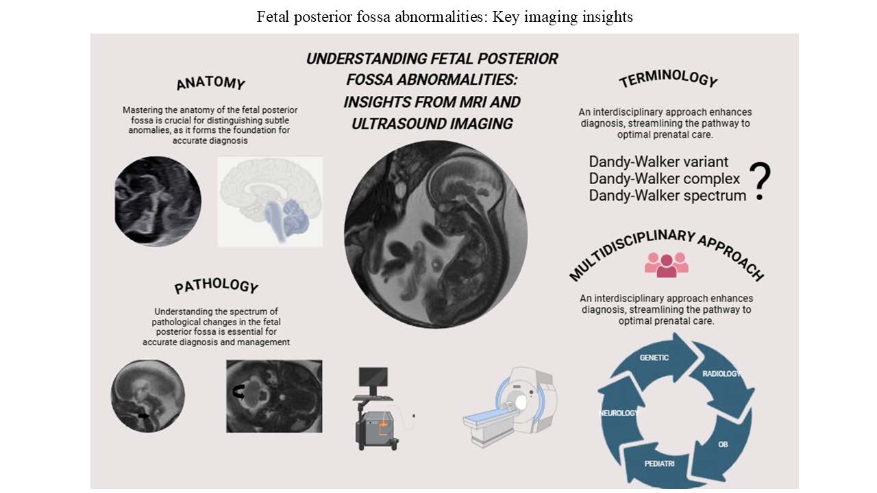
Background: The posterior fossa is a critical brain region housing essential structures such as the cerebellum and brainstem, crucial for coordination, balance, and autonomic functions. Abnormalities in this area significantly impact fetal development and postnatal outcomes, necessitating accurate diagnosis and characterization of fetal posterior fossa abnormalities. Aims: This review aims to analyze the diagnostic role of ultrasound, the primary imaging modality due to its accessibility and safety, and magnetic resonance imaging, which provides enhanced diagnostic accuracy in identifying central nervous system anomalies, particularly those involving the posterior fossa. A comprehensive approach is proposed to address the diagnostic complexities of cerebellar malformations, including ambiguous terminology and overlapping clinical features, which pose notable challenges. Relevance for patients: The complexity and lack of consensus regarding cerebellar malformations across medical disciplines can hinder accurate diagnosis and timely intervention. By advancing interdisciplinary research and refining diagnostic approaches, this study aims to enhance clinical management strategies and improve patient outcomes in cases of posterior fossa pathologies, ultimately fostering a more integrated and effective approach to diagnosis and treatment.
- Moore KL, Persaud TVN, Torchia MG. The Developing Human: Clinically Oriented Embryology. 9th ed. Philadelphia, PA: Elsevier; 2019. p. 404-406.
- Larsen WJ, Schoenwolf GC, Bleyl SB, Brauer PR. Larsen’s Human Embryology. 5th ed. Philadelphia, PA: Churchill Livingstone/Elsevier; 2018. p. 95-9.
- American College of Obstetricians and Gynecologists. ACOG practice bulletin No. 101: Ultrasonography in pregnancy. Obstet Gynecol. 2009;113(2 Pt 1):451-461. doi: 10.1097/AOG.0b013e31819930b0
- American Institute of Ultrasound in Medicine. Case Study Requirements for Accreditation. Available from: https://www. aium.org/docs/default-source/accreditation/case-study-requirements/76811.pdf [Last accessed on 2024 Jan 19].
- Australasian Society for Ultrasound in Medicine. Guidelines for the Performance of Second (mid) Trimester Ultrasound. Available from: https://www.asum.com.au/files/public/ sop/curver/obs-gynae/guidelines-for-the-performance-of-second-mid-trimester-ultrasound.pdf [Last accessed on 2024 Jan 19].
- UK Government. Fetal Anomaly Screening Programme Handbook: 20-Week Screening Scan. Available from: https:// www.gov.uk/government/publications/fetal-anomaly-screening-programme-handbook/20-week-screening-scan [Last accessed on 2024 Jan 19].
- International Society of Ultrasound in Obstetrics and Gynecology. ISUOG Practice Guidelines: Routine Mid-trimester Fetal Ultrasound. Available from: https://www. isuog.org/static/4e2ed89e-fa8a-42c2-9c0929cd89cb58ff/ isuog-practice-guidelines-routine-mid-trimester-fetal-ultrasound.pdf [Last accessed on 2024 Jan 19].
- Fileva N, Severino M, Tortora D, Ramaglia A, Paladini D, Rossi A. Second trimester fetal MRI of the brain: Through the ground glass. J Clin Ultrasound. 2023;51(2):283-299. doi: 10.1002/jcu.23423
- Chaoui R, Benoit B, Mitkowska-Wozniak H, Heling KS, Nicolaides KH. Assessment of intracranial translucency (IT) in the detection of spina bifida at the 11-13-week scan. Ultrasound Obstet Gynecol. 2009;34(3):249-252. doi: 10.1002/uog.7329
- International Society of Ultrasound in Obstetrics and Gynecology. ISUOG practice guidelines (updated): Sonographic examination of the fetal central nervous system. Part 1: Performance of screening examination and indications for targeted neurosonography. Ultrasound Obstet Gynecol. 2020;56(3):476-484. doi: 10.1002/uog.22145
- Robinson AJ, Blaser S, Toi A, et al. The fetal cerebellar vermis: Assessment for abnormal development by ultrasonography and magnetic resonance imaging. Ultrasound Q. 2007;23(3):211-223. doi: 10.1097/RUQ.0b013e31814b162c
- Malinger G, Lev D, Lerman-Sagie T. Is fetal magnetic resonance imaging superior to neurosonography for detection of brain anomalies? Ultrasound Obstet Gynecol. 2002;20(4):317-321. doi: 10.1046/j.1469-0705.2002.00825.x
- International Society of Ultrasound in Obstetrics and Gynecology. ISUOG practice guidelines: Performance of fetal magnetic resonance imaging. Ultrasound Obstet Gynecol. 2017;49(5):671-680. doi: 10.1002/uog.17412
- Barkovich AJ, Raybaud CA, editors. Congenital malformations of the brain and skull. In: Pediatric Neuroimaging. 6th ed. Philadelphia, PA: Wolters Kluwer; 2019. p. 724.
- Whitehead MT, Barkovich MJ, Sidpra J, et al. Refining the neuroimaging definition of the dandy-walker phenotype. AJNR Am J Neuroradiol. 2022;43(10):1488-1493. doi: 10.3174/ajnr.A7659
- Aldinger KA, Lehmann OJ, Hudgins L, et al. FOXC1 is required for normal cerebellar development and is a major contributor to chromosome 6p25.3 Dandy-Walker malformation. Nat Genet. 2009;41(9):1037-1042. doi: 10.1038/ng.422
- Grinberg I, Northrup H, Ardinger H, et al. Heterozygous deletion of the linked genes ZIC1 and ZIC4 is involved in Dandy-Walker malformation. Nat Genet. 2004;36(10): 1053-1055. doi: 10.1038/ng1420
- Alsamal M, Zitoun OA, Abdulghani EA, Sula I. Meckel- Gruber syndrome together with Dandy-Walker malformation: An atypical case report of a 2nd recurrence in a consanguine marriage. Childs Nerv Syst. 2024;40(1): 257-261. doi: 10.1007/s00381-023-06104-x
- Spennato P, Mirone G, Nastro A, et al. Hydrocephalus in Dandy-Walker malformation. Childs Nerv Syst. 2011;27(10):1665-1681. doi: 10.1007/s00381-011-1544-4
- Bolduc ME, Limperopoulos C. Neurodevelopmental outcomes in children with cerebellar malformations: A systematic review. Dev Med Child Neurol. 2009;51(4): 256-267. doi: 10.1111/j.1469-8749.2008.03224.x
- Venkatesan C, Kline-Fath B, Horn PS, Poisson KE, Hopkin R, Nagaraj UD. Short- and long-term outcomes of prenatally diagnosed Dandy-Walker malformation, vermian hypoplasia, and Blake pouch cyst. J Child Neurol. 2021;36(12):1111-1119. doi: 10.1177/08830738211049115
- Parisi MA, Dobyns WB. Human malformations of the midbrain and hindbrain: Review and proposed classification scheme. Mol Genet Metab. 2003;80(1-2):36-53. doi: 10.1016/j.ymgme.2003.08.010
- Wakeling EL, Jolly M, Fisk NM, Gannon C, Holder SE. X-linked inheritance of Dandy-Walker variant. Clin Dysmorphol. 2002;11(1):15-18. doi: 10.1097/00019605-200201000-00003
- Cesaroni E, Matricardi S, Cappanera S, Marini C. First reported case of an inherited PACS2 pathogenic variant with variable expression. Epileptic Disord. 2022;24(3):572-576. doi: 10.1684/epd.2022.1417
- Bosemani T, Orman G, Boltshauser E, Tekes A, Huisman TA, Poretti A. Congenital abnormalities of the posterior fossa. Radiographics. 2015;35(1):200-220. doi: 10.1148/rg.351140038
- Patek KJ, Kline-Fath BM, Hopkin RJ, Pilipenko VV, Crombleholme TM, Spaeth CG. Posterior fossa anomalies diagnosed with fetal MRI: Associated anomalies and neurodevelopmental outcomes. Prenat Diagn. 2012;32(1):75-82. doi: 10.1002/pd.2911
- Tarui T, Limperopoulos C, Sullivan NR, Robertson RL, Du Plessis AJ. Long-term developmental outcome of children with a fetal diagnosis of isolated inferior vermian hypoplasia. Arch Dis Child Fetal Neonatal Ed. 2014;99(1):F54-F58. doi: 10.1136/archdischild-2013-304678
- Poretti A, Boltshauser E, Doherty D. Cerebellar hypoplasia: Differential diagnosis and diagnostic approach. Am J Med Genet C Semin Med Genet. 2014;166(2):211-226. doi: 10.1002/ajmg.c.31398
- Poretti A, Boltshauser E, Huisman TA. Cerebellar and brainstem malformations. Neuroimaging Clin N Am. 2016;26(3):341-357. doi: 10.1016/j.nic.2016.03.005
- Clement E, Mercuri E, Godfrey C, et al. Brain involvement in muscular dystrophies with defective dystroglycan glycosylation. Ann Neurol. 2008;64(5):573-582. doi: 10.1002/ana.21482
- Shekdar K. Posterior fossa malformations. Semin Ultrasound CT MR. 2011;32(3):228-241. doi: 10.1053/j.sult.2011.02.003
- Whitehead MT, Vezina G, Schlatterer SD, Mulkey SB, Du Plessis AJ. Taenia-Tela choroidea complex and choroid plexus location help distinguish Dandy-Walker malformation and Blake pouch cysts. Pediatr Radiol. 2021;51(8):1457-1470. doi: 10.1007/s00247-021-04991-3
- Gandolfi Colleoni G, Contro E, Carletti A, et al. Prenatal diagnosis and outcome of fetal posterior fossa fluid collections. Ultrasound Obstet Gynecol. 2012;39(6):625-631. doi: 10.1002/uog.11071
- Barkovich AJ, Raybaud CA, editors. Congenital malformations of the brain and skull. In: Pediatric Neuroimaging. 6th ed. Philadelphia, PA: Wolters Kluwer; 2019. p. 798.
- Osborn AG, Preece MT. Intracranial cysts: Radiologic-pathologic correlation and imaging approach. Radiology. 2006;239(3):650-664. doi: 10.1148/radiol.2393050823
- Qureshi HM, Mekbib KY, Allington G, et al. Familial and syndromic forms of arachnoid cyst implicate genetic factors in disease pathogenesis. Cereb Cortex. 2023;33(6):3012-3025. doi: 10.1093/cercor/bhac257
- Mahony BS, Callen PW, Filly RA, Hoddick WK. The fetal cisterna magna. Radiology. 1984;153(3):773-776. doi: 10.1148/radiology.153.3.6387792
- Khan AN, Smirniotopoulos JG. Arachnoid Cyst Imaging. eMedicine; 2021. Available from: https://emedicine. medscape.com/article/336489-overview [Last accessed on 2024 Mar 03].
- Osborn AG, Linscott LL, Salzman KL. Osborn’s Brain: Imaging, Pathology, and Anatomy. Netherlands: Elsevier Health Sciences; 2024.
- Bedei I, Krispin E, Sanz Cortes M, et al. Prenatal diagnosis and postnatal outcome of closed spinal dysraphism. Prenat Diagn. 2023;43(12):1521-1532. doi: 10.1002/pd.6454
- Bulas D. Fetal evaluation of spine dysraphism. Pediatr Radiol. 2010;40(6):1029-1037. doi: 10.1007/s00247-010-1583-0
- Kunpalin Y, Richter J, Mufti N, et al. Cranial findings detected by second-trimester ultrasound in fetuses with myelomeningocele: A systematic review. BJOG. 2021;128(2):366-374. doi: 10.1111/1471-0528.16496
- Khalaveh F, Seidl R, Czech T, et al. Myelomeningocele- Chiari II malformation-neurological predictability based on fetal and postnatal magnetic resonance imaging. Prenat Diagn. 2021;41(8):922-932. doi: 10.1002/pd.5987
- Talamonti G, Marcati E, Mastino L, Meccariello G, Picano M, D’Aliberti G. Surgical management of Chiari malformation type II. Childs Nerv Syst. 2020;36(8):1621-1634. doi: 10.1007/s00381-020-04675-7
- Osborn AG. Osborn’s Brain: Imaging, Pathology, and Anatomy. 2nd ed. Netherlands: Elsevier; 2018. p. 1298.
- Thompson DN. Postnatal management and outcome for neural tube defects including spina bifida and encephalocoeles. Prenat Diagn. 2009;29(4):412-419. doi: 10.1002/pd.2199
- Kasprian GJ, Paldino MJ, Mehollin-Ray AR, et al. Prenatal imaging of occipital encephaloceles. Fetal Diagn Ther. 2015;37(3):241-248. doi: 10.1159/000366159
- Markovic I, Bosnjakovic P, Milenkovic Z. Occipital encephalocele: Cause, incidence, neuroimaging and surgical management. Curr Pediatr Rev. 2020;16(3):200-205. doi: 10.2174/1573396315666191018161535
- Demaerel P, Morel C, Lagae L, Wilms G. Partial rhombencephalosynapsis. AJNR Am J Neuroradiol. 2004;25(1):29-31.
- Schell-Apacik CC, Cohen M, Vojta S, et al. Gomez-Lopez- Hernandez syndrome (cerebello-trigeminal-dermal dysplasia): Description of an additional case and review of the literature. Eur J Pediatr. 2008;167(1):123-126. doi: 10.1007/s00431-007-0478-z
- Ishak GE, Dempsey JC, Shaw DW, et al. Rhombencephalosynapsis: A hindbrain malformation associated with incomplete separation of midbrain and forebrain, hydrocephalus and a broad spectrum of severity. Brain. 2012;135(Pt 5):1370-1386. doi: 10.1093/brain/aws065
- Guibaud L. Practical approach to prenatal posterior fossa abnormalities using MRI. Pediatr Radiol. 2004;34(9): 700-711. doi: 10.1007/s00247-004-1248-y
- Massoud M, Guibaud L. Prenatal imaging of posterior fossa disorders: A review. Eur J Paediatr Neurol. 2018;22(6): 972-988. doi: 10.1016/j.ejpn.2018.07.007
- Khaladkar SM, Jhala NA, Shukla A, Shah R, Durgi EC. Rhombencephalosynapsis: A rare hindbrain malformation. Cureus. 2024;16(7):e65400. doi: 10.7759/cureus.65400
- Stone SS, Warf BC. Combined endoscopic third ventriculostomy and choroid plexus cauterization as primary treatment for infant hydrocephalus: A prospective North American series. J Neurosurg Pediatr. 2014;14(5):439-446. doi: 10.3171/2014.7.PEDS14152
- Adle-Biassette H, Saugier-Veber P, Fallet-Bianco C, et al. Neuropathological review of 138 cases genetically tested for X-linked hydrocephalus: Evidence for closely related clinical entities of unknown molecular bases. Acta Neuropathol. 2013;126(3):427-442. doi: 10.1007/s00401-013-1146-1
- Emery SP, Narayanan S, Greene S. Fetal aqueductal stenosis: Prenatal diagnosis and intervention. Prenat Diagn. 2020;40(1):58-65. doi: 10.1002/pd.5527
- Peiro JL, Fabbro MD. Fetal therapy for congenital hydrocephalus-where we came from and where we are going. Childs Nerv Syst. 2020;36(8):1697-1712. doi: 10.1007/s00381-020-04738-9
- Hildebrandt F, Benzing T, Katsanis N. Ciliopathies. N Engl J Med. 2011;364(16):1533-1543. doi: 10.1056/NEJMra1010172
- Surisetti BK, Holla VV, Prasad S, et al. Clinical and imaging profile of patients with Joubert syndrome. J Mov Disord. 2021;14(3):231-235. doi: 10.14802/jmd.21066
- Al-Smair A, Younes S, Saadeh A, Kaoukji AR, Jaber O. Joubert-Plus syndrome with an atretic cephalocele: A case report. Radiol Case Rep. 2022;17(10):3630-3634. doi: 10.1016/j.radcr.2022.07.038
- Poretti A, Singhi S, Huisman TA, et al. Tecto-cerebellar dysraphism with occipital encephalocele: Not a distinct disorder, but part of the Joubert syndrome spectrum? Neuropediatrics. 2011;42(4):170-174. doi: 10.1055/s-0031-1287763
- Van De Weghe JC, Gomez A, Doherty D. The Joubert- Meckel-Nephronophthisis spectrum of ciliopathies. Annu Rev Genomics Hum Genet. 2022;23:301-329. doi: 10.1146/annurev-genom-121321-093528
- Online Mendelian Inheritance in Man. Pontocerebellar Hypoplasia Search Results. Available from: https://www. omim.org/search?index=entry&search=%22pontocer ebellar+hypoplasia%22 [Last accessed on 2024 Apr 21.
- Rüsch CT, Bölsterli BK, Kottke R, Steinfeld R, Boltshauser E. Pontocerebellar hypoplasia: A pattern recognition approach. Cerebellum. 2020;19(4):569-582. doi: 10.1007/s12311-020-01135-5
- Accogli A, Addour-Boudrahem N, Srour M. Diagnostic approach to cerebellar hypoplasia. Cerebellum. 2021;20(4):631-658. doi: 10.1007/s12311-020-01224-5
- Zago S, Silvestri E, Arcangeli T, et al. Fetal presentation of Walker-Warburg syndrome with compound heterozygous POMT2 missense mutations. Fetal Pediatr Pathol. 2023;42(2):334-341. doi: 10.1080/15513815.2022.2116620
- Osborn AG. Osborn’s Brain: Imaging, Pathology, and Anatomy. 2nd ed. Netherlands: Elsevier; 2018. p. 1218.
- Scelsa B, Cutillo G, Lanna MM, et al. Prenatal diagnosis and neurodevelopmental outcome in isolated cerebellar hypoplasia of suspected hemorrhagic etiology: A retrospective cohort study. Cerebellum. 2022;21(6):944-953. doi: 10.1007/s12311-021-01341-9
- Simonazzi G, Bernabini D, Curti A, et al. Fetal cerebellar damage in fetuses with severe anemia undergoing intrauterine transfusions. J Matern Fetal Neonatal Med. 2016;29(3):389-392. doi: 10.3109/14767058.2014.1001973
- Feygin T, Khalek N, Moldenhauer JS. Fetal brain, head, and neck tumors: Prenatal imaging and management. Prenat Diagn. 2020;40(10):1203-1219. doi: 10.1002/pd.5722
- Barkovich AJ, Raybaud CA, editors. Congenital malformations of the brain and skull. In: Pediatric Neuroimaging. 6th ed. Netherlands: Wolters Kluwer; 2019. p. 1094.
- Meoded A, Turan S, Harman C, et al. Pre- and postnatal ultrasound and magnetic resonance imaging of intracranial extra-axial glioneuronal heterotopia. Fetal Diagn Ther. 2011;30(4):314-316. doi: 10.1159/000330859
- Russler-Germain E, Majumdar S, Nguyen T, Hirose K, Yang PH, Dahiya S. Glioneuronal heterotopia in the right middle cranial fossa. Free Neuropathol. 2024;5:28. doi: 10.17879/freeneuropathology-2024-5848