

Pfirrmann grading of intervertebral disc degeneration: Morphological assessment, treatment response, and emerging quantitative MRI approaches

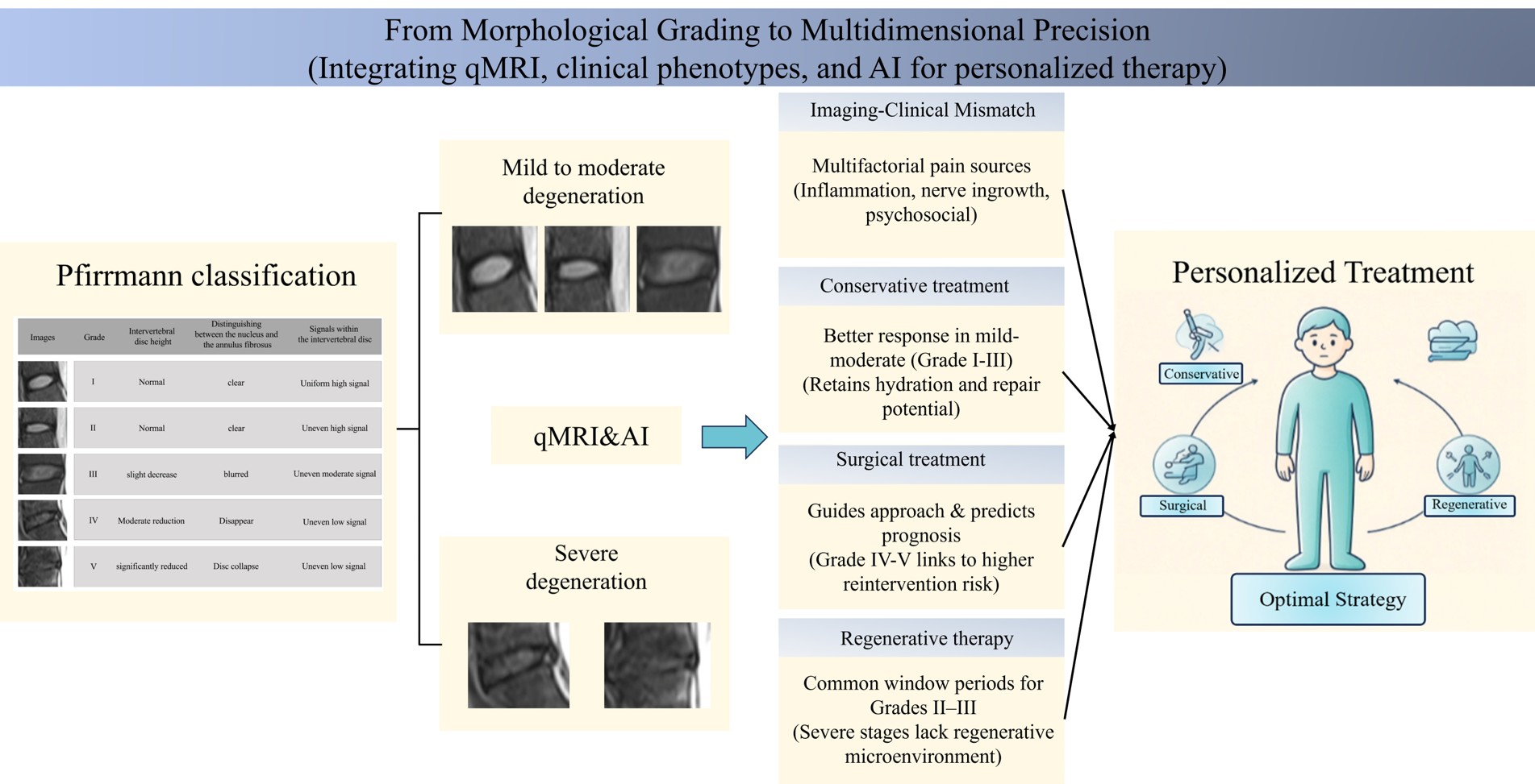
Introduction: Intervertebral disc degeneration (IVDD) is a major cause of low back pain, and magnetic resonance imaging (MRI) is the primary tool for its assessment. Among these, the Pfirrmann classification is widely used in clinical practice and research due to its standardization and practicality.
Objective: This paper reviews MRI-based methods for assessing IVDD, focusing on the relationship between the Pfirrmann classification and the efficacy of conservative treatment, surgical treatment, and regenerative therapy.
Methods: This narrative review searched PubMed, Web of Science, and Embase for literature published from January 2000 to June 2026 using terms including “disc degeneration,” “Pfirrmann classification,” and “quantitative magnetic resonance imaging,” with inclusion criteria applied.
Results: The Pfirrmann classification reflects changes in intervertebral disc morphology and signal intensity; however, it does not fully correlate with clinical symptoms or treatment response, suggesting that pain and treatment efficacy are influenced by multiple factors, including inflammation, nerve root involvement, and endplate changes. In clinical practice, patients with mild to moderate degeneration typically respond better to conservative treatment, and pre-operative grading aids in surgical decision-making. Advances in quantitative MRI techniques—such as T2 mapping, T1ρ, and diffusion kurtosis imaging—along with developments in artificial intelligence, have provided new tools for early identification of degeneration and improved accuracy in predicting treatment outcomes.
Conclusion: The Pfirrmann grading system remains the foundation for IVDD assessment; however, future work will need to integrate quantitative imaging, biomarkers, and clinical phenotypes to establish a comprehensive, multi-parameter model that enables more precise treatment outcome prediction and personalized therapy.
- Pang H, Chen S, Klyne DM, et al. Low back pain and osteoarthritis pain: a perspective of estrogen. Bone Res. 2023;11(1):42. doi: 10.1038/s41413-023-00280-x
- Knezevic NN, Candido KD, Vlaeyen JWS, Van Zundert J, Cohen SP. Low back pain. Lancet. 2021;398(10294):78-92. doi: 10.1016/S0140-6736(21)00733-9
- Ząbek Z, Wyczałkowska-Tomasik A, Poboży K, et al. Understanding the microenvironment of intervertebral disc degeneration: a comprehensive review of pathophysiological insights and therapeutic implications. Int J Mol Sci. 2025;26(20):9938. doi: 10.3390/ijms26209938
- Liu S, Hu Y, Xu W, et al. Restoration of lysosomal function attenuates autophagic flux impairment in nucleus pulposus cells and protects against mechanical overloading-induced intervertebral disc degeneration. Autophagy. 2025;21(5):979-995. doi: 10.1080/15548627.2024.2440844
- Bu W, Shi Y, Huang X, et al. Rescue of nucleus pulposus cells from an oxidative stress microenvironment via glutathione-derived carbon dots to alleviate intervertebral disc degeneration. J Nanobiotechnology. 2024;22(1):412. doi: 10.1186/s12951-024-02683-2
- Xia Q, Zhao Y, Dong H, et al. Progress in the study of molecular mechanisms of intervertebral disc degeneration. Biomed Pharmacother. 2024;174:116593. doi: 10.1016/j.biopha.2024.116593
- Zhu D, Liang H, Du Z, et al. Altered metabolism and inflammation driven by post-translational modifications in intervertebral disc degeneration. Research (Wash D C). 2024;7:0350. doi: 10.34133/research.0350
- Zàaba NF, Ogaili RH, Ahmad F, Mohd Isa IL. Neuroinflammation and nociception in intervertebral disc degeneration: a review of precision medicine perspective. Spine J. 2025;25(6):1139-1153. doi: 10.1016/j.spinee.2024.12.033
- Yu LP, Qian WW, Yin GY, Ren YX, Hu ZY. MRI assessment of lumbar intervertebral disc degeneration with lumbar degenerative disease using the Pfirrmann grading systems. PLoS One. 2012;7(12):e48074. doi: 10.1371/journal.pone.0048074
- Griffith JF, Wang YX, Antonio GE, et al. Modified Pfirrmann grading system for lumbar intervertebral disc degeneration. Spine (Phila Pa 1976). 2007;32(24):E708-E712. doi: 10.1097/BRS.0b013e31815a59a0
- Bouhsina N, Decante C, Hardel JB, et al. Comparison of MRI T1, T2, and T2* mapping with histology for assessment of intervertebral disc degeneration in an ovine model. Sci Rep. 2022;12(1):5398. doi: 10.1038/s41598-022-09348-w
- Blumenkrantz G, Zuo J, Li X, Kornak J, Link TM, Majumdar S. In vivo 3.0-tesla magnetic resonance T1rho and T2 relaxation mapping in subjects with intervertebral disc degeneration and clinical symptoms. Magn Reson Med. 2010;63(5):1193-1200. doi: 10.1002/mrm.22362
- Togao O, Hiwatashi A, Wada T, et al. A qualitative and quantitative correlation study of lumbar intervertebral disc degeneration using glycosaminoglycan chemical exchange saturation transfer, Pfirrmann grade, and T1-ρ. AJNR Am J Neuroradiol. 2018;39(7):1369-1375. doi: 10.3174/ajnr.A5657
- Rahyussalim AJ, Zufar MLL, Kurniawati T. Significance of the association between disc degeneration changes on imaging and low back pain: a review article. Asian Spine J. 2020;14(2):245-257. doi: 10.31616/asj.2019.0046
- Pfirrmann CW, Metzdorf A, Zanetti M, Hodler J, Boos N. Magnetic resonance classification of lumbar intervertebral disc degeneration. Spine (Phila Pa 1976). 2001;26(17):1873-1878. doi: 10.1097/00007632-200109010-00011
- Kettler A, Wilke HJ. Review of existing grading systems for cervical or lumbar disc and facet joint degeneration. Eur Spine J. 2006;15(6):705-718. doi: 10.1007/s00586-005-0954-y
- Patil S, Gandhi OH, Karabacak M, Carr MT, Margetis K. MRI-based deep learning radiomics model for automated classification of disc degeneration in the lumbar spine. Sci Rep. 2026;16(1):24695. doi: 10.1038/s41598-026-56056-w
- Stelzeneder D, Welsch GH, Kovács BK, et al. Quantitative T2 evaluation at 3.0T compared to morphological grading of the lumbar intervertebral disc: a standardized evaluation approach in patients with low back pain. Eur J Radiol. 2012;81(2):324-330. doi: 10.1016/j.ejrad.2010.12.093
- Feng F, Wang W, Liu S, et al. T2 mapping and q-Dixon for assessment of intervertebral disc degeneration in lower back pain. BMC Musculoskelet Disord. 2025;26(1):234. doi: 10.1186/s12891-025-08453-x
- Chen C, Huang M, Han Z, et al. Quantitative T2 magnetic resonance imaging compared to morphological grading of the early cervical intervertebral disc degeneration: an evaluation approach in asymptomatic young adults. PLoS One. 2014;9(2):e87856. doi: 10.1371/journal.pone.0087856
- Raudner M, Schreiner MM, Juras V, et al. Prediction of lumbar disk herniation and clinical outcome using quantitative magnetic resonance imaging: a 5-year follow-up study. Invest Radiol. 2019;54(3):183-189. doi: 10.1097/RLI.0000000000000527
- Yang L, Sun C, Gong T, Li Q, Chen X, Zhang X. T1ρ, T2 and T2* mapping of lumbar intervertebral disc degeneration: a comparison study. BMC Musculoskelet Disord. 2022;23(1):1135. doi: 10.1186/s12891-022-06040-y
- Chavhan GB, Babyn PS, Thomas B, Shroff MM, Haacke EM. Principles, techniques, and applications of T2*-based MR imaging and its special applications. Radiographics. 2009;29(5):1433-1449. doi: 10.1148/rg.295095034
- Paul CPL, Smit TH, de Graaf M, et al. Quantitative MRI in early intervertebral disc degeneration: T1rho correlates better than T2 and ADC with biomechanics, histology and matrix content. PLoS One. 2018;13(1):e0191442. doi: 10.1371/journal.pone.0191442
- Falck AL, Buko EO, Chase KL, et al. Relationship between quantitative MRI and radiological, histological, and biochemical measures of intervertebral disc health in client-owned, nonchondrodystrophic-breed dogs. JOR Spine. 2025;8(3):e70105. doi: 10.1002/jsp2.70105
- Barnes NA, Dkhar W, Kadavigere R, et al. Quantitative assessment of early intervertebral disc degeneration with MR diffusion kurtosis imaging: a radiologic correlation with Pfirrmann grade. Magn Reson Imaging. 2026;127:110592. doi: 10.1016/j.mri.2025.110592
- Barnes NA, Dkhar W, Kadavigere R, Sukumar S, Vaishali K, Pradhan A. A narrative review of diffusion kurtosis MRI parameters in diagnosing degenerative spine diseases in animal models. Open Vet J. 2024;14(12):3181-3188. doi: 10.5455/OVJ.2024.v14.i12.3
- Barnes NA, Dkhar W, Kadavigere R, et al. Exploring mean kurtosis in MR diffusion kurtosis imaging for early detection of lumbar spine degeneration: a systematic review. F1000Res. 2025;14:440. doi: 10.12688/f1000research.163638.2
- Chen R, Wang H, Li Y, et al. CEST MRI: translational prospects for intervertebral disc degeneration—from basic research to clinical applications. J Orthop Translat. 2026;58:101131. doi: 10.1016/j.jot.2026.101131
- Deng M, Yuan J, Chen WT, Chan Q, Griffith JF, Wang YX. Evaluation of glycosaminoglycan in the lumbar disc using chemical exchange saturation transfer MR at 3.0 Tesla: reproducibility and correlation with disc degeneration. Biomed Environ Sci. 2016;29(1):47-55. doi: 10.3967/bes2016.005
- Zuo J, Saadat E, Romero A, et al. Assessment of intervertebral disc degeneration with magnetic resonance single-voxel spectroscopy. Magn Reson Med. 2009;62(5):1140-1146. doi: 10.1002/mrm.22093
- Bhari Thippeswamy P, Rajasekaran S, Ramachandran K, et al. Role of magnetic resonance spectroscopy and T2 relaxometry as imaging biomarker of early lumbar intervertebral disc degeneration. Global Spine J. 2025;15(5):2736-2743. doi: 10.1177/21925682241311515
- Tekin A, Çelik U, Öztürk A, et al. The association between lumbar disc degeneration and paraspinal muscle morphology: the role of age in a quantitative magnetic resonance imaging-based study. Biomedicines. 2026;14(8):1673. doi: 10.3390/biomedicines14081673
- Suryadevara M, Mishra GV, Parihar P, et al. Role of end plate changes and paraspinal muscle pathology in lower back pain: a narrative review. Cureus. 2024;16(5):e61319. doi: 10.7759/cureus.61319
- Natalia F, Sudirman S, Ruslim D, Al-Kafri A. Lumbar spine MRI annotation with intervertebral disc height and Pfirrmann grade predictions. PLoS One. 2024;19(5):e0302067. doi: 10.1371/journal.pone.0302067
- Liawrungrueang W, Cholamjiak W, Sarasombath P, Jitpakdee K, Kotheeranurak V. Artificial intelligence classification for detecting and grading lumbar intervertebral disc degeneration. Spine Surg Relat Res. 2024;8(6):552-559. doi: 10.22603/ssrr.2024-0154
- Baur D, Bieck R, Berger J, et al. Automated three-dimensional imaging and Pfirrmann classification of intervertebral disc using a graphical neural network in sagittal magnetic resonance imaging of the lumbar spine. J Imaging Inform Med. 2025;38(2):979-987. doi: 10.1007/s10278-024-01251-2
- Minin A, Leonova O, Krutko A, et al. SpineScan: a deep learning model for lumbar spine MRI annotation and Pfirrmann grading assessment. Eur Spine J. 2026;35(3):1261-1269. doi: 10.1007/s00586-025-09537-x
- Zhao P, Zhu S. Advances and challenges in AI-assisted MRI for lumbar disc degeneration detection and classification. Eur Spine J. 2026;35(3):1291-1300. doi: 10.1007/s00586-025-09179-z
- Shi Y, Wang Y. The AI-driven blueprint: decoding intervertebral disc repair mechanisms for intelligent biomaterial design. Front Cell Dev Biol. 2025;13:1751851. doi: 10.3389/fcell.2025.1751851
- De Simone M, Choucha A, Ciaglia E, et al. Discogenic low back pain: anatomic and pathophysiologic characterization, clinical evaluation, biomarkers, AI, and treatment options. J Clin Med. 2024;13(19):5915. doi: 10.3390/jcm13195915
- Lama P, Le Maitre CL, Harding IJ, Dolan P, Adams MA. Nerves and blood vessels in degenerated intervertebral discs are confined to physically disrupted tissue. J Anat. 2018;233(1):86-97. doi: 10.1111/joa.12817
- Lama P, Le Maitre CL, Dolan P, Tarlton JF, Harding IJ, Adams MA. Do intervertebral discs degenerate before they herniate, or after? Bone Joint J. 2013;95-B(8):1127-1133. doi: 10.1302/0301-620X.95B8.31660
- Kaneda G, Zila L, Wechsler JT, et al. What a pain in the back: etiology, diagnosis and future treatment directions for discogenic low back pain. Bone Res. 2025;13(1):89. doi: 10.1038/s41413-025-00472-7
- Mohd Isa IL, Abbah SA, Kilcoyne M, et al. Implantation of hyaluronic acid hydrogel prevents the pain phenotype in a rat model of intervertebral disc injury. Sci Adv. 2018;4(4):eaaq0597. doi: 10.1126/sciadv.aaq0597
- Peng BG, Li YC, Yang L. Role of neurogenic inflammation in intervertebral disc degeneration. World J Orthop. 2025;16(1):102120. doi: 10.5312/wjo.v16.i1.102120
- Rajasekaran S, Bt P, Murugan C, et al. The disc-endplate-bone-marrow complex classification: progress in our understanding of Modic vertebral endplate changes and their clinical relevance. Spine J. 2024;24(1):34-45. doi: 10.1016/j.spinee.2023.09.002
- Hartvigsen J, Hancock MJ, Kongsted A, et al. What low back pain is and why we need to pay attention. Lancet. 2018;391(10137):2356-2367. doi: 10.1016/S0140-6736(18)30480-X
- Mertimo T, Heikkala E, Niinimäki J, et al. The role of co-occurring insomnia and mental distress in the association between lumbar disc degeneration and low back pain related disability. BMC Musculoskelet Disord. 2023;24(1):293. doi: 10.1186/s12891-023-06365-2
- Jahn A, Andersen JH, Seidler A, Christiansen DH, Dalbøge A. Occupational psychosocial exposures and chronic low-back pain: a systematic review and meta-analysis. Scand J Work Environ Health. 2024;50(5):329-340. doi: 10.5271/sjweh.4165
- Abdollah V, Parent EC, Su A, Wachowicz K, Battié MC. The effects of axial loading on the morphometric and T2 characteristics of lumbar discs in relation to disc degeneration. Clin Biomech (Bristol). 2021;83:105291. doi: 10.1016/j.clinbiomech.2021.105291
- Liu ZZ, Wen HQ, Zhu YQ, et al. Short-term effect of lumbar traction on intervertebral discs in patients with low back pain: correlation between the T2 value and ODI/VAS score. Cartilage. 2021;13(1 suppl):414S-423S. doi: 10.1177/1947603521996793
- Nakajima H, Watanabe S, Honjoh K, Kubota A, Matsumine A. Indication and limitation of intradiscal condoliase injection for patients with lumbar disc herniation: literature review and meta-analysis. Spine Surg Relat Res. 2024;8(4):362-372. doi: 10.22603/ssrr.2023-0294
- Wang M, Huang JB, Zou J, Huang GF. Electroacupuncture attenuates intervertebral disc degeneration by upregulating aquaporins via the cAMP/PKA pathway. J Orthop Surg Res. 2025;20(1):310. doi: 10.1186/s13018-025-05729-9
- Shi L, Chen L, Li C. Effect of balanced acupuncture in traditional Chinese medicine for chronic low back pain and leg pain base on magnetic resonance imaging evaluation. J Radiat Res Appl Sci. 2026;19(1):102194. doi: 10.1016/j.jrras.2026.102194
- Leonova O, Elgaeva E, Berdnikova A, Tsepilov Y, Krutko A. Lumbar MRI and back pain after failed conservative treatment: the RuDDS study. Global Spine J. 2026;16(2):1126-1134. doi: 10.1177/21925682251373046
- Aguiar PV, Afonso DB, Pereira PMDS, Silva PS. Impact of degenerative disc changes on the early outcomes in patients undergoing full-endoscopic lumbar discectomy—a prospective study. Porto Biomed J. 2025;10(6):e313. doi: 10.1097/j.pbj.0000000000000313
- Cheng XL, Qu Y, Dong RP, Kang MY, Zhao JW. A comparison of long-term efficacy of K-rod-assisted non-fusion operation and posterior lumbar interbody fusion for single-segmental lumbar disc herniation. J Clin Neurosci. 2022;95:1-8. doi: 10.1016/j.jocn.2021.11.025
- Wang F, Li J, Li J, et al. Bone anchoring annular suture technique for repairing annular defects at vertebral body edge following lumbar discectomy. Sci Rep. 2025;15(1):5047. doi: 10.1038/s41598-025-89179-7
- Sakai D, Mochida J, Yamamoto Y, et al. Transplantation of mesenchymal stem cells embedded in Atelocollagen gel to the intervertebral disc: a potential therapeutic model for disc degeneration. Biomaterials. 2003;24(20):3531-3541. doi: 10.1016/S0142-9612(03)00222-9
- Masuda K. Biological repair of the degenerated intervertebral disc by the injection of growth factors. Eur Spine J. 2008;17(suppl 4):441-451. doi: 10.1007/s00586-008-0749-z
- Mohd Isa IL, Mokhtar SA, Abbah SA, Fauzi MB, Devitt A, Pandit A. Intervertebral disc degeneration: biomaterials and tissue engineering strategies toward precision medicine. Adv Healthc Mater. 2022;11(13):e2102530. doi: 10.1002/adhm.202102530
- Mochida J, Sakai D, Nakamura Y, Watanabe T, Yamamoto Y, Kato S. Intervertebral disc repair with activated nucleus pulposus cell transplantation: a three-year, prospective clinical study of its safety. Eur Cell Mater. 2015;29:202-212. doi: 10.22203/ecm.v029a15
- Vadalà G, Russo F, Papalia GF, et al. Mesenchymal stromal cells for the treatment of discogenic low back pain: a systematic review of clinical studies. Neurospine. 2025;22(4):998-1011. doi: 10.14245/ns.2551046.523
- Noriega DC, Ardura F, Hernández-Ramajo R, et al. Treatment of degenerative disc disease with allogeneic mesenchymal stem cells: long-term follow-up results. Transplantation. 2021;105(2):e25-e27. doi: 10.1097/TP.0000000000003471
- Randy R, Yosua K, Guntara A, Hardiansyah NP. Stem cells therapy as a treatment for discogenic low back pain: a systematic review. Int J Spine Surg. 2025;19(2):156-167. doi: 10.14444/86717
- Conza G, Trotta MC, Mastronardi C, et al. Current perspective on orthobiology applications for the treatment of intervertebral disc degeneration (IDD)—a narrative review. Medicina (Kaunas). 2026;62(4):758. doi: 10.3390/medicina62040758
- Xu Y, Xu X, Chai R, Wu X. Targeting ferroptosis to enhance the efficacy of mesenchymal stem cell-based treatments for intervertebral disc degeneration. Int J Biol Sci. 2025;21(3):1222-1241. doi: 10.7150/ijbs.107021
- Hong Y, Duan Y, Zhu Z, et al. IL-1ra loaded chondroitin sulfate-functionalized microspheres for minimally invasive treatment of intervertebral disc degeneration. Acta Biomater. 2024;185:336-349. doi: 10.1016/j.actbio.2024.06.048
- Wang D, Lu K, Zou G, Wu D, Cheng Y, Sun Y. Attenuating intervertebral disc degeneration through spermidine-delivery nanoplatform based on polydopamine for persistent regulation of oxidative stress. Int J Biol Macromol. 2024;274:132881. doi: 10.1016/j.ijbiomac.2024.132881
- Diwan AD, Melrose J. Intervertebral disc degeneration and how it leads to low back pain. JOR Spine. 2023;6(1):e1231. doi: 10.1002/jsp2.1231
- Qiu Z, Chen S, Feng N, et al. Is there a positive or negative correlation between cervical vertebral bone quality and intervertebral disc degeneration by Pfirrmann grading. JOR Spine. 2025;8(1):e70038. doi: 10.1002/jsp2.70038