

Fortified trabeculae-like biomimetic bone-filling material organoids for repair of weight-bearing bone defects

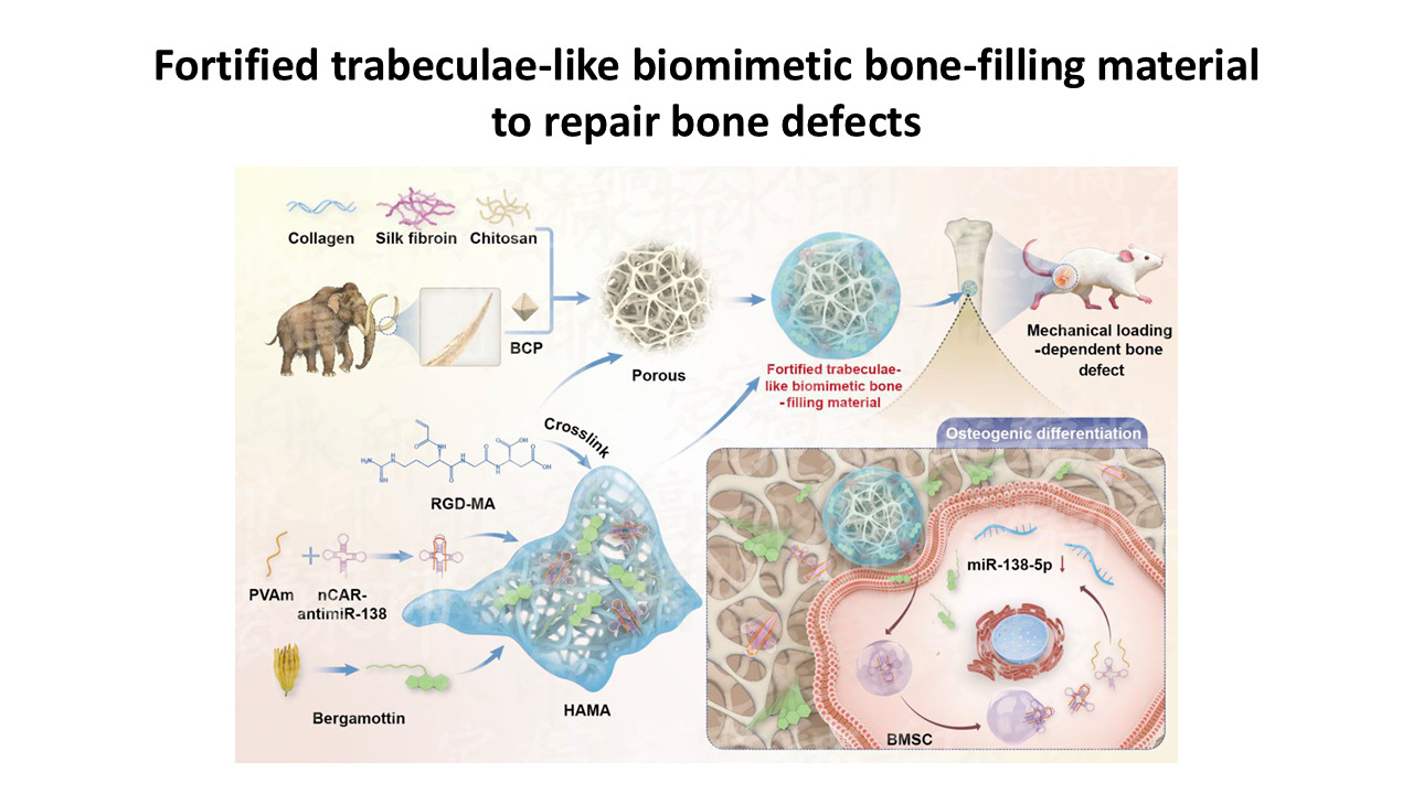
Repairing critical-sized bone defects remains challenging, and many synthetic fillers trade mechanical stability against bioactivity. In this study, a novel bone tissue-filling material was designed and synthesized. Based on a previously described trabeculae‑like biomimetic bone-filling material (TBM), mammoth tusk dentin was incorporated into this material, which endowed it with enhanced mechanical properties. We designated this material as fortified TBM (FTBM). FTBM demonstrated favorable mechanical strength, biocompatibility, and sustained drug-release capacity, thereby improving osteogenesis. In addition, human mesenchymal stem cells were encapsulated within the scaffold, resulting in a cell-laden biomaterial. Its efficacy in repairing bone defects was superior to the original TBM. This high‑hardness bone-filling material offers a strategy to enhance the mechanical stability of trabeculae-like fillers, warranting evaluation in load-bearing models.
- Sparks DS, Saifzadeh S, Savi FM, et al. A preclinical large-animal model for the assessment of critical-size load-bearing bone defect reconstruction. Nat Protoc. 2020;15(3):877-924. doi: 10.1038/s41596-019-0271-2
- Meng F, Yu Y, Yu J, et al. A trabeculae-like biomimetic bone-filling material as a potential cell-laden scaffold for bone defect treatment. Cell-Laden Scaffold Res. 2025;1(2):025040003. doi: 10.36922/OR025040003
- De La Vega RE, van Griensven M, Zhang W, et al. Efficient healing of large osseous segmental defects using optimized chemically modified messenger RNA encoding BMP-2. Sci Adv. 2022;8(7):eabl6242. doi: 10.1126/sciadv.abl6242
- Wang Z, Lv ZH, Cai XJ, et al. Sculpting the Future of Bone: The Evolution of Absorbable Materials in Orthopedics. Adv Mater. 2026;38(9):e2510848. doi: 10.1002/adma.202510848
- Yin C, Deng M, Yu J, et al. An Andrias davidianus derived composite hydrogel with enhanced antibacterial and bone repair properties for osteomyelitis treatment. Sci Rep. 2024;14(1):24626. doi: 10.1038/s41598-024-75997-8
- Salamanca E, Hsu C, Huang H, et al. Bone regeneration using a porcine bone substitute collagen composite in vitro and in vivo. Sci Rep. 2018;8(1):984. doi: 10.1038/s41598-018-19629-y
- Zhang J, Zhang W, Yue W, Qin W, Zhao Y, Xu G. Research Progress of Bone Grafting: A Comprehensive Review. Int J Nanomed. 2025;20:4729-4757. doi: 10.2147/IJN.S510524
- Gui X, Peng W, Xu X, et al. Synthesis and application of nanometer hydroxyapatite in biomedicine. Nanotechnol Rev. 2022;11(1):2154-2168. doi: 10.1515/ntrev-2022-0127
- Dec P, Modrzejewski A, Pawlik A. Existing and novel biomaterials for bone tissue engineering. Int J Mol Sci. 2023;24(1):529. doi: 10.3390/ijms24010529
- Duan H, Cao C, Wang X, et al. Magnesium-alloy rods reinforced bioglass bone cement composite scaffolds with cortical bone-matching mechanical properties for load-bearing bone in vivo regeneration. Sci Rep. 2020;10(1):18193. doi: 10.1038/s41598-020-75328-7
- Xu Y, Sheng L, Zhu M, et al. From niche to organoid: Engineering bone tissues through microenvironmental insights. J Tissue Eng. 2025;16:1-25. doi: 10.1177/20417314251358567
- Lin S, Narayanan K, Vashishth D. Bone Organoids: A Novel Tool for Modeling and Managing Skeletal Disorders in Diabetes. Adv Sci. 2026;38:e2518788. doi: 10.1002/advs.202518788
- Wei HP, Cui JJ, Lin KL, et al. Recent advances in smart stimuli-responsive biomaterials for bone therapeutics and regeneration[J]. Bone Res. 2022,10(1):58. doi: 10.1038/s41413-021-00180-y
- Taherian MH, Le TA, Le AVT, et al. Controlled microstructural evolution of hydroxyapatite–Bioglass® nanocomposites via two-step sintering. J Mech Behav Biomed Mater. 2026;175:107329. doi: 10.1016/j.jmbbm.2025.107329
- Arantes Fernandes Vieira L, Marinho JPN, de Sousa RG, et al. Development of a composite based on hydroxyapatite, carboxymethyl cellulose, and boron nitride nanotubes as a potential material for bone graft. ACS Appl Eng Mater. 2025;3(11):3964-3975. doi: 10.1021/acsaenm.5c00679
- Qi L, Zhao T, Yan J, et al. Advances in magnesium-containing bioceramics for bone repair. Biomater Transl. 2024;5(1):3-20. doi: 10.12336/biomatertransl.2024.01.002
- Liang J, Lu X, Zheng X, et al. Modification of titanium orthopedic implants with bioactive glass: a systematic review of in vivo and in vitro studies. Front Bioeng Biotechnol. 2023;11:1269223. doi: 10.3389/fbioe.2023.1269223
- Mani N, Sola A, Trinchi A, Fox K. Is there a future for additive manufactured titanium bioglass composites in biomedical application? A perspective. Biointerphases. 2020;15(6):068501. doi: 10.1116/6.0000557
- Fu Q, Saiz E, Rahaman MN, et al. Bioactive glass scaffolds for bone tissue engineering: state of the art and future perspectives. Mater Sci Eng C Mater Biol Appl. 2011;31(7):1245-1256. doi: 10.1016/j.msec.2011.04.022
- Oliver-Urrutia C, Kashimbetova A, Slámečka K, et al. Porous titanium/hydroxyapatite interpenetrating phase composites with optimal mechanical and biological properties for personalized bone repair. Biomater Adv. 2025;166:214079. doi: 10.1016/j.bioadv.2024.214079
- Shen M, Lu Z, Xu Y, et al. Vivianite and its oxidation products in mammoth ivory and their implications to the burial process. ACS Omega. 2021;6(34):22284–22291. doi: 10.1021/acsomega.1c02964
- van der Valk T, Pečnerová P, Díez-Del-Molino D, et al. Million-year-old DNA sheds light on the genomic history of mammoths. Nature. 2021;591(7849):265-269. doi: 10.1038/s41586-021-03224-9
- Zhao L, Li X, Zhao J, et al. A novel smart injectable hydrogel prepared by microbial transglutaminase and human-like collagen: its characterization and biocompatibility. Mater Sci Eng C Mater Biol Appl. 2016;68(1):317-326. doi: 10.1016/j.msec.2016.05.108
- Tian Y, Zhao Y, Yin C, et al. Polyvinylamine with moderate binding affinity as a highly effective vehicle for RNA delivery. J Control Release. 2022;345:20-37. doi: 10.1016/j.jconrel.2022.03.003
- Hu L, Su P, Li R, et al. Knockdown of microtubule actin crosslinking factor 1 inhibits cell proliferation in MC3T3-E1 osteoblastic cells. BMB Rep. 2015;48(10):583-588. doi: 10.5483/bmbrep.2015.48.10.098
- Liu G, Wang L, He Y, et al. Polydopamine nanosheets doped injectable hydrogel with nitric oxide release and photothermal effects for bacterial ablation and wound healing. Adv Healthc Mater. 2021;10(23):e2101476. doi: 10.1002/adhm.202101476
- Hu L, Yin C, Chen D, et al. MACF1 promotes osteoblast differentiation by sequestering repressors in cytoplasm. Cell Death Differ. 2021;28(7):2160-2178. doi: 10.1038/s41418-021-00744-9
- Li K, Chen Y, Lin Y, et al. PD-1/PD-L1 blockade is a potent adjuvant in treatment of Staphylococcus aureus osteomyelitis in mice. Mol Ther. 2023;31(1):174-192. doi: 10.1016/j.ymthe.2022.09.006
- Ushiku C, Adams DJ, Jiang X, et al. Long bone fracture repair in mice harboring GFP reporters for cells within the osteoblastic lineage. J Orthop Res. 2010;28(10):1338-1347. doi: 10.1002/jor.21105
- Yin C, Tian Y, Li D, et al. Long noncoding RNA Lnc-DIF inhibits bone formation by sequestering miR-489-3p. iScience. 2022;25(3):103949. doi: 10.1016/j.isci.2022.103949
- Wang X, Tian Y, Liang X, et al. Bergamottin promotes osteoblast differentiation and bone formation via activating the Wnt/β-catenin signaling pathway. Food Funct. 2022;13(5):2913-2924. doi: 10.1039/d1fo02755g
- Chen Z, Zhao F, Liang C, et al. Silencing of miR-138-5p sensitizes bone anabolic action to mechanical stimuli. Theranostics. 2020;10(26):12263-12278. doi: 10.7150/thno.53009
- Kuczkowski M, Kotwica K. Biocompatible thermoplastics in additive manufacturing of bone defect fillers: state-of-the-art and future prospects. Materials. 2025;18(16):3723. doi: 10.3390/ma18163723
- Naini A, Rachmawati D, Cholid Z, et al. Enhanced osteogenic marker expression in alveolar bone via hydroxyapatite gypsum puger-cassava starch scaffold: an in vivo study. J Indian Prosthodont Soc. 2025;25(3):258-265. doi: 10.4103/jips.jips_97_25
- Fernandez de Grado G, Keller L, Idoux-Gillet Y, et al. Bone substitutes: a review of their characteristics, clinical use, and perspectives for large bone defects management. J Tissue Eng. 2018;9:2041731418776819. doi: 10.1177/2041731418776819
- Pérez-Lloret M, Erxleben A. Improved and highly reproducible synthesis of methacrylated hyaluronic acid with tailored degrees of substitution. ACS Omega. 2024;9(24):25914-25921. doi: 10.1021/acsomega.4c00372
- Pittenger MF, Mackay AM, Beck SC, et al. Multilineage potential of adult human mesenchymal stem cells. Science. 1999;284(5411):143-147. doi: 10.1126/science.284.5411.143
- Lee M, Jeong SY, Ha J, et al. Low immunogenicity of allogeneic human umbilical cord blood-derived mesenchymal stem cells in vitro and in vivo. Biochem Biophys Res Commun. 2014;446(4):983-989. doi: 10.1016/j.bbrc.2014.03.0510