

Metabolomics of healthy hematopoietic stem cells and leukemia stem cells

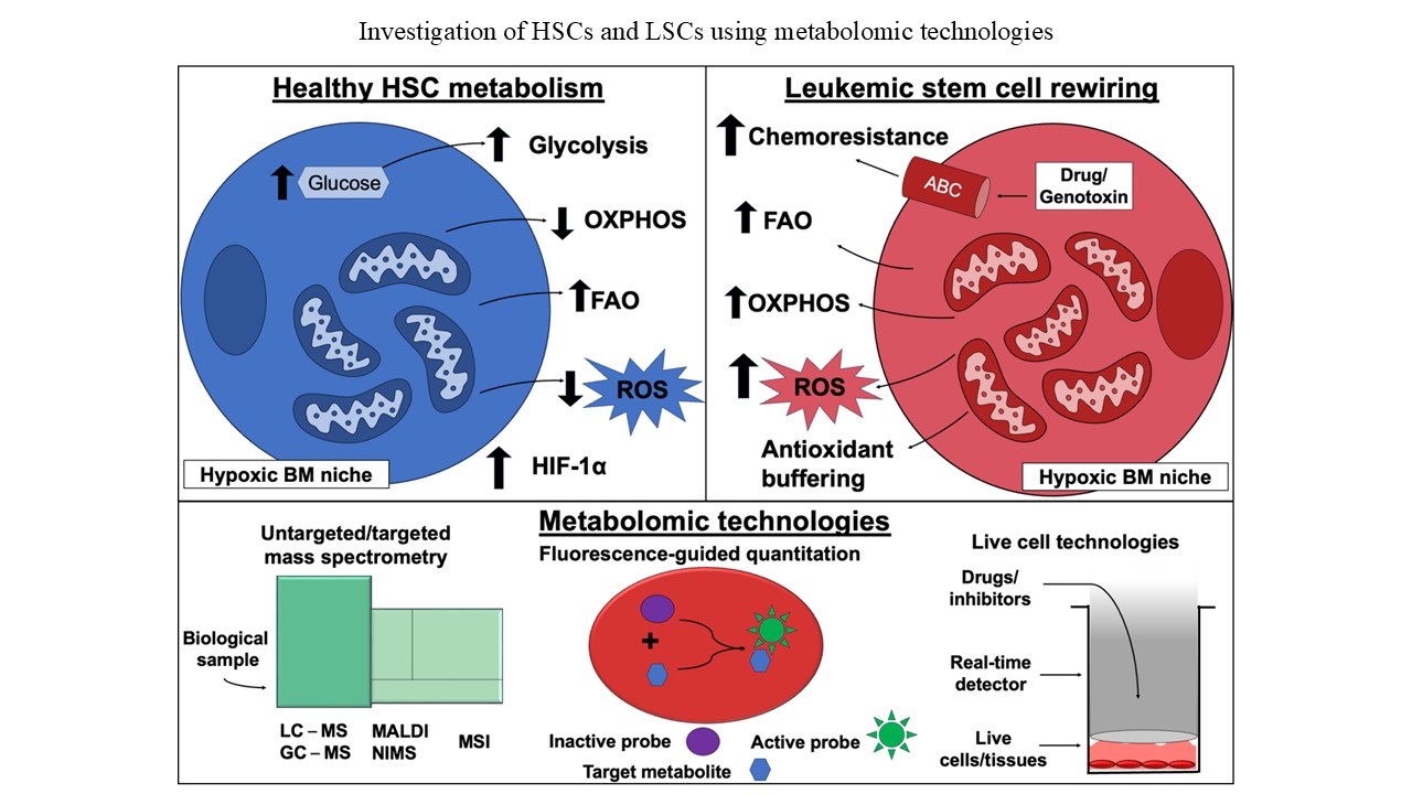
Background: Hematopoietic stem cells (HSCs) reside in the bone marrow (BM) and sustain life-long hematopoiesis by balancing quiescence, self-renewal, and differentiation. A key feature distinguishing quiescent HSCs from their activated counterparts is a shift in their metabolic profile, including changes in glycolytic flux and mitochondrial oxidative metabolism. Disruption of HSC homeostasis can lead to hematologic diseases such as BM failure, clonal hematopoiesis, or oncogenic transformation into leukemia stem cells (LSCs). Similar to HSCs, LSCs retain stem-like characteristics but acquire malignant features, including drug resistance and a reprogrammed metabolism, which results in distinct metabolic profiles that contribute to their pathogenesis. Aim: This review summarizes the key metabolic characteristics distinguishing healthy quiescent and active HSCs from oncogenic LSCs. In addition, we highlight modern tools for investigating the metabolome, which enable the identification of novel metabolites, metabolic interactions, pathways, and potential targets for diagnosis or therapeutic intervention in hematologic diseases. Conclusion: Metabolic regulation is essential for maintaining HSC quiescence, self-renewal, and lineage commitment, whereas its disruption often underlies oncogenic transformation into LSCs. Advances in metabolic profiling reveal key differences between healthy HSCs and LSCs and identify LSC vulnerabilities that sustain survival and therapeutic resistance. Targeting these hijacked metabolic pathways may facilitate the development of LSC-specific treatment while preserving normal hematopoiesis. Further investigation of stem cell metabolism will be critical for translating these insights into effective treatments for hematologic malignancies. Relevance for patients: Understanding the metabolic profiles of healthy HSCs and LSCs can facilitate the development of innovative techniques, technologies, and therapeutics. These advances can be applied to the identification, treatment, and prevention of hematologic disease. By elucidating the metabolome of LSCs, therapies can be designed to selectively target their unique metabolic pathways, dependencies, and resistance mechanisms.
- Olson OC, Kang YA, Passegue E. Normal hematopoiesis is a balancing act of self-renewal and regeneration. Cold Spring Harb Perspect Med. 2020;10(12):a035519. doi: 10.1101/cshperspect.a035519
- Morrison SJ, Scadden DT. The bone marrow niche for haematopoietic stem cells. Nature. 2014;505(7483):327-334. doi: 10.1038/nature12984
- Barreto IV, Pessoa F, Machado CB, et al. Leukemic stem cell: A mini-review on clinical perspectives. Front Oncol. 2022;12:931050. doi: 10.3389/fonc.2022.931050
- Seita J, Weissman IL. Hematopoietic stem cell: Self-renewal versus differentiation. Wiley Interdiscip Rev Syst Biol Med. 2010;2(6):640-653. doi: 10.1002/wsbm.86
- Morganti C, Cabezas-Wallscheid N, Ito K. Metabolic regulation of hematopoietic stem cells. Hemasphere. 2022;6(7):e740. doi: 10.1097/HS9.0000000000000740
- Mann Z, Sengar M, Verma YK, Rajalingam R, Raghav PK. Hematopoietic stem cell factors: Their functional role in self-renewal and clinical aspects. Front Cell Dev Biol. 2022;10:664261. doi: 10.3389/fcell.2022.664261
- Zhang CC, Lodish HF. Cytokines regulating hematopoietic stem cell function. Curr Opin Hematol. 2008;15(4):307-311. doi: 10.1097/MOH.0b013e3283007db5
- Simsek T, Kocabas F, Zheng J, et al. The distinct metabolic profile of hematopoietic stem cells reflects their location in a hypoxic niche. Cell Stem Cell. 2010;7(3):380-390. doi: 10.1016/j.stem.2010.07.011
- Papa L, Djedaini M, Hoffman R. Mitochondrial role in stemness and differentiation of hematopoietic stem cells. Stem Cells Int. 2019;2019:4067162. doi: 10.1155/2019/4067162
- Liao Y, Octaviani S, Tian Z, Wang SR, Huang C, Huang J. Mitochondrial quality control in hematopoietic stem cells: Mechanisms, implications, and therapeutic opportunities. Stem Cell Res Ther. 2025;16(1):180. doi: 10.1186/s13287-025-04304-7
- Hira VVV, Van Noorden CJF, Carraway HE, Maciejewski JP, Molenaar RJ. Novel therapeutic strategies to target leukemic cells that hijack compartmentalized continuous hematopoietic stem cell niches. Biochim Biophys Acta Rev Cancer. 2017;1868(1):183-198. doi: 10.1016/j.bbcan.2017.03.010
- Mesbahi Y, Trahair TN, Lock RB, Connerty P. Exploring the metabolic landscape of AML: From haematopoietic stem cells to myeloblasts and leukaemic stem cells. Front Oncol. 2022;12:807266. doi: 10.3389/fonc.2022.807266
- O’Reilly E, Zeinabad HA, Szegezdi E. Hematopoietic versus leukemic stem cell quiescence: Challenges and therapeutic opportunities. Blood Rev. 2021;50:100850. doi: 10.1016/j.blre.2021.100850
- Man CH, Li C, Xu X, Zhao M. Metabolic regulation in normal and leukemic stem cells. Trends Pharmacol Sci. 2024;45(10):919-930. doi: 10.1016/j.tips.2024.08.004
- Patti GJ, Yanes O, Siuzdak G. Innovation: Metabolomics: The apogee of the omics trilogy. Nat Rev Mol Cell Biol. 2012;13(4):263-269. doi: 10.1038/nrm3314
- DeBerardinis RJ, Keshari KR. Metabolic analysis as a driver for discovery, diagnosis, and therapy. Cell. 2022;185(15):2678-2689. doi: 10.1016/j.cell.2022.06.029
- Song BH, Son SY, Kim HK, et al. Profiling of metabolic differences between hematopoietic stem cells and acute/ chronic myeloid leukemia. Metabolites. 2020;10(11):427. doi: 10.3390/metabo10110427
- Zhao X, Zhang C, Cui X, Liang Y. Interactions of hematopoietic stem cells with bone marrow niche. Methods Mol Biol. 2021;2346:21-34. doi: 10.1007/7651_2020_298
- Cheung TH, Rando TA. Molecular regulation of stem cell quiescence. Nat Rev Mol Cell Biol. 2013;14(6):329-340. doi: 10.1038/nrm3591
- Chotinantakul K, Leeanansaksiri W. Hematopoietic stem cell development, niches, and signaling pathways. Bone Marrow Res. 2012;2012:270425. doi: 10.1155/2012/270425
- Huang X, Trinh T, Aljoufi A, Broxmeyer HE. Hypoxia signaling pathway in stem cell regulation: Good and evil. Curr Stem Cell Rep. 2018;4(2):149-157. doi: 10.1007/s40778-018-0127-7
- Mistry JJ, Bowles K, Rushworth SA. HSC-derived fatty acid oxidation in steady-state and stressed hematopoiesis. Exp Hematol. 2023;117:1-8. doi: 10.1016/j.exphem.2022.10.003
- Mohrin M, Chen D. The mitochondrial metabolic checkpoint and aging of hematopoietic stem cells. Curr Opin Hematol. 2016;23(4):318-324. doi: 10.1097/MOH.0000000000000244
- Zhao T, Zhang J, Lei H, et al. NRF1-mediated mitochondrial biogenesis antagonizes innate antiviral immunity. EMBO J. 2023;42(16):e113258. doi: 10.15252/embj.2022113258
- Peng M, Huang Y, Zhang L, Zhao X, Hou Y. Targeting mitochondrial oxidative phosphorylation eradicates acute myeloid leukemic stem cells. Front Oncol. 2022;12:899502. doi: 10.3389/fonc.2022.899502
- De Beauchamp L, Himonas E, Helgason GV. Mitochondrial metabolism as a potential therapeutic target in myeloid leukaemia. Leukemia. 2022;36(1):1-12. doi: 10.1038/s41375-021-01416-w
- Nwajei F, Konopleva M. The bone marrow microenvironment as niche retreats for hematopoietic and leukemic stem cells. Adv Hematol. 2013;2013:953982. doi: 10.1155/2013/953982
- Yao Y, Li F, Huang J, Jin J, Wang H. Leukemia stem cell-bone marrow microenvironment interplay in acute myeloid leukemia development. Exp Hematol Oncol. 2021;10(1):39. doi: 10.1186/s40164-021-00233-2
- Yamazaki S, Iwama A, Takayanagi S, Eto K, Ema H, Nakauchi H. TGF-beta as a candidate bone marrow niche signal to induce hematopoietic stem cell hibernation. Blood. 2009;113(6):1250-1256. doi: 10.1182/blood-2008-04-146480
- Vaidya A, Kale VP. TGF-β signaling and its role in the regulation of hematopoietic stem cells. Syst Synth Biol. 2015;9(1-2):1-10. doi: 10.1007/s11693-015-9161-2
- Blank U, Karlsson S. TGF-β signaling in the control of hematopoietic stem cells. Blood. 2015;125(23):3542-3550. doi: 10.1182/blood-2014-12-618090
- Naka K, Hirao A. Regulation of hematopoiesis and hematological disease by TGF-β family signaling molecules. Cold Spring Harb Perspect Biol. 2017;9(9):a027987. doi: 10.1101/cshperspect.a027987
- Arai F, Hirao A, Ohmura M, et al. Tie2/angiopoietin-1 signaling regulates hematopoietic stem cell quiescence in the bone marrow niche. Cell. 2004;118(2):149-161. doi: 10.1016/j.cell.2004.07.004
- Nagasawa T. The chemokine CXCL12 and regulation of HSC and B lymphocyte development in the bone marrow niche. Adv Exp Med Biol. 2007;602:69-75. doi: 10.1007/978-0-387-72009-8_9
- Zhang Y, Depond M, He L, et al. CXCR4/CXCL12 axis counteracts hematopoietic stem cell exhaustion through selective protection against oxidative stress. Sci Rep. 2016;6:37827. doi: 10.1038/srep37827
- Driessen RL, Johnston HM, Nilsson SK. Membrane-bound stem cell factor is a key regulator in the initial lodgment of stem cells within the endosteal marrow region. Exp Hematol. 2003;31(12):1284-1291. doi: 10.1016/j.exphem.2003.08.015
- Li J. Quiescence regulators for hematopoietic stem cell. Exp Hematol. 2011;39(5):511-520. doi: 10.1016/j.exphem.2011.01.008
- Zhou BO, Yu H, Yue R, et al. Bone marrow adipocytes promote the regeneration of stem cells and haematopoiesis by secreting SCF. Nat Cell Biol. 2017;19(8):891-903. doi: 10.1038/ncb3570
- Qian H, Buza-Vidas N, Hyland CD, et al. Critical role of thrombopoietin in maintaining adult quiescent hematopoietic stem cells. Cell Stem Cell. 2007;1(6):671-684. doi: 10.1016/j.stem.2007.10.008
- Nilsson SK, Johnston HM, Whitty GA, et al. Osteopontin, a key component of the hematopoietic stem cell niche and regulator of primitive hematopoietic progenitor cells. Blood. 2005;106(4):1232-1239. doi: 10.1182/blood-2004-11-4422
- Cao H, Cao B, Heazlewood CK, et al. Osteopontin is an important regulative component of the fetal bone marrow hematopoietic stem cell niche. Cells. 2019;8(9):985. doi: 10.3390/cells8090985
- De Graaf CA, Metcalf D. Thrombopoietin and hematopoietic stem cells. Cell Cycle. 2011;10(10):1582-1589. doi: 10.4161/cc.10.10.15619
- Sage J. The retinoblastoma tumor suppressor and stem cell biology. Genes Dev. 2012;26(13):1409-1420. doi: 10.1101/gad.193730.112
- Viatour P, Somervaille TC, Venkatasubrahmanyam S, et al. Hematopoietic stem cell quiescence is maintained by compound contributions of the retinoblastoma gene family. Cell Stem Cell. 2008;3(4):416-428. doi: 10.1016/j.stem.2008.07.009
- Kim E, Cheng Y, Bolton-Gillespie E, et al. Rb family proteins enforce the homeostasis of quiescent hematopoietic stem cells by repressing Socs3 expression. J Exp Med. 2017;214(7):1901-1912. doi: 10.1084/jem.20160719
- Bigarella CL, Li J, Rimmele P, Liang R, Sobol RW, Ghaffari S. FOXO3 transcription factor is essential for protecting hematopoietic stem and progenitor cells from oxidative DNA damage. J Biol Chem. 2017;292(7):3005-3015. doi: 10.1074/jbc.M116.769455
- Tothova Z, Kollipara R, Huntly BJ, et al. FoxOs are critical mediators of hematopoietic stem cell resistance to physiologic oxidative stress. Cell. 2007;128(2):325-339. doi: 10.1016/j.cell.2007.01.003
- Johnson C, Belluschi S, Laurenti E. Beyond “to divide or not to divide”: Kinetics matters in hematopoietic stem cells. Exp Hematol. 2020;92:1-10.e2. doi: 10.1016/j.exphem.2020.11.003
- Maurer B, Brandstoetter T, Kollmann S, Sexl V, Prchal- Murphy M. Inducible deletion of CDK4 and CDK6 - deciphering CDK4/6 inhibitor effects in the hematopoietic system. Haematologica. 2021;106(10):2624-2632. doi: 10.3324/haematol.2020.256313
- Asai T, Liu Y, Bae N, Nimer SD. The p53 tumor suppressor protein regulates hematopoietic stem cell fate. J Cell Physiol. 2011;226(9):2215-2221. doi: 10.1002/jcp.22561
- Matsumoto A, Takeishi S, Kanie T, et al. p57 is required for quiescence and maintenance of adult hematopoietic stem cells. Cell Stem Cell. 2011;9(3):262-271. doi: 10.1016/j.stem.2011.06.014
- Zou P, Yoshihara H, Hosokawa K, et al. p57(Kip2) and p27(Kip1) cooperate to maintain hematopoietic stem cell quiescence through interactions with Hsc70. Cell Stem Cell. 2011;9(3):247-261. doi: 10.1016/j.stem.2011.07.003
- Takam Kamga P, Bazzoni R, Dal Collo G, et al. The role of notch and wnt signaling in MSC communication in normal and leukemic bone marrow niche. Front Cell Dev Biol. 2020;8:599276. doi: 10.3389/fcell.2020.599276
- Yu M, Qin K, Fan J, et al. The evolving roles of Wnt signaling in stem cell proliferation and differentiation, the development of human diseases, and therapeutic opportunities. Genes Dis. 2024;11(3):101026. doi: 10.1016/j.gendis.2023.04.042
- Evans AG, Calvi LM. Notch signaling in the malignant bone marrow microenvironment: Implications for a niche-based model of oncogenesis. Ann N Y Acad Sci. 2015;1335(1):63-77. doi: 10.1111/nyas.12562
- Ge Y, Wang J, Zhang H, Li J, Ye M, Jin X. Fate of hematopoietic stem cells determined by notch1 signaling (review). Exp Ther Med. 2022;23(2):170. doi: 10.3892/etm.2021.11093
- Chen J, Sun Y, Chi Z. Regulation of hematopoiesis by hedgehog signaling (review). Mol Med Rep. 2023;27(5):100. doi: 10.3892/mmr.2023.12987
- Cain CJ, Manilay JO. Hematopoietic stem cell fate decisions are regulated by Wnt antagonists: Comparisons and current controversies. Exp Hematol. 2013;41(1):3-16. doi: 10.1016/j.exphem.2012.09.006
- Merchant A, Joseph G, Wang Q, Brennan S, Matsui W. Gli1 regulates the proliferation and differentiation of HSCs and myeloid progenitors. Blood. 2010;115(12):2391-2396. doi: 10.1182/blood-2009-09-241703
- Chandel NS. Glycolysis. Cold Spring Harb Perspect Biol. 2021;13(5):a040535. doi: 10.1101/cshperspect.a040535
- Spencer JA, Ferraro F, Roussakis E, et al. Direct measurement of local oxygen concentration in the bone marrow of live animals. Nature. 2014;508(7495):269-273. doi: 10.1038/nature13034
- Cullen SC, Cook EV. Normal human arterial oxygen tension. Am J Physiol Legacy Content. 1942;137(1):238-241. doi: 10.1152/ajplegacy.1942.137.1.238
- Takubo K, Goda N, Yamada W, et al. Regulation of the HIF- 1alpha level is essential for hematopoietic stem cells. Cell Stem Cell. 2010;7(3):391-402. doi: 10.1016/j.stem.2010.06.020
- Du J, Chen Y, Li Q, et al. HIF-1α deletion partially rescues defects of hematopoietic stem cell quiescence caused by cited2 deficiency. Blood. 2012;119(12):2789-2798. doi: 10.1182/blood-2011-10-387902
- Zhou MY, Cheng ML, Huang T, et al. Transforming growth factor beta-1 upregulates glucose transporter 1 and glycolysis through canonical and noncanonical pathways in hepatic stellate cells. World J Gastroenterol. 2021;27(40):6908-6926. doi: 10.3748/wjg.v27.i40.6908
- Lane AN, Fan TW. Regulation of mammalian nucleotide metabolism and biosynthesis. Nucleic Acids Res. 2015;43(4):2466-2485. doi: 10.1093/nar/gkv047
- Chandel NS. Lipid metabolism. Cold Spring Harb Perspect Biol. 2021;13(9):a040576. doi: 10.1101/cshperspect.a040576
- Chandel NS. Carbohydrate metabolism. Cold Spring Harb Perspect Biol. 2021;13(1):a040568. doi: 10.1101/cshperspect.a040568
- Mohrin M, Shin J, Liu Y, et al. Stem cell aging. A mitochondrial UPR-mediated metabolic checkpoint regulates hematopoietic stem cell aging. Science. 2015;347(6228):1374-1377. doi: 10.1126/science.aaa2361
- Lin YF, Haynes CM. Metabolism and the UPR(mt). Mol Cell. 2016;61(5):677-682. doi: 10.1016/j.molcel.2016.02.004
- Ocampo A, Izpisua Belmonte JC. Stem cells. Holding your breath for longevity. Science. 2015;347(6228):1319-1320. doi: 10.1126/science.aaa9608
- Vannini N, Girotra M, Naveiras O, et al. Specification of haematopoietic stem cell fate via modulation of mitochondrial activity. Nat Commun. 2016;7:13125. doi: 10.1038/ncomms13125
- Houten SM, Wanders RJ. A general introduction to the biochemistry of mitochondrial fatty acid β-oxidation. J Inherit Metab Dis. 2010;33(5):469-477. doi: 10.1007/s10545-010-9061-2
- Ito K, Carracedo A, Weiss D, et al. A PML-PPAR-δ pathway for fatty acid oxidation regulates hematopoietic stem cell maintenance. Nat Med. 2012;18(9):1350-1358. doi: 10.1038/nm.2882
- Bonora M, Morganti C, Van Gastel N, et al. A mitochondrial NADPH-cholesterol axis regulates extracellular vesicle biogenesis to support hematopoietic stem cell fate. Cell Stem Cell. 2024;31(3):359-377.e10. doi: 10.1016/j.stem.2024.02.004
- Tiwari SK, Toshniwal AG, Mandal S, Mandal L. Fatty acid β-oxidation is required for the differentiation of larval hematopoietic progenitors in Drosophila. Elife. 2020;9:e53247. doi: 10.7554/eLife.53247
- Jackson BT, Finley LWS. Metabolic regulation of the hallmarks of stem cell biology. Cell Stem Cell. 2024;31(2):161-180. doi: 10.1016/j.stem.2024.01.003
- Harayama T, Riezman H. Understanding the diversity of membrane lipid composition. Nat Rev Mol Cell Biol. 2018;19(5):281-296. doi: 10.1038/nrm.2017.138
- Chen W, Zhao H, Li Y. Mitochondrial dynamics in health and disease: Mechanisms and potential targets. Signal Transduct Target Ther. 2023;8(1):333. doi: 10.1038/s41392-023-01547-9
- Zhou D, Shao L, Spitz DR. Reactive oxygen species in normal and tumor stem cells. Adv Cancer Res. 2014;122:1-67. doi: 10.1016/B978-0-12-420117-0.00001-3
- Reuter S, Gupta SC, Chaturvedi MM, Aggarwal BB. Oxidative stress, inflammation, and cancer: How are they linked? Free Radic Biol Med. 2010;49(11):1603-1616. doi: 10.1016/j.freeradbiomed.2010.09.006
- Pickles S, Vigie P, Youle RJ. Mitophagy and quality control mechanisms in mitochondrial maintenance. Curr Biol. 2018;28(4):R170-R185. doi: 10.1016/j.cub.2018.01.004
- Jomova K, Raptova R, Alomar SY, et al. Reactive oxygen species, toxicity, oxidative stress, and antioxidants: Chronic diseases and aging. Arch Toxicol. 2023;97(10):2499-2574. doi: 10.1007/s00204-023-03562-9
- Kulkarni CA, Brookes PS. Cellular compartmentation and the redox/nonredox functions of NAD. Antioxid Redox Signal. 2019;31(9):623-642. doi: 10.1089/ars.2018.7722
- Ye H, Adane B, Khan N, et al. Leukemic stem cells evade chemotherapy by metabolic adaptation to an adipose tissue niche. Cell Stem Cell. 2016;19(1):23-37. doi: 10.1016/j.stem.2016.06.001
- Griessinger E, Pereira-Martins D, Nebout M, et al. Oxidative phosphorylation fueled by fatty acid oxidation sensitizes leukemic stem cells to cold. Cancer Res. 2023;83(15):2461-2470. doi: 10.1158/0008-5472.CAN-23-1006
- Okoye CN, Koren SA, Wojtovich AP. Mitochondrial complex I ROS production and redox signaling in hypoxia. Redox Biol. 2023;67:102926. doi: 10.1016/j.redox.2023.102926
- Lagadinou ED, Sach A, Callahan K, et al. BCL-2 inhibition targets oxidative phosphorylation and selectively eradicates quiescent human leukemia stem cells. Cell Stem Cell. 2013;12(3):329-341. doi: 10.1016/j.stem.2012.12.013
- Umemoto T, Hashimoto M, Matsumura T, Nakamura-Ishizu A, Suda T. Ca2+-mitochondria axis drives cell division in hematopoietic stem cells. J Exp Med. 2018;215(8):2097-2113. doi: 10.1084/jem.20180421
- Kuntz EM, Baquero P, Michie AM, et al. Targeting mitochondrial oxidative phosphorylation eradicates therapy-resistant chronic myeloid leukemia stem cells. Nat Med. 2017;23(10):1234-1240. doi: 10.1038/nm.4399
- Liu L, Wise DR, Diehl JA, Simon MC. Hypoxic reactive oxygen species regulate the integrated stress response and cell survival. J Biol Chem. 2008;283(45):31153-31162. doi: 10.1074/jbc.M805056200
- Nwosu GO, Powell JA, Pitson SM. Targeting the integrated stress response in hematologic malignancies. Exp Hematol Oncol. 2022;11(1):94. doi: 10.1186/s40164-022-00348-0
- Takao S, Morell V, Uni M, et al. Epigenetic mechanisms controlling human leukemia stem cells and therapy resistance. Nat Commun. 2025;16(1):3196. doi: 10.1038/s41467-025-58370-9
- Le HT, Yu J, Ahn HS, et al. eIF2α phosphorylation-ATF4 axis-mediated transcriptional reprogramming mitigates mitochondrial impairment during ER stress. Mol Cells. 2025;48(2):100176. doi: 10.1016/j.mocell.2024.100176
- Shi X, Jiang Y, Kitano A, et al. Nuclear NAD+ homeostasis governed by NMNAT1 prevents apoptosis of acute myeloid leukemia stem cells. Sci Adv. 2021;7(30):eabf3895. doi: 10.1126/sciadv.abf3895
- Amaya ML, Inguva A, Pei S, et al. The STAT3-MYC axis promotes survival of leukemia stem cells by regulating SLC1A5 and oxidative phosphorylation. Blood. 2022;139(4):584-596. doi: 10.1182/blood.2021013201
- Rodriguez-Zabala M, Ramakrishnan R, Reinbach K, et al. Combined GLUT1 and OXPHOS inhibition eliminates acute myeloid leukemia cells by restraining their metabolic plasticity. Blood Adv. 2023;7(18):5382-5395. doi: 10.1182/bloodadvances.2023009967
- Jones CL, Inguva A, Jordan CT. Targeting energy metabolism in cancer stem cells: Progress and challenges in leukemia and solid tumors. Cell Stem Cell. 2021;28(3):378-393. doi: 10.1016/j.stem.2021.02.013
- Ochocki JD, Simon MC. Nutrient-sensing pathways and metabolic regulation in stem cells. J Cell Biol. 2013;203(1):23-33. doi: 10.1083/jcb.201303110
- Gan B, DePinho RA. mTORC1 signaling governs hematopoietic stem cell quiescence. Cell Cycle. 2009;8(7):1003-1006. doi: 10.4161/cc.8.7.8045
- Garcia D, Shaw RJ. AMPK: Mechanisms of cellular energy sensing and restoration of metabolic balance. Mol Cell. 2017;66(6):789-800. doi: 10.1016/j.molcel.2017.05.032
- Torrence ME, MacArthur MR, Hosios AM, et al. The mTORC1-mediated activation of ATF4 promotes protein and glutathione synthesis downstream of growth signals. Elife. 2021;10:e63326. doi: 10.7554/eLife.63326
- Ben-Sahra I, Hoxhaj G, Ricoult SJH, Asara JM, Manning BD. mTORC1 induces purine synthesis through control of the mitochondrial tetrahydrofolate cycle. Science. 2016;351(6274):728-733. doi: 10.1126/science.aad0489
- Hardie DG. AMPK--sensing energy while talking to other signaling pathways. Cell Metab. 2014;20(6):939-952. doi: 10.1016/j.cmet.2014.09.013
- Krastinaite I, Charkavliuk S, Navakauskiene R, Borutinskaite VV. Metformin as an enhancer for the treatment of chemoresistant CD34+ acute myeloid leukemia cells. Genes (Basel). 2024;15(5):648. doi: 10.3390/genes15050648
- Bao B, Wang Z, Ali S, et al. Metformin inhibits cell proliferation, migration and invasion by attenuating CSC function mediated by deregulating miRNAs in pancreatic cancer cells. Cancer Prev Res (Phila). 2012;5(3):355-364. doi: 10.1158/1940-6207.Capr-11-0299
- Farge T, Saland E, De Toni F, et al. Chemotherapy-resistant human acute myeloid leukemia cells are not enriched for leukemic stem cells but require oxidative metabolism. Cancer Discov. 2017;7(7):716-735. doi: 10.1158/2159-8290.CD-16-0441
- De Jonge-Peeters SD, Kuipers F, De Vries EG, Vellenga E. ABC transporter expression in hematopoietic stem cells and the role in AML drug resistance. Crit Rev Oncol Hematol. 2007;62(3):214-226. doi: 10.1016/j.critrevonc.2007.02.003
- Porro A, Iraci N, Soverini S, et al. c-MYC oncoprotein dictates transcriptional profiles of ATP-binding cassette transporter genes in chronic myelogenous leukemia CD34+ hematopoietic progenitor cells. Mol Cancer Res. 2011;9(8):1054-1066. doi: 10.1158/1541-7786.MCR-10-0510
- Rozovski U, Hazan-Halevy I, Barzilai M, Keating MJ, Estrov Z. Metabolism pathways in chronic lymphocytic leukemia. Leuk Lymphoma. 2016;57(4):758-765. doi: 10.3109/10428194.2015.1106533
- Takubo K, Nagamatsu G, Kobayashi CI, et al. Regulation of glycolysis by Pdk functions as a metabolic checkpoint for cell cycle quiescence in hematopoietic stem cells. Cell Stem Cell. 2013;12(1):49-61. doi: 10.1016/j.stem.2012.10.011
- Testa U, Labbaye C, Castelli G, Pelosi E. Oxidative stress and hypoxia in normal and leukemic stem cells. Exp Hematol. 2016;44(7):540-560. doi: 10.1016/j.exphem.2016.04.012
- Velasco-Hernandez T, Soneji S, Hidalgo I, Erlandsson E, Cammenga J, Bryder D. Hif-1α deletion may lead to adverse treatment effect in a mouse model of MLL-AF9-driven AML. Stem Cell Reports. 2019;12(1):112-121. doi: 10.1016/j.stemcr.2018.11.023
- Qiu S, Cai Y, Wang Z, Xie Y, Zhang A. Decoding functional significance of small molecule metabolites. Biomed Pharmacother. 2023;158:114188. doi: 10.1016/j.biopha.2022.114188
- Cajka T, Fiehn O. Toward merging untargeted and targeted methods in mass spectrometry-based metabolomics and lipidomics. Anal Chem. 2016;88(1):524-545. doi: 10.1021/acs.analchem.5b04491
- Roberts LD, Souza AL, Gerszten RE, Clish CB. Targeted metabolomics. Curr Protoc Mol Biol. 2012;98:30.2.1-30.2.24. doi: 10.1002/0471142727.mb3002s98
- Greving MP, Patti GJ, Siuzdak G. Nanostructure-initiator mass spectrometry metabolite analysis and imaging. Anal Chem. 2011;83(1):2-7. doi: 10.1021/ac101565f
- Niehaus M, Soltwisch J, Belov ME, Dreisewerd K. Transmission-mode MALDI-2 mass spectrometry imaging of cells and tissues at subcellular resolution. Nat Methods. 2019;16(9):925-931. doi: 10.1038/s41592-019-0536-2
- Molenaar MR, Shahraz M, Delafiori J, et al. Increasing quantitation in spatial single-cell metabolomics by using fluorescence as ground truth. Front Mol Biosci. 2022;9:1021889. doi: 10.3389/fmolb.2022.1021889
- Schmidt CA, Fisher-Wellman KH, Neufer PD. From OCR and ECAR to energy: Perspectives on the design and interpretation of bioenergetics studies. J Biol Chem. 2021;297(4):101140. doi: 10.1016/j.jbc.2021.101140
- Chen L, Zhong F, Zhu J. Bridging targeted and untargeted mass spectrometry-based metabolomics via hybrid approaches. Metabolites. 2020;10(9):348. doi: 10.3390/metabo10090348
- Zhang X, Tong X, Chen Y, et al. A metabolomics study on carcinogenesis of ground-glass nodules. Cytojournal. 2024;21:12. doi: 10.25259/Cytojournal_68_2023
- Li H, Braunig S, Dhapolar P, Karlsson G, Lang S, Scheding S. Identification of phenotypically, functionally, and anatomically distinct stromal niche populations in human bone marrow based on single-cell RNA sequencing. Elife. 2023;12:e81656. doi: 10.7554/eLife.81656
- Zeijlemaker W, Kelder A, Oussoren-Brockhoff YJ, et al. A simple one-tube assay for immunophenotypical quantification of leukemic stem cells in acute myeloid leukemia. Leukemia. 2016;30(2):439-446. doi: 10.1038/leu.2015.252
- Schmidt JR, Rucker-Braun E, Heidrich K, et al. Pilot study on mass spectrometry-based analysis of the proteome of CD34+CD123+ progenitor cells for the identification of potential targets for immunotherapy in acute myeloid leukemia. Proteomes. 2018;6(1):11. doi: 10.3390/proteomes6010011
- Tautenhahn R, Patti GJ, Rinehart D, Siuzdak G. XCMS online: A web-based platform to process untargeted metabolomic data. Anal Chem. 2012;84(11):5035-5039. doi: 10.1021/ac300698c
- Wang F, Zhang Z, Li Q, Yu T, Ma C. Untargeted LC-MS/MS analysis reveals metabolomics feature of osteosarcoma stem cell response to methotrexate. Cancer Cell Int. 2020;20:269. doi: 10.1186/s12935-020-01356-y
- Chen X, Peng Z, Yang Z. Metabolomics studies of cell-cell interactions using single cell mass spectrometry combined with fluorescence microscopy. Chem Sci. 2022;13(22):6687-6695. doi: 10.1039/d2sc02298b
- Rappez L, Stadler M, Triana S, et al. Spacem reveals metabolic States of single cells. Nat Methods. 2021;18(7):799-805. doi: 10.1038/s41592-021-01198-0
- Wei D, Xu M, Wang Z, Tong J. The development of single-cell metabolism and its role in studying cancer emergent properties. Front Oncol. 2021;11:814085. doi: 10.3389/fonc.2021.814085
- Parmar K, Mauch P, Vergilio JA, Sackstein R, Down JD. Distribution of hematopoietic stem cells in the bone marrow according to regional hypoxia. Proc Natl Acad Sci U S A. 2007;104(13):5431-5436. doi: 10.1073/pnas.0701152104
- Mendez LM, Posey RR, Pandolfi PP. The interplay between the genetic and immune landscapes of AML: Mechanisms and implications for risk stratification and therapy. Front Oncol. 2019;9:1162. doi: 10.3389/fonc.2019.01162
- Cairns JL, Huber J, Lewen A, et al. Mass-guided single-cell MALDI imaging of low-mass metabolites reveals cellular activation markers. Adv Sci (Weinh). 2025;12(5):e2410506. doi: 10.1002/advs.202410506
- O’Brien PJ, Lee M, Spilker ME, et al. Monitoring metabolic responses to chemotherapy in single cells and tumors using nanostructure-initiator mass spectrometry (NIMS) imaging. Cancer Metab. 2013;1(1):4. doi: 10.1186/2049-3002-1-4
- Liu R, Li J, Lan Y, Nguyen TD, Chen YA, Yang Z. Quantifying cell heterogeneity and subpopulations using single cell metabolomics. Anal Chem. 2023;95(18):7127-7133. doi: 10.1021/acs.analchem.2c05245
- Comi TJ, Neumann EK, Do TD, Sweedler JV. microMS: A python platform for image-guided mass spectrometry profiling. J Am Soc Mass Spectrom. 2017;28(9):1919-1928. doi: 10.1007/s13361-017-1704-1
- Liu R, Zhang G, Yang Z. Towards rapid prediction of drug-resistant cancer cell phenotypes: Single cell mass spectrometry combined with machine learning. Chem Commun (Camb). 2019;55(5):616-619. doi: 10.1039/c8cc08296k
- Sun M, Yang Z. Metabolomic studies of live single cancer stem cells using mass spectrometry. Anal Chem. 2019;91(3):2384-2391. doi: 10.1021/acs.analchem.8b05166
- Chimge NO, Chen MH, Nguyen C, et al. A deeply quiescent subset of CML LSC depend on FAO yet avoid deleterious ROS by suppressing mitochondrial complex I. Curr Mol Pharmacol. 2024;17(1):e060923220758. doi: 10.2174/1874467217666230906092236
- Walsh MA, Musci RV, Jacobs RA, Hamilton KL. A practical perspective on how to develop, implement, execute, and reproduce high-resolution respirometry experiments: The physiologist’s guide to an Oroboros O2k. Faseb J. 2023;37(12):e23280. doi: 10.1096/fj.202301644RR
- Acin-Perez R, Benador IY, Petcherski A, et al. A novel approach to measure mitochondrial respiration in frozen biological samples. Embo J. 2020;39(13):e104073. doi: 10.15252/embj.2019104073
- Hagen JT, Montgomery MM, Aruleba RT, et al. Acute myeloid leukemia mitochondria hydrolyze ATP to support oxidative metabolism and resist chemotherapy. Sci Adv. 2025;11(15):eadu5511. doi: 10.1126/sciadv.adu5511
- Nelson MA, McLaughlin KL, Hagen JT, et al. Intrinsic OXPHOS limitations underlie cellular bioenergetics in leukemia. Elife. 2021;10:e63104. doi: 10.7554/eLife.63104
- Bednarski TK, Rahim M, Young JD. In vivo 2H/13C flux analysis in metabolism research. Curr Opin Biotechnol. 2021;71:1-8. doi: 10.1016/j.copbio.2021.04.005
- O’Brien C, Ling T, Berman JM, et al. Simultaneous inhibition of Sirtuin 3 and cholesterol homeostasis targets acute myeloid leukemia stem cells by perturbing fatty acid β-oxidation and inducing lipotoxicity. Haematologica. 2023;108(9):2343-2357. doi: 10.3324/haematol.2022.281894