

Network pharmacology and bioinformatics reveal the multi-target mechanisms of the Qiang-gu-jian-shen formula osteoporosis treatment

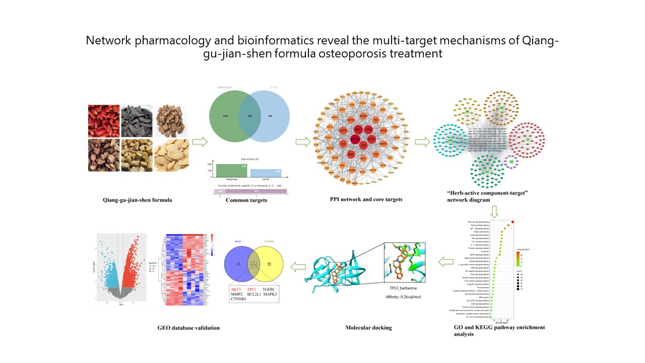
Introduction: Osteoporosis (OP) is a systemic skeletal disease characterized by reduced bone mass and deteriorated bone microstructure, significantly increasing fracture risk. As global aging intensifies, OP has become a significant public health issue. Present pharmacological interventions, such as bisphosphonates and selective estrogen receptor modulators, are associated with side effects and limitations, highlighting the need for safe and effective alternatives. Objective: This study investigates the potential mechanisms of the Qiang-gu-jian-shen formula (QGJSF), a traditional Chinese medicine (TCM) compound, in treating OP using network pharmacology and bioinformatics. Methods: A total of 1,395 potential targets for QGJSF were identified by querying the TCMSP and BATMAN-TCM databases and converting targets through UniProt. Cross-referencing with OP-related targets from GeneCards, OMIM, and DisGeNET yielded 500 mapped targets. Protein-protein interaction network constructed through the STRING database led to the identification of 69 core targets. An “herb-active component-target” network was built using Cytoscape 3.9.0. Gene ontology functional annotation and Kyoto Encyclopedia of Genes and Genomes pathway enrichment analyses highlighted key pathways, including the PI3K/AKT and FoxO. Results: Molecular docking showed that key components, such as quercetin, dioscin, genistein, calycosin, and berberine, bind favorably to core targets (binding energies < −5 kcal/mol). GEO dataset (GSE5958) analysis identified seven common core genes, including TGFB1, MMP2, BCL2L1, MAPK3, AKT1, CTNNB1, and TP53. The findings suggest that QGJSF may improve OP through multiple components that regulate osteoblast differentiation, osteoclastogenesis, and activate key pathways, such as Wnt/β-catenin, PI3K/AKT, and JAK/STAT, thereby enhancing bone formation and reducing resorption. Core targets such as ESR1, STAT3, AKT1, and TP53 regulate bone metabolism by modulating osteoblasts, osteoclasts, and their interactions with immune and hematopoietic cells to maintain bone remodeling. Conclusion: This study advances understanding of QGJSF’s mechanisms and provides a foundation for novel OP therapies. Future validation and exploration of additional therapeutic targets and mechanisms are needed.
- International Osteoporosis Foundation. Worldwide Incidence of HIP Fracture Increase from 1990 to 2050 [EB/ OL]. Available from: https//www.osteoporosisfoundation/ policy-makers#policy-reports-audits [Last accessed on 2025 Jun 16].
- World Health Organization. Musculoskeletal Conditions Affect Millions [EB/OL]. Available from: https://www.who. int/news/item/27-10-2003musculoskeletal-conditions-affect-millions [Last accessed on 2025 Jun 16].
- Schroeder RJ, Staszkiewicz J, O’Quin C, et al. Oral therapeutics post-menopausal osteoporosis. Cureus. 2023;15(8):e42870. doi: 10.7759/cureus.42870
- Wang L, Huang X, Qin J, et al. The role of traditional Chinese medicines in the treatment of osteoporosis. Am J Chin Med. 2024;52(4):949-986. doi: 10.1142/S0192415X24500393
- Xiong M, Chen X, Wang H, et al. Combining transcriptomics and network pharmacology to reveal the mechanism of zuojin capsule improving spasmolytic polypeptide-expressing metaplasia. J Ethnopharmacol. 2024;318(Pt B):117075. doi: 10.1016/j.jep.2023.117075
- Yu W, Li X, Sun Q, et al. Metabolomics and network pharmacology reveal the mechanism of Castanopsis honey against Streptococcus pyogenes. Food Chem. 2024;441:138388. doi: 10.1016/j.foodchem.2024.138388
- Zhu H, Wang S, Shan C, et al. Mechanism of protective effect of xuan-bai-cheng-qi decoction on LPS-induced acute lung injury based on an integrated network pharmacology and RNA-sequencing approach. Respir Res. 2021;22(1):188. doi: 10.1186/s12931-021-01781-1
- Deng TT, Ding WY, Lu XX, et al. Pharmacological and mechanistic aspects of quercetin in osteoporosis. Front Pharmacol. 2024;15:1338951. doi: 10.3389/fphar.2024.1338951
- Xiong Y, Huang CW, Shi C, et al. Corrigendum: Quercetin suppresses ovariectomy-induced osteoporosis in rat mandibles by regulating autophagy and the NLRP3 pathway. Exp Biol Med (Maywood). 2024;249:10149. doi: 10.3389/ebm.2024.10149
- Wu S, Zhao F, Zhao J, et al. Dioscin improves postmenopausal osteoporosis through inducing bone formation and inhibiting apoptosis in ovariectomized rats. Biosci Trends. 2019;13(5):394-401. doi: 10.5582/bst.2019.01186
- Tao X, Qi Y, Xu L, et al. Dioscin reduces ovariectomy-induced bone loss by enhancing osteoblastogenesis and inhibiting osteoclastogenesis Pharmacol Res. 2020;151:104397. doi: 10.1016/j.phrs.2019.104397
- Li M, Yu Y, Xue K, et al. Genistein mitigates senescence of bone marrow mesenchymal stem cells via ERRα-mediated mitochondrial biogenesis and mitophagy in ovariectomized rats. Redox Biol. 2023;61:102649. doi: 10.1016/j.redox.2023.102649
- Wu Z, Liu L. The protective activity of genistein against bone and cartilage diseases. Front Pharmacol. 2022;13:1016981. doi: 10.3389/fphar.2022.1016981
- Quan GH, Wang H, Cao J, et al. Calycosin suppresses RANKL-mediated osteoclastogenesis through inhibition of MAPKs and NF-κB. Int J Mol Sci. 2015;16(12):29496-29507. doi: 10.3390/ijms161226179
- Li N, Tu Y, Shen Y, Qin Y, Lei C, Liu X. Calycosin attenuates osteoporosis and regulates the expression of OPG/RANKL in ovariectomized rats via MAPK signaling. Pharmazie. 2016;71(10):607-612. doi: 10.1691/ph.2016.6627
- Duan X, Meng Q, Wang C, et al. Calycosin attenuates triglyceride accumulation and hepatic fibrosis in murine model of non-alcoholic steatohepatitis via activating farnesoid X receptor. Phytomedicine. 2017;25:83-92. doi: 10.1016/j.phymed.2016.12.006
- Li Z, Geng YN, Jiang JD, Kong WJ. Antioxidant and anti-inflammatory activities of berberine in the treatment of diabetes mellitus. Evid Based Complement Alternat Med. 2014;2014:289264. doi: 10.1155/2014/289264
- Lee JW, Mase N, Yonezawa T, et al. Palmatine attenuates osteoclast differentiation and function through inhibition of receptor activator of nuclear factor-κb ligand expression in osteoblast cells. Biol Pharm Bull. 2010;33(10):1733-1739. doi: 10.1248/bpb.33.1733
- Chen ZZ. Berberine induced apoptosis of human osteosarcoma cells by inhibiting phosphoinositide 3 kinase/ protein kinase B (PI3K/Akt) signal pathway activation. Iran J Public Health. 2016;45(5):578-585.
- Li H, Miyahara T, Tezuka Y, Tran QL, Seto H, Kadota S. Effect of berberine on bone mineral density in SAMP6 as a senile osteoporosis model. Biol Pharm Bull. 2003;26(1):110-111. doi: 10.1248/bpb.26.110
- Matsushita K, Itoh S, Ikeda S, Yamamoto Y, Yamauchi Y, Hayashi M. LIF/STAT3/SOCS3 signaling pathway in murine bone marrow stromal cells suppresses osteoblast differentiation. J Cell Biochem. 2014;115(7):1262-1268. doi: 10.1002/jcb.24777
- Vimalraj S, Arumugam B, Miranda PJ, Selvamurugan N. Runx2: Structure, function, and phosphorylation in osteoblast differentiation. Int J Biol Macromol. 2015;78:202-208. doi: 10.1016/j.ijbiomac.2015.04.008
- Wang L, Yang H, Huang J, et al. Targeted ptpn11 deletion in mice reveals the essential role of SHP2 in osteoblast differentiation and skeletal homeostasis. Bone Res. 2021;9(1):6. doi: 10.1038/s41413-020-00129-7
- Zhou S, Dai Q, Huang X, et al. STAT3 is critical for skeletal development and bone homeostasis by regulating osteogenesis. Nat Commun. 2021;12(1):6891. doi: 10.1038/s41467-021-27273-w
- Joung YH, Darvin P, Kang DY, et al. Methylsulfonylmethane Inhibits RANKL-induced osteoclastogenesis in BMMs by suppressing NF-κB and STAT3 activities. PLoS One. 2016;11(7):e0159891. doi: 10.1371/journal.pone.0159891
- Zhang B, Qu Z, Hui H, et al. Exploring the therapeutic potential of isoorientin in the treatment of osteoporosis: A study using network pharmacology and experimental validation. Mol Med. 2024;30(1):27. doi: 10.1186/s10020-024-00799-7
- Chen X, Chen W, Aung ZM, Han W, Zhang Y, Chai G. LY3023414 inhibits both osteogenesis and osteoclastogenesis through the PI3K/Akt/GSK3 signalling pathway. Bone Joint Res. 2021;10(4):237-249. doi: 10.1302/2046-3758.104.BJR-2020-0255.R2
- Kawamura N, Kugimiya F, Oshima Y, et al. Akt1 in osteoblasts and osteoclasts controls bone remodeling. PLoS One. 2007;2(10):e1058. doi: 10.1371/journal.pone.0001058
- Vaddavalli PL, Schumacher B. The p53 network: Cellular and systemic DNA damage responses in cancer and aging. Trends Genet. 2022;38(6):598-612. doi: 10.1016/j.tig.2022.02.010
- Wang B, Wang J. Research progress on MDM2/MDMX heterodimer and MDMX phosphorylation in regulating p53. Chin Bull Life Sci. 2020;32(5):446-452. doi: 10.13376/j.cbls/2020056
- Hojo H, Ohba S, He X, Lai LP, McMahon AP. Sp7/osterix is restricted to bone-forming vertebrates where it acts as a dlx co-factor in osteoblast specification. Dev Cell. 2016;37(3):238-253. doi: 10.1016/j.devcel.2016.04.002
- Kawane T, Komori H, Liu W, et al. Dlx5 and mef2 regulate a novel runx2 enhancer for osteoblast-specific expression. J Bone Miner Res. 2014;29(9):1960-1969. doi: 10.1002/jbmr.2240
- Zhen YF, Wang GD, Zhu LQ, et al. P53 dependent mitochondrial permeability transition pore opening is required for dexamethasone-induced death of osteoblasts. J Cell Physiol. 2014;229(10):1475-1483. doi: 10.1002/jcp.24589
- Zhang F, Peng W, Zhang J, et al. P53 and Parkin co-regulate mitophagy in bone marrow mesenchymal stem cells to promote the repair of early steroid-induced osteonecrosis of the femoral head. Cell Death Dis. 2020;11(1):42. doi: 10.1038/s41419-020-2238-1
- Zauli G, Rimondi E, Corallini F, Fadda R, Capitani S, Secchiero P. MDM2 antagonist nutlin-3 suppresses the proliferation and differentiation of human pre-osteoclasts through a p53-dependent pathway. J Bone Miner Res. 2007;22(10):1621-1630. doi: 10.1359/jbmr.070618
- Tong X, Gu J, Chen M, et al. P53 positively regulates osteoprotegerin-mediated inhibition of osteoclastogenesis by downregulating TSC2-induced autophagy in vitro. Differentiation. 2020;114:58-66. doi: 10.1016/j.diff.2020.06.002
- Qu L, Zhao M, Wang D, Song L, Zhou K. Research progress and mechanism of estrogen receptor α in postmenopausal osteoporosis. Chin J Osteoporos. 2024;30(7):1021-1027.
- Feng C, Xu Z, Tang X, Cao H, Zhang G, Tan J. Estrogen-related receptor α: A significant regulator and promising target in bone homeostasis and bone metastasis. Molecules. 2022;27(13):3976. doi: 10.3390/molecules27133976
- Moon YJ, Zhang Z, Bang IH, et al. Sirtuin 6 in preosteoclasts suppresses age- and estrogen deficiency-related bone loss by stabilizing estrogen receptor α. Cell Death Differ. 2019;26(11):2358-2370. doi: 10.1038/s41418-019-0306-9
- Kim SJ, Piao Y, Lee MG, et al. Loss of sirtuin 6 in osteoblast lineage cells activates osteoclasts, resulting in osteopenia. Bone. 2020;138:115497. doi: 10.1016/j.bone.2020.115497
- Melville KM, Kelly NH, Surita G, et al. Effects of deletion of ERα in osteoblast-lineage cells on bone mass and adaptation to mechanical loading differ in female and male mice. J Bone Miner Res. 2015;30(8):1468-1480. doi: 10.1002/jbmr.2488
- Yang Y, Feng N, Liang L, et al. Progranulin, a moderator of estrogen/estrogen receptor α binding, regulates bone homeostasis through PERK/p-eIF2 signaling pathway. J Mol Med (Berl). 2022;100(8):1191-1207. doi: 10.1007/s00109-022-02233-z
- León-Reyes G, Argoty-Pantoja AD, Becerra-Cervera A, López-Montoya P, Rivera-Paredez B, Velázquez-Cruz R. Oxidative-stress-related genes in osteoporosis: A systematic review. Antioxidants (Basel). 2023;12(4):915. doi: 10.3390/antiox12040915
- Knowles HJ, Cleton-Jansen AM, Korsching E, Athanasou NA. Hypoxia-inducible factor regulates osteoclast-mediated bone resorption: Role of angiopoietin-like 4. FASEB J. 2010;24(12):4648-4659.doi: 10.1096/fj.10-162230
- Yao Z, Getting SJ, Locke IC. Regulation of TNF-induced osteoclast differentiation. Cells. 2021;11(1):132. doi: 10.3390/cells11010132
- Miyauchi Y, Sato Y, Kobayashi T, et al. HIF1α is required for osteoclast activation by estrogen deficiency in postmenopausal osteoporosis. Proc Natl Acad Sci U S A. 2013;110(41):16568-16573. doi: 10.1073/pnas.1308755110
- Wan C, Shao J, Gilbert SR, et al. Role of HIF-1α in skeletal development. Ann N Y Acad Sci. 2010;1192:322-326. doi: 10.1111/j.1749-6632.2009.05238.x
- Medema RH, Kops GJ, Bos JL, Burgering BM. AFX-like forkhead transcription factors mediate cell-cycle regulation by ras and PKB through p27kip1. Nature. 2000;404(6779):782-787. doi: 10.1038/35008115
- Tran H, Brunet A, Grenier JM, et al. DNA repair pathway stimulated by the forkhead transcription factor FOXO3a through the gadd45 protein. Science. 2002;296(5567):530-534. doi: 10.1126/science.1068712
- Kops GJ, Dansen TB, Polderman PE, et al. Forkhead transcription factor FOXO3a protects quiescent cells from oxidative stress. Nature. 2002;419(6904):316-321. doi: 10.1038/nature01036
- Brunet A, Bonni A, Zigmond MJ, et al. Akt promotes cell survival by phosphorylating and inhibiting a forkhead transcription factor. Cell. 1999;96(6):857-868. doi: 10.1016/s0092-8674(00)80595-4
- Nemoto S, Finkel T. Redox regulation of forkhead proteins through a p66shc-dependent signaling pathway. Science. 2002;295(5564):2450-2452. doi: 10.1126/science.1069004
- Speckmann B, Walter PL, Alili L, et al. Selenoprotein P expression is controlled through interaction of the coactivator PGC-1alpha with FoxO1a and hepatocyte nuclear factor 4alpha transcription factors. Hepatology. 2008;48(6):1998-2006. doi: 10.1002/hep.22526.
- Sidhu A, Miller PJ, Hollenbach AD. FOXO1 stimulates ceruloplasmin promoter activity in human hepatoma cells treated with IL-6. Biochem Biophys Res Commun. 2011;404(4):963-967. doi: 10.1016/j.bbrc.2010.12.089
- Almeida M. Unraveling the role of FoxOs in bone-insights from mouse models. Bone. 2011;49(3):319-327. doi: 10.1016/j.bone.2011.05.023
- Y Ye C, Zhang W, Hang K, et al. Extracellular IL-37 promotes osteogenic differentiation of human bone marrow mesenchymal stem cells via activation of the PI3K/AKT signaling pathway. Cell Death Dis. 2019;10(10):753. doi: 10.1038/s41419-019-1904-7
- Zhou H, Jiao G, Dong M, et al. Orthosilicic acid accelerates bone formation in human osteoblast-like cells through the PI3K-Akt-mTOR pathway. Biol Trace Elem Res. 2019;190(2):327-335. doi: 10.1007/s12011-018-1574-9
- Bartell SM, Kim HN, Ambrogini E, et al. FoxO proteins restrain osteoclastogenesis and bone resorption by attenuating H2O2 accumulation. Nat Commun. 2014;5:3773. doi: 10.1038/ncomms4773
- Feng Y. Ph.D. Thesis. China: Lanzhou University; 2018.
- Sugatani T, Alvarez U, Hruska KA. PTEN regulates RANKL- and osteopontin-stimulated signal transduction during osteoclast differentiation and cell motility. J Biol Chem. 2003;278(7):5001-5008. doi: 10.1074/jbc.M209299200
- Stein GH, Drullinger LF, Soulard A, Dulić V. Differential roles for cyclin-dependent kinase inhibitors p21 and p16 in the mechanisms of senescence and differentiation in human fibroblasts. Mol Cell Biol. 1999;19(3):2109-2117. doi: 10.1128/MCB.19.3.2109
- Xia C, Jiang T, Wang Y, Chen X, Hu Y, Gao Y. The p53/ miR-145a axis promotes cellular senescence and inhibits osteogenic differentiation by targeting cbfb in mesenchymal stem cells. Front Endocrinol (Lausanne). 2021;11:609186. doi: 10.3389/fendo.2020.609186
- Kim HJ, Kim WJ, Shin HR, et al. ROS-induced PADI2 downregulation accelerates cellular senescence via the stimulation of SASP production and NFκB activation. Cell Mol Life Sci. 2022;79(3):155. doi: 10.1007/s00018-022-04186-5
- Lian WS, Wu RW, Chen YS, et al. MicroRNA-29a mitigates osteoblast senescence and counteracts bone loss through oxidation resistance-1 Control of FoxO3 methylation. Antioxidants (Basel). 2021;10(8):1248. doi: 10.3390/antiox10081248
- Raynaud-Messina B, Verollet C, Maridonneau-Parini I. The osteoclast, a target cell for microorganisms. Bone. 2019;127:315-323. doi: 10.1016/j.bone.2019.06.023
- Tokunaga T, Mokuda S, Kohno H, et al. TGFβ1 regulates human RANKL-induced osteoclastogenesis via suppression of NFATc1 expression. Int J Mol Sci. 2020;21(3):800.doi: 10.3390/ijms21030800
- Hu F, Jiang C, Bu G, Fu Y, Yu Y. Silencing long noncoding RNA colon cancer-associated transcript-1 upregulates microRNA-34a-5p to promote proliferation and differentiation of osteoblasts in osteoporosis. Cancer Gene Ther. 2021;28(10-11):1150-1161. doi: 10.1038/s41417-020-00264-7
- John AA, Xie J, Yang YS, et al. AAV-mediated delivery of osteoblast/osteoclast-regulating miRNAs for osteoporosis therapy. Mol Ther Nucleic Acids. 2022;29:296-311. doi: 10.1016/j.omtn.2022.07.008
- Li D, Liu J, Guo B, et al. Osteoclast-derived exosomal miR- 214-3p inhibits osteoblastic bone formation. Nat Commun. 2016;7:10872. doi: 10.1038/ncomms10872
- Ohnuma K, Kasagi S, Uto K, et al. MicroRNA-124 inhibits TNF-α- and IL-6-induced osteoclastogenesis. Rheumatol Int. 2019;39(4):689-695. doi: 10.1007/s00296-018-4218-7
- Liu C, Cao Z, Bai Y, et al. LncRNA AK077216 promotes RANKL-induced osteoclastogenesis and bone resorption via NFATc1 by inhibition of NIP45. J Cell Physiol. 2019;234(2):1606-1617. doi: 10.1002/jcp.27031