

Self-organizing vascularized subchondral bone organoids from stromal vascular fraction enable functional osteochondral interface regeneration

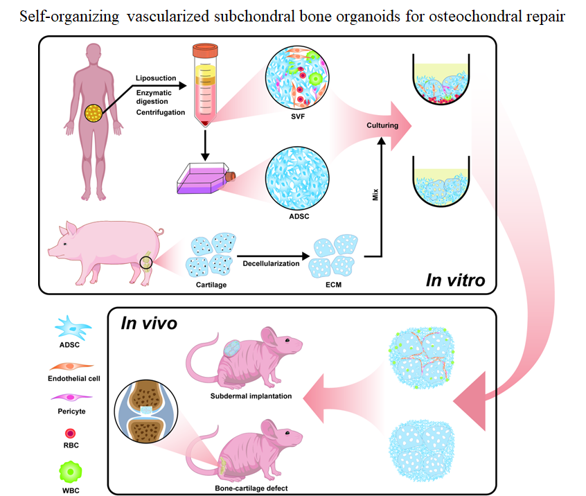
Osteoarthritis is closely associated with subchondral bone (SCB) degeneration; however, current models fail to adequately mimic its complex microenvironment. Here, we developed a self-organizing SCB organoid (SSBO) by co-culturing stromal vascular fraction (SVF) cells with decellularized cartilage extracellular matrix (CECM). SVF provided cellular heterogeneity, including adipose-derived stem cells (ADSCs), endothelial cells, pericytes, and macrophages, while CECM served as a native scaffold with tissue-specific cues. SSBO exhibited spontaneous spheroid formation, active cellular infiltration, and dynamic matrix remodeling. Compared to ADSC-only controls, SSBO showed enhanced cell viability, vascularization, collagen remodeling, and spatial organization. Immunostaining and quantitative real-time polymerase chain reaction analyses confirmed an endochondral ossification-like process, characterized by the sequential expression of SOX9, COL2A1, RUNX2, COL1A1, and OCN. In vivo implantation into immunodeficient mice demonstrated robust angiogenesis, bone-like tissue formation, and integration with host vasculature. Furthermore, in a mouse osteochondral defect model, SSBO significantly promoted repair, as evidenced by increased bone volume, improved trabecular architecture, and enhanced cartilage regeneration. Collectively, this study presents a novel strategy for constructing vascularized, immunomodulatory, and osteogenic SCB organoids, offering a promising platform for regenerative medicine and bone–cartilage interface repair.
- Bijlsma JW, Berenbaum F, Lafeber FP. Osteoarthritis: An update with relevance for clinical practice. Lancet. 2011;377(9783):2115-2126. doi: 10.1016/s0140-6736(11)60243-2
- James SL, Abate D, Abate KH, et al. Global, regional, and national incidence, prevalence, and years lived with disability for 354 diseases and injuries for 195 countries and territories, 1990–2017: a systematic analysis for the Global Burden of Disease Study 2017. The Lancet. 2018;392(10159):1789-1858. doi: 10.1016/s0140-6736(18)32279-7
- Reichenbach S, Felson DT, Hincapié CA, et al. Effect of biomechanical footwear on knee pain in people with knee osteoarthritis: The BIOTOK randomized clinical trial. JAMA. 2020;323(18):1802-1812. doi: 10.1001/jama.2020.3565
- Martel-Pelletier J, Barr AJ, Cicuttini FM, et al. Osteoarthritis. Nat Rev Dis Primers. 2016;2:16072. doi: 10.1038/nrdp.2016.72
- Grynpas MD, Alpert B, Katz I, Lieberman I, Pritzker KP. Subchondral bone in osteoarthritis. Calcif Tissue Int. 1991;49(1):20-26. doi: 10.1007/bf02555898
- Burr DB, Gallant MA. Bone remodelling in osteoarthritis. Nat Rev Rheumatol. 2012;8(11):665-673. doi: 10.1038/nrrheum.2012.130 7. Suri S, Walsh DA. Osteochondral alterations in osteoarthritis. Bone. 2012;51(2):204-211. doi: 10.1016/j.bone.2011.10.010
- Goldring SR, Goldring MB. Changes in the osteochondral unit during osteoarthritis: Structure, function and cartilage-bone crosstalk. Nat Rev Rheumatol. 2016;12(11):632-644. doi: 10.1038/nrrheum.2016.148
- Karsdal MA, Bay-Jensen AC, Lories RJ, et al. The coupling of bone and cartilage turnover in osteoarthritis: Opportunities for bone antiresorptives and anabolics as potential treatments? Ann Rheum Dis. 2014;73(2):336-348. doi: 10.1136/annrheumdis-2013-204111
- Zhang H, Wang L, Cui J, et al. Maintaining hypoxia environment of subchondral bone alleviates osteoarthritis progression. Sci Adv. 2023;9(14):eabo7868. doi: 10.1126/sciadv.abo7868
- Chen Z, Bo Q, Wang C, Xu Y, Fei X, Chen R. Single BMSC-derived cartilage organoids for gradient heterogeneous osteochondral regeneration by leveraging native vascular microenvironment. J Nanobiotechnol. 2025;23(1):325 doi: 10.1186/s12951-025-03403-0
- Lyu X, Wang J, Su J. Intelligent manufacturing for osteoarthritis organoids. Cell Prolif. 2025;58(7):e70043. doi: 10.1111/cpr.70043
- Day JS, Ding M, Van Der Linden JC, Hvid I, Sumner DR, Weinans H. A decreased subchondral trabecular bone tissue elastic modulus is associated with pre-arthritic cartilage damage. J Orthop Res. 2001;19(5):914-918. doi: 10.1016/s0736-0266(01)00012-2
- Hu W, Chen Y, Dou C, Dong S. Microenvironment in subchondral bone: Predominant regulator for the treatment of osteoarthritis. Ann Rheum Dis. 2021;80(4):413-422. doi: 10.1136/annrheumdis-2020-218089
- Hu Y, Chen X, Wang S, Jing Y, Su J. Subchondral bone microenvironment in osteoarthritis and pain. Bone Res. 2021;9(1):20. doi: 10.1038/s41413-021-00147-z
- Song H, Li X, Zhao Z, et al. Reversal of osteoporotic activity by endothelial cell-secreted bone targeting and biocompatible exosomes. Nano Lett. 2019;19(5):3040-3048. doi: 10.1021/acs.nanolett.9b00287
- Castañeda S, Roman-Blas JA, Largo R, Herrero-Beaumont G. Subchondral bone as a key target for osteoarthritis treatment. Biochem Pharmacol. 2012;83(3):315-323. doi: 10.1016/j.bcp.2011.09.018
- Li G, Yin J, Gao J, et al. Subchondral bone in osteoarthritis: Insight into risk factors and microstructural changes. Arthritis Res Ther. 2013;15(6):223. doi: 10.1186/ar4405
- Henrotin Y, Pesesse L, Sanchez C. Subchondral bone and osteoarthritis: Biological and cellular aspects. Osteoporos Int. 2012;23(Suppl 8):847-851. doi: 10.1007/s00198-012-2162-z
- Kim W, Gwon Y, Park S, Kim H, Kim J. Therapeutic strategies of three-dimensional stem cell spheroids and organoids for tissue repair and regeneration. Bioact Mater. 2023;19:50-74. doi: 10.1016/j.bioactmat.2022.03.039
- Hofer M, Lutolf MP. Engineering organoids. Nat Rev Mater. 2021;6(5):402-420. doi: 10.1038/s41578-021-00279-y
- Takebe T, Sekine K, Enomura M, et al. Vascularized and functional human liver from an iPSC-derived organ bud transplant. Nature. 2013;499(7459):481-484. doi: 10.1038/nature12271
- Lancaster MA, Renner M, Martin CA, et al. Cerebral organoids model human brain development and microcephaly. Nature. 2013;501(7467):373-379. doi: 10.1038/nature12517
- Takasato M, Er PX, Chiu HS, et al. Kidney organoids from human iPS cells contain multiple lineages and model human nephrogenesis. Nature. 2015;526(7574):564-568. doi: 10.1038/nature15695
- Zhang C, Jing Y, Wang J, et al. Skeletal organoids. Biomater Transl. 2024;5(4):390-410. doi: 10.12336/biomatertransl.2024.04.005
- Zakhari JS, Zabonick J, Gettler B, Williams SK. Vasculogenic and angiogenic potential of adipose stromal vascular fraction cell populations in vitro. In Vitro Cell Dev Biol Anim. 2018;54(1):32-40. doi: 10.1007/s11626-017-0213-7
- Reid G, Cerino G, Melly L, Fusco D, Zhang C, Reuthebuch O, et al. Harnessing the angiogenic potential of adipose-derived stromal vascular fraction cells with perfusion cell seeding. Stem Cell Res Ther. 2025;16(1):220. doi: 10.1186/s13287-025-04286-6
- Moreira HR, Rodrigues DB, Freitas-Ribeiro S, et al. Spongy-like hydrogels prevascularization with the adipose tissue vascular fraction delays cutaneous wound healing by sustaining inflammatory cell influx. Mater Today Bio. 2022;17:100496. doi: 10.1016/j.mtbio.2022.100496
- Liu W, Jiang H, Chen J, et al. High paracrine activity of hADSCs cartilage microtissues inhibits extracellular matrix degradation and promotes cartilage regeneration. Mater Today Bio. 2025;30:101372. doi: 10.1016/j.mtbio.2024.101372
- Kim YS, Majid M, Melchiorri AJ, Mikos AG. Applications of decellularized extracellular matrix in bone and cartilage tissue engineering. Bioeng Transl Med. 2019;4(1):83-95. doi: 10.1002/btm2.10110
- Morris AH, Stamer DK, Kyriakides TR. The host response to naturally-derived extracellular matrix biomaterials. Semin Immunol. 2017;29:72-91. doi: 10.1016/j.smim.2017.01.002
- Liu C, Pei M, Li Q, Zhang Y. Decellularized extracellular matrix mediates tissue construction and regeneration. Front Med. 2022;16(1):56-82. doi: 10.1007/s11684-021-0900-3
- Zhang X, Chen X, Hong H, Hu R, Liu J, Liu C. Decellularized extracellular matrix scaffolds: Recent trends and emerging strategies in tissue engineering. Bioact Mater. 2022;10:15-31. doi: 10.1016/j.bioactmat.2021.09.014
- Guo X, Liu B, Zhang Y, et al. Decellularized extracellular matrix for organoid and engineered organ culture. J Tissue Eng. 2024;15:1-31. doi: 10.1177/20417314241300386
- Pan J, Zhou X, Li W, Novotny JE, Doty SB, Wang L. In situ measurement of transport between subchondral bone and articular cartilage. J Orthop Res. 2009;27(10):1347-1352. doi: 10.1002/jor.20883
- Verdugo-Avello F, Wychowaniec JK, Villacis-Aguirre CA, D’Este M, Toledo JR. Bone microphysiological models for biomedical research. Lab Chip. 2025;25(5):806-836. doi: 10.1039/d4lc00762j
- Wang J, Wu Y, Li G, et al. Engineering large-scale self-mineralizing bone organoids with bone matrix-inspired hydroxyapatite hybrid bioinks. Adv Mater. 2024;36(30):e2309875. doi: 10.1002/adma.202309875
- Zhang X, Jiang W, Wu X, Xie C, Zhang Y, Li L, et al. Divide-and-conquer strategy with engineered ossification center organoids for rapid bone healing through developmental cell recruitment. Nat Commun. 2025;16(1):6200. doi: 10.1038/s41467-025-61619-y
- Bora P, Majumdar AS. Adipose tissue-derived stromal vascular fraction in regenerative medicine: A brief review on biology and translation. Stem Cell Res Ther. 2017;8(1):145. doi: 10.1186/s13287-017-0598-y
- Rehman J, Traktuev D, Li J, et al. Secretion of angiogenic and antiapoptotic factors by human adipose stromal cells. Circulation. 2004;109(10):1292-1298. doi: 10.1161/01.Cir.0000121425.42966.F1
- Wu J, He Y, Qian T, et al. Stromal vascular fraction self-assembles vascularized osteogenic organoids with immunomodulatory functions. Bioact Mater. 2026;57:323-343. doi: 10.1016/j.bioactmat.2025.10.030
- Ahmad N, Anker A, Klein S, et al. Autologous fat grafting-a panacea for scar tissue therapy? Cells. 2024;13(16):1384. doi: 10.3390/cells13161384
- Airuddin SS, Halim AS, Wan Sulaiman WA, Kadir R, Nasir NAM. Adipose-derived stem cell: “Treat or trick.” Biomedicines. 2021;9(11):1624. doi: 10.3390/biomedicines9111624
- Guan F, Wang R, Yi Z, et al. Tissue macrophages: Origin, heterogeneity, biological functions, diseases and therapeutic targets. Signal Transduct Target Ther. 2025;10(1):93. doi: 10.1038/s41392-025-02124-y
- Uribe-Querol E, Rosales C. Phagocytosis: Our current understanding of a universal biological process. Front Immunol. 2020;11:1066. doi: 10.3389/fimmu.2020.01066
- Blanchard L, Girard JP. High endothelial venules (HEVs) in immunity, inflammation and cancer. Angiogenesis. 2021;24(4):719-753. doi: 10.1007/s10456-021-09792-8
- Mohan SP, Priya SP, Tawfig N, et al. The potential role of adipose-derived stem cells in regeneration of peripheral nerves. Neurol Int. 2025;17(2):23. doi: 10.3390/neurolint17020023
- Xiong J, Qiang H, Li T, et al. Human adipose-derived stem cells promote seawater-immersed wound healing via proangiogenic effects. Aging (Albany NY). 2021;13(13):17118-17136. doi: 10.18632/aging.202773
- Li J, Liu Y, Zhang R, et al. Insights into the role of mesenchymal stem cells in cutaneous medical aesthetics: From basics to clinics. Stem Cell Res Ther. 2024;15(1):169. doi: 10.1186/s13287-024-03774-5
- Vanderstichele S, Vranckx JJ. Anti-fibrotic effect of adipose-derived stem cells on fibrotic scars. World J Stem Cells. 2022;14(2):200-213. doi: 10.4252/wjsc.v14.i2.200