

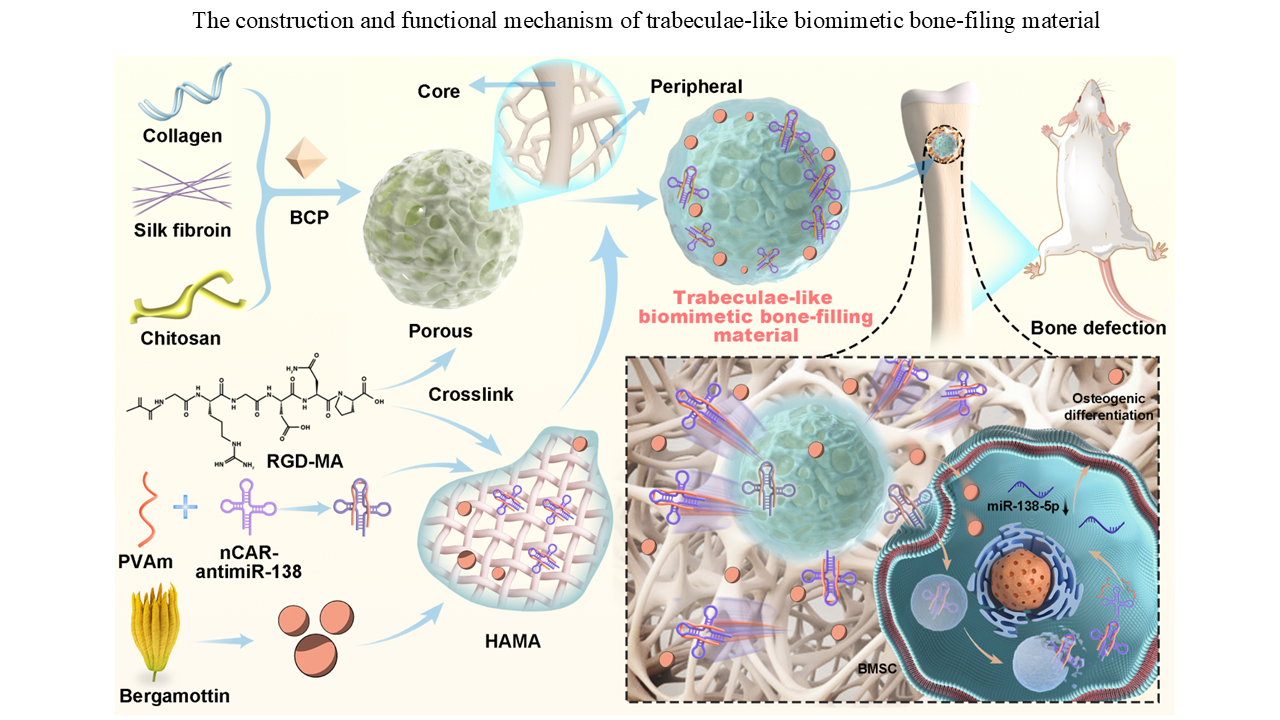
A trabeculae-like biomimetic bone-filling material as a potential organoid for bone defect treatment

Bone-filling materials are critical tools for the treatment of bone defects. However, existing materials require improvements in tissue compatibility and drug-loading capacity. In this study, we designed and synthesized a novel trabeculae-like biomimetic bone-filling material (TBM) that mimics the composition and structure of natural bone trabecular tissue. This TBM demonstrated high mechanical strength and excellent biocompatibility. It effectively embedded osteogenic cells and potentially functioned as an organoid. We demonstrated that the TBM exhibited therapeutic efficacy in treating various bone defects and fractures by filling defect regions and enabling sustained release of small-molecule and nucleic acid drugs. Based on these findings, we propose TBM as a promising candidate for the treatment of bone defects and provide innovative insights for the development of bone-filling materials.
- Sparks DS, Saifzadeh S, Savi FM, et al. A preclinical large-animal model for the assessment of critical-size load-bearing bone defect reconstruction. Nat Protoc. 2020;15(3):877-924. doi: 10.1038/s41596-019-0271-2
- El-Rashidy A, Roether JA, Harhaus L, Kneser U, Boccaccini AR. Regenerating bone with bioactive glass scaffolds: A review of in vivo studies in bone defect models. Acta Biomater. 2017;62:1-28. doi: 10.1016/j.actbio.2017.08.030
- De La Vega RE, van Griensven M, Zhang W, Coenen MJ, Nagelli CV, Panos JA, et al. Efficient healing of large osseous segmental defects using optimized chemically modified messenger RNA encoding BMP-2. Sci Adv. 2022;8(7):eabl6242. doi: 10.1126/sciadv.abl6242
- Yang J, Chen Z, Gao C, et al. A mechanical-assisted post-bioprinting strategy for challenging bone defects repair. Nat Commun. 2024;15(1):3565. doi: 10.1038/s41467-024-48023-8
- Reichert JC, Saifzadeh S, Wullschleger ME, et al. The challenge of establishing preclinical models for segmental bone defect research. Biomaterials. 2009;30(12):2149-2163. doi: 10.1016/j.biomaterials.2008.12.050.
- Zhang L, Yang G, Johnson BN, Jia X. Three-dimensional (3D) printed scaffold and material selection for bone repair. Acta Biomater. 2019;84:16-33. doi: 10.1016/j.actbio.2018.11.039
- Jariwala SH, Lewis GS, Bushman ZJ, Adair JH, Donahue HJ. 3D printing of personalized artificial bone scaffolds. 3D Print Addit Manuf. 2015;2(2):56-64. doi: 10.1089/3dp.2015.0001
- Chen Y, Sheng W, Lin J, et al. Magnesium oxide nanoparticle coordinated phosphate-functionalized chitosan injectable hydrogel for osteogenesis and angiogenesis in bone regeneration. ACS Appl Mater Interfaces. 2022;14(6):7592-7608. doi: 10.1021/acsami.1c21260
- McNeill EP, Zeitouni S, Pan S, et al. Characterization of a pluripotent stem cell-derived matrix with powerful osteoregenerative capabilities. Nat Commun. 2020;11(1):3025. doi: 10.1038/s41467-020-16646-2
- Mitra D, Whitehead J, Yasui OW, Leach JK. Bioreactor culture duration of engineered constructs influences bone formation by mesenchymal stem cells. Biomaterials. 2017;146:29-39. doi: 10.1016/j.biomaterials.2017.08.044
- Qin L, Yang S, Zhao C, et al. Prospects and challenges for the application of tissue engineering technologies in the treatment of bone infections. Bone Res. 2024;12(1):28. doi: 10.1038/s41413-024-00332-w
- Su J, Xu H, Sun J, Gong X, Zhao H. Dual delivery of BMP-2 and bFGF from a new nano-composite scaffold, loaded with vascular stents for large-size mandibular defect regeneration. Int J Mol Sci. 2013;14(6):12714-12728. doi: 10.3390/ijms140612714
- Davison N, Yuan H, De Bruijn JD, Barrere-de Groot F. In vivo performance of microstructured calcium phosphate formulated in novel water-free carriers. Acta Biomater. 2012;8(7):2759-2769. doi: 10.1016/j.actbio.2012.04.007
- Wang W, Yeung KWK. Bone grafts and biomaterials substitutes for bone defect repair: A review. Bioact Mater. 2017;2(4):224-247. doi: 10.1016/j.bioactmat.2017.05.007
- Xie C, Liang R, Ye J, et al. High-efficient engineering of osteo-callus organoids for rapid bone regeneration within one month. Biomaterials. 2022;288:121741. doi: 10.1016/j.biomaterials.2022.121741
- Li S, Yang H, Qu X, et al. Multiscale architecture design of 3D printed biodegradable Zn-based porous scaffolds for immunomodulatory osteogenesis. Nat Commun. 2024;15(1):3131. doi: 10.1038/s41467-024-47189-5
- Mao Z, Bi X, Yu C, et al. Mechanically robust and personalized silk fibroin-magnesium composite scaffolds with water-responsive shape-memory for irregular bone regeneration. Nat Commun. 2024;15(1):4160. doi: 10.1038/s41467-024-48417-8
- Fan S, Zeng Z. Research progress of bone grafting material in interbody fusion. J Spinal Surg. 2018;16(1):57-61. doi: 10.3969/j.issn.1672-2957.2018.01.012
- Motasadizadeh H, Tavakoli M, Damoogh S, et al. Dual drug delivery system of teicoplanin and phenamil based on pH-sensitive silk fibroin/sodium alginate hydrogel scaffold for treating chronic bone infection. Biomater Adv. 2022;139:213032. doi: 10.1016/j.bioadv.2022.213032
- Zhao L, Li X, Zhao J, et al. A novel smart injectable hydrogel prepared by microbial transglutaminase and human-like collagen: Its characterization and biocompatibility. Mater Sci Eng C Mater Biol Appl. 2016;68:317-326. doi: 10.1016/j.msec.2016.05.108
- Tian Y, Zhao Y, Yin C, et al. Polyvinylamine with moderate binding affinity as a highly effective vehicle for RNA delivery. J Control Release. 2022;345:20-37. doi: 10.1016/j.jconrel.2022.03.003
- Hu L, Su P, Li R, et al. Knockdown of microtubule actin crosslinking factor 1 inhibits cell proliferation in MC3T3-E1 osteoblastic cells. BMB Rep. 2015;48(10):583-588. doi: 10.5483/bmbrep.2015.48.10.098
- Liu G, Wang L, He Y, et al. Polydopamine nanosheets doped injectable hydrogel with nitric oxide release and photothermal effects for bacterial ablation and wound healing. Adv Healthc Mater. 2021;10(23):e2101476. doi: 10.1002/adhm.202101476
- Yin C, Tian Y, Yu Y, et al. Long noncoding RNA AK039312 and AK079370 inhibits bone formation via miR-199b-5p. Pharmacol Res. 2021;163:105230. doi: 10.1016/j.phrs.2020.105230
- Li X, Xu H, Li C, et al. Biological characteristics of tissue engineered-nerve grafts enhancing peripheral nerve regeneration. Stem Cell Res Ther. 2024;15(1):215. doi: 10.1186/s13287-024-03827-9
- Hu L, Yin C, Chen D, et al. MACF1 promotes osteoblast differentiation by sequestering repressors in cytoplasm. Cell Death Differ. 2021;28(7):2160-2178. doi: 10.1038/s41418-021-00744-9
- Li K, Chen Y, Lin Y, et al. PD-1/PD-L1 blockade is a potent adjuvant in treatment of Staphylococcus aureus osteomyelitis in mice. Mol Ther. 2023;31(1):174-192. doi: 10.1016/j.ymthe.2022.09.006
- Yin C, Tian Y, Yu Y, et al. A novel long noncoding RNA AK016739 inhibits osteoblast differentiation and bone formation. J Cell Physiol. 2019;234(7):11524-11536. doi: 10.1002/jcp.27815
- Ushiku C, Adams DJ, Jiang X, Wang L, Rowe DW. Long bone fracture repair in mice harboring GFP reporters for cells within the osteoblastic lineage. J Orthop Res. 2010;28(10):1338-1347. doi: 10.1002/jor.21105
- Yin C, Deng M, Yu J, et al. An Andrias davidianus derived composite hydrogel with enhanced antibacterial and bone repair properties for osteomyelitis treatment. Sci Rep. 2024;14:24626. doi: 10.1038/s41598-024-75997-8
- Yin C, Tian Y, Hu L, et al. MACF1 alleviates aging-related osteoporosis via HES1. J Cell Mol Med. 2021;25(13):6242-6257. doi: 10.1111/jcmm.16579
- Wang X, Tian Y, Liang X, et al. Bergamottin promotes osteoblast differentiation and bone formation via activating the Wnt/β-catenin signaling pathway. Food Funct. 2022;13(5):2913-2924. doi: 10.1039/d1fo02755g
- Chen Z, Zhao F, Liang C, et al. Silencing of miR-138-5p sensitizes bone anabolic action to mechanical stimuli. Theranostics. 2020;10(26):12263-12278. doi: 10.7150/thno.53009
- Burwell RG. Studies in the transplantation of bone. 8. Treated composite homograft-autografts of cancellous bone: An analysis of inductive mechanisms in bone transplantation. J Bone Joint Surg Br. 1966;48(3):532-566.
- Filippi M, Born G, Chaaban M, Scherberich A. Natural polymeric scaffolds in bone regeneration. Front Bioeng Biotechnol. 2020;8:474. doi: 10.3389/fbioe.2020.00474
- Daculsi G, LeGeros RZ, Nery E, Lynch K, Kerebel B. Transformation of biphasic calcium phosphate ceramics in vivo: Ultrastructural and physicochemical characterization. J Biomed Mater Res. 1989;23(8):883-894. doi: 10.1002/jbm.820230806
- Vallet‐Regí M, Ruiz‐Hernández E. Bioceramics: From bone regeneration to cancer nanomedicine. Adv Mater. 2011;23(44):5177-5218. doi: 10.1002/adma.201101586
- Fereshteh Z. Freeze-drying technologies for 3D scaffold engineering. In: Functional 3D Tissue Engineering Scaffolds. Elsevier; 2018:151-174. doi: 10.1016/B978-0-08-100979-6.00007-0
- Isaac AH, Recalde Phillips SY, Ruben E, et al. Impact of PEG sensitization on the efficacy of PEG hydrogel-mediated tissue engineering. Nat Commun. 2024;15(1):3283. doi: 10.1038/s41467-024-46327-3
- Li S, Dong S, Xu W, et al. Antibacterial hydrogels. Adv Sci (Weinh). 2018;5(5):1700527. doi: 10.1002/advs.201700527
- Hao L, Tao X, Feng M, et al. Stepwise multi-cross-linking bioink for 3D embedded bioprinting to promote full-thickness wound healing. ACS Appl Mater Interfaces. 2023;15(20):24034-24046. doi: 10.1021/acsami.3c00688
- Zhan Y, Yang K, Zhao J, et al. Injectable and in situ formed dual-network hydrogel reinforced by mesoporous silica nanoparticles and loaded with BMP-4 for the closure and repair of skull defects. ACS Biomater Sci Eng. 2024;10(4):2414-2425. doi: 10.1021/acsbiomaterials.3c01685
- Pérez-Lloret M, Erxleben A. Improved and highly reproducible synthesis of methacrylated hyaluronic acid with tailored degrees of substitution. ACS Omega. 2024;9(24):25914-25921. doi: 10.1021/acsomega.4c00372
- Aouabdi S, Nedjadi T, Alsiary R, Mouffouk F, Ansari HR. Transcriptomics demonstrates significant biological effect of growing stem cells on RGD-cotton scaffold. Tissue Eng Part A. 2024;30(15-16):485-498. doi: 10.1089/ten.TEA.2023.0333
- Moghaddam AS, Khonakdar HA, Arjmand M, et al. Review of bioprinting in regenerative medicine: Naturally derived bioinks and stem cells. ACS Appl Bio Mater. 2021;4(5):4049-4070. doi: 10.1021/acsabm.1c00219