

Bioengineered materials-driven construction of musculoskeletal organoids in aging research: Strategies, applications, and future perspectives

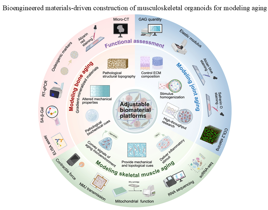
Age-related deterioration of the musculoskeletal (MSK) system drives loss of mobility, lower quality of life, and escalates healthcare costs in older adults. Organoids are three-dimensional, self-organizing constructs that recapitulate key aspects of tissue architecture and function, providing a promising platform to model human MSK aging with higher physiological relevance than conventional two-dimensional cultures and many animal models. Current reliance on decellularized extracellular matrices as scaffolds constrains reproducibility and limits the ability to tune biochemical and biophysical cues. In contrast, engineered matrices allow for precise control over composition, mechanics, and degradability, thereby supporting organoid formation, maturation, and the induction of aging-related phenotypes. This review specifically focuses on MSK organoids and presents a conceptual synthesis linking biomaterial parameters to core MSK aging hallmarks and functional validation assays. We synthesize current strategies for constructing aging MSK organoids and delineate biomaterials design principles to recapitulate aged MSK microenvironments. We examine the key structural, mechanical, and biochemical material properties that influence organoid formation and the establishment of an aging-related microenvironment. Finally, we discuss smart material platforms and strategies for multi-tissue integration, assessing their potential to facilitate the exploration of mechanistic insights and therapeutic testing, as well as to enhance the accuracy and translational relevance of in vitro aging models.
- Cruz-Jentoft AJ, Sayer AA. Sarcopenia. The Lancet. 2019;393(10191):2636-2646. doi: 10.1016/S0140-6736(19)31138-9
- Liu Y, Zhou Z, Lu G, et al. Musculoskeletal organoids: An emerging toolkit for establishing personalized models of musculoskeletal disorders and developing regenerative therapies. Acta Biomater. 2025;200:158-186. doi: 10.1016/j.actbio.2025.05.037
- Yilmaz D, Mathavan N, Wehrle E, Kuhn GA, Muller R. Mouse models of accelerated aging in musculoskeletal research for assessing frailty, sarcopenia, and osteoporosis - A review. Ageing Res Rev. 2024;93:102118. doi: 10.1016/j.arr.2023.102118
- Pitrez PR, Monteiro LM, Borgogno O, Nissan X, Mertens J, Ferreira L. Cellular reprogramming as a tool to model human aging in a dish. Nat Commun. 2024;15(1):1816. doi: 10.1038/s41467-024-46004-5
- Jothi D, Kulka LAM. Strategies for modeling aging and age-related diseases. NPJ Aging. 2024;10(1):32. doi: 10.1038/s41514-024-00161-5
- Fischer NG. Extracellular matrix guides the fate of organoids. Organoid Res. 2025;1(3) doi: 10.36922/or025360028
- Hu JL, Todhunter ME, LaBarge MA, Gartner ZJ. Opportunities for organoids as new models of aging. J. Cell Biol. 2017;217(1):39-50. doi: 10.1083/jcb.201709054
- Torrens-Mas M, Perelló-Reus C, Navas-Enamorado C, et al. Organoids: An Emerging Tool to Study Aging Signature across Human Tissues. Modeling Aging with Patient- Derived Organoids. Int. J. Mol. Sci. 2021;22(19):10547. doi: 10.3390/ijms221910547
- Chen W, Liu D, Lu K, et al. Organoids of Musculoskeletal System for Disease Modeling, Drug Screening, and Regeneration. Adv. Healthc. Mater. 2025;14(9):2402444. doi: 10.1002/adhm.202402444
- Roberts S, Colombier P, Sowman A, et al. Ageing in the musculoskeletal system. Acta Orthop. 2016;87(sup363):15-25. doi: 10.1080/17453674.2016.1244750
- López-Otín C, Blasco MA, Partridge L, Serrano M, Kroemer G. The hallmarks of aging. Cell. 2013;153(6):1194-217. doi: 10.1016/j.cell.2013.05.039
- Li K, Hu S, Chen H. Cellular senescence and other age-related mechanisms in skeletal diseases. Bone Res. 2025;13(1):68. doi: 10.1038/s41413-025-00448-7
- Krenning L, Feringa Femke M, Shaltiel Indra A, van den Berg J, Medema René H. Transient Activation of p53 in G2 Phase Is Sufficient to Induce Senescence. Mol. Cell. 2014;55(1):59-72. doi: 10.1016/j.molcel.2014.05.007
- Mijit M, Caracciolo V, Melillo A, Amicarelli F, Giordano A. Role of p53 in the Regulation of Cellular Senescence. Biomolecules. 2020;10(3) doi: 10.3390/biom10030420
- Xu X, Pang Y, Fan X. Mitochondria in oxidative stress, inflammation and aging: from mechanisms to therapeutic advances. Signal Transduct. Target. Ther. 2025;10(1):190. doi: 10.1038/s41392-025-02253-4
- Guo C, Sun L, Chen X, Zhang D. Oxidative stress, mitochondrial damage and neurodegenerative diseases. Neural Regen Res. 2013;8(21):2003-14. doi: 10.3969/j.issn.1673-5374.2013.21.009
- Garcia-Castorena JM, Riester R, Gamino-Ornelas M, Ada N, Guilak F, Danalache M. PIEZO1-mediated calcium influx transiently alters nuclear mechanical properties via actin remodeling in chondrocytes. Biochem Biophys Res Commun. 2025;742:151135. doi: 10.1016/j.bbrc.2024.151135
- Liu Y, Zhang Z, Li T, Xu H, Zhang H. Senescence in osteoarthritis: from mechanism to potential treatment. Arthritis Res Ther. 2022;24(1):174. doi: 10.1186/s13075-022-02859-x
- Diekman BO, Loeser RF. Aging and the emerging role of cellular senescence in osteoarthritis. Osteoarthritis and Cartilage. 2024;32(4):365-371. doi: 10.1016/j.joca.2023.11.018
- Callaway DA, Jiang JX. Reactive oxygen species and oxidative stress in osteoclastogenesis, skeletal aging and bone diseases. J Bone Miner Metab. 2015;33(4):359-70. doi: 10.1007/s00774-015-0656-4
- Dao T, Green AE, Kim YA, et al. Sarcopenia and Muscle Aging: A Brief Overview. Endocrinol Metab (Seoul). 2020;35(4):716-732. doi: 10.3803/EnM.2020.405
- Chen Y, Wu J. Aging-Related Sarcopenia: Metabolic Characteristics and Therapeutic Strategies. Aging Dis. 2024;16(2):1003-1022. doi: 10.14336/AD.2024.0407
- Hu W, Chen S, Zou X, et al. Oral microbiome, periodontal disease and systemic bone-related diseases in the era of homeostatic medicine. J. Adv. Res. 2025;73:443-458. doi: 10.1016/j.jare.2024.08.019
- García-Prat L, Sousa-Victor P, Muñoz-Cánoves P. Functional dysregulation of stem cells during aging: a focus on skeletal muscle stem cells. FEBS J. 2013;280(17):4051-4062. doi: 10.1111/febs.12221
- Tylutka A, Walas Ł, Zembron-Lacny A. Level of IL-6, TNF, and IL-1β and age-related diseases: a systematic review and meta-analysis. Front Immunol. 2024;15:1330386. doi: 10.3389/fimmu.2024.1330386
- Kwan KYC, Ng KWK, Rao Y, et al. Effect of Aging on Tendon Biology, Biomechanics and Implications for Treatment Approaches. Int J Mol Sci. 2023;24(20). doi: 10.3390/ijms242015183
- Snedeker JG, Gautieri A. The role of collagen crosslinks in ageing and diabetes - the good, the bad, and the ugly. Muscle Ligaments Tendons J. 2019;04(03):303. doi: 10.32098/mltj.03.2014.07
- Carvalho MS, Alves L, Bogalho I, Cabral JMS, da Silva CL. Impact of Donor Age on the Osteogenic Supportive Capacity of Mesenchymal Stromal Cell-Derived Extracellular Matrix. Front Cell Dev Biol. 2021;9:747521. doi: 10.3389/fcell.2021.747521
- Pääsuke R, Eimre M, Piirsoo A, et al. Proliferation of Human Primary Myoblasts Is Associated with Altered Energy Metabolism in Dependence on Ageing In Vivo and In Vitro. Oxid Med Cell Longev. 2016:8296150. doi: 10.1155/2016/8296150
- Caobi A, Dutta RK, Garbinski LD, et al. The Impact of CRISPR-Cas9 on Age-related Disorders: From Pathology to Therapy. Aging Dis. 2020;11(4):895-915. doi: 10.14336/ad.2019.0927
- Lin Z, Li Z, Li EN, et al. Osteochondral Tissue Chip Derived From iPSCs: Modeling OA Pathologies and Testing Drugs. Front Bioeng Biotechnol. 2019;7:411. doi: 10.3389/fbioe.2019.00411
- Menche C, Farin HF. Strategies for genetic manipulation of adult stem cell-derived organoids. Exp Mol Med. 2021;53(10):1483-1494. doi: 10.1038/s12276-021-00609-8
- Qin H, Xu F, Li J, Ding Y. Construction of gelatin-alginate scaffolds containing chondrocytes using 3D bioprinting technology for the study of in vitro cartilage senescence. Int. J. Bioprinting. 2025;0(0) doi: 10.36922/ijb025150136
- Herr LM, Schaffer ED, Fuchs KF, Datta A, Brosh RM. Replication stress as a driver of cellular senescence and aging. Commun. Biol. 2024;7(1):616. doi: 10.1038/s42003-024-06263-w
- Rajabian N, Shahini A, Asmani M, et al. Bioengineered Skeletal Muscle as a Model of Muscle Aging and Regeneration. Tissue Eng Part A. 2021;27(1-2):74-86. doi: 10.1089/ten.TEA.2020.0005
- Mitra SK, Schlaepfer DD. Integrin-regulated FAK-Src signaling in normal and cancer cells. Curr Opin Cell Biol. 2006;18(5):516-23. doi: 10.1016/j.ceb.2006.08.011
- Lai W, Geliang H, Bin X, Wang W. Effects of hydrogel stiffness and viscoelasticity on organoid culture: a comprehensive review. Mol Med. 2025;31(1):83. doi: 10.1186/s10020-025-01131-7
- Donges L, Damle A, Mainardi A, et al. Engineered human osteoarthritic cartilage organoids. Biomaterials. 2024;308:122549. doi: 10.1016/j.biomaterials.2024.122549
- Mu X, Tseng C, Hambright WS, et al. Cytoskeleton stiffness regulates cellular senescence and innate immune response in Hutchinson–Gilford Progeria Syndrome. Aging Cell. 2020;19(8):e13152. doi: 10.1111/acel.13152
- Hussain Z, Mehmood S, Liu X, Liu Y, Wang G, Pei R. Decoding bone-inspired and cell-instructive cues of scaffolds for bone tissue engineering. Engineered Regeneration. 2024;5(1):21-44. doi: 10.1016/j.engreg.2023.10.003
- Wang X, Ayati BP, Brouillete MJ, Graham JM, Ramakrishnan PS, Martin JA. Modeling and simulation of the effects of cyclic loading on articular cartilage lesion formation. Int J Numer Method Biomed Eng. 2014;30(10):927-41. doi: 10.1002/cnm.2636
- Li B, Ming H, Qin S, et al. Redox regulation: mechanisms, biology and therapeutic targets in diseases. Signal Transduct. Target. Ther. 2025;10(1):72. doi: 10.1038/s41392-024-02095-6
- Pantelis P, Theocharous G, Lagopati N, et al. The Dual Role of Oxidative-Stress-Induced Autophagy in Cellular Senescence: Comprehension and Therapeutic Approaches. Antioxidants. 2023;12(1):169. doi: 10.3390/antiox12010169
- Yang YK. Aging of mesenchymal stem cells: Implication in regenerative medicine. Regen Ther. 2018;9:120-122. doi: 10.1016/j.reth.2018.09.002
- Zupan J, Strazar K, Kocijan R, Nau T, Grillari J, Marolt Presen D. Age-related alterations and senescence of mesenchymal stromal cells: Implications for regenerative treatments of bones and joints. Mech Ageing Dev. 2021;198:111539. doi: 10.1016/j.mad.2021.111539
- Molla A. Environmental influences on genetic aging processes: Experimental evidence from model systems. Eur J Clin Biomed Sci. 2025;11(4):49-59. doi: 10.11648/j.ejcbs.20251104.12
- Ryan P, Lee J. In vitro senescence and senolytic functional assays. Biomater. Sci. 2025;13(13):3509-3531. doi: 10.1039/D4BM01684J
- Rossi M, Abdelmohsen K. The Emergence of Senescent Surface Biomarkers as Senotherapeutic Targets. Cells. 2021;10(7). doi: 10.3390/cells10071740
- Wu G, Li S, Qu G, et al. Genistein alleviates H(2)O(2)- induced senescence of human umbilical vein endothelial cells via regulating the TXNIP/NLRP3 axis. Pharm Biol. 2021;59(1):1388-1401. doi: 10.1080/13880209.2021.1979052
- Coryell PR, Diekman BO, Loeser RF. Mechanisms and therapeutic implications of cellular senescence in osteoarthritis. Nat. Rev. Rheumatol. 2021;17(1):47-57. doi: 10.1038/s41584-020-00533-7
- Salameh Y, Bejaoui Y, El Hajj N. DNA Methylation Biomarkers in Aging and Age-Related Diseases. Front Genet. 2020;11:171. doi: 10.3389/fgene.2020.00171
- Liang R, Tang Q, Chen J, Zhu L. Epigenetic Clocks: Beyond Biological Age, Using the Past to Predict the Present and Future. Aging Dis. 2024;16(6):3520-3545. doi: 10.14336/ad.2024.1495
- He X, Hu W, Zhang Y, et al. Cellular senescence in skeletal disease: mechanisms and treatment. Cell. Mol. Biol. Lett. 2023;28(1):88. doi: 10.1186/s11658-023-00501-5
- Kong Y, Yang Y, Hou Y, Wang Y, Li W, Song Y. Advance in the application of organoids in bone diseases. Front Cell Dev Biol. 2024;12:1459891. doi: 10.3389/fcell.2024.1459891
- Zhang T, Ma L, Ling S, et al. Protocol for measuring the Young’s modulus of organoids using atomic force microscopy. STAR Protoc. 2025;6(2):103825. doi: 10.1016/j.xpro.2025.103825
- Lin W, Wang M, Xu L, Tortorella M, Li G. Cartilage organoids for cartilage development and cartilage-associated disease modeling. Front Cell Dev Biol. 2023;11:1125405. doi: 10.3389/fcell.2023.1125405
- Xiu J, Xue R, Duan X, et al. Mechanical characterization of nonlinear elasticity of growing intestinal organoids with a microinjection method. Acta Biomater. 2025;196:271-280. doi: 10.1016/j.actbio.2025.02.054
- Schini M, Vilaca T, Gossiel F, Salam S, Eastell R. Bone Turnover Markers: Basic Biology to Clinical Applications. Endocr. Rev. 2022;44(3):417-473. doi: 10.1210/endrev/bnac031
- Nagy EE, Nagy-Finna C, Popoviciu H, Kovács B. Soluble Biomarkers of Osteoporosis and Osteoarthritis, from Pathway Mapping to Clinical Trials: An Update. Clin Interv Aging. 2020;15:501-518. doi: 10.2147/cia.S242288
- Jeffries AM, Yu T, Ziegenfuss JS, et al. Single-cell transcriptomic and genomic changes in the ageing human brain. Nature. 2025;646:657-666. doi: 10.1038/s41586-025-09435-8
- Zhang T, Yuan X, Jiang M, et al. Proteomic analysis reveals the aging-related pathways contribute to pulmonary fibrogenesis. Aging (Albany NY). 2023;15(24):15382-15401. doi: 10.18632/aging.205355
- Oh HS-H, Rutledge J, Nachun D, et al. Organ aging signatures in the plasma proteome track health and disease. Nature. 2023;624(7990):164-172. doi: 10.1038/s41586-023-06802-1
- Cerantonio A, Greco BM, Citrigno L, et al. Epigenetic Clocks and Their Prospective Application in the Complex Landscape of Aging and Alzheimer’s Disease. Genes (Basel). 2025;16(6). doi: 10.3390/genes16060679
- Ecker S, Beck S. The epigenetic clock: a molecular crystal ball for human aging? Aging (Albany NY). 2019;11(2):833-835. doi: 10.18632/aging.101712
- Sprio S, Panseri S, Montesi M, et al. Hierarchical porosity inherited by natural sources affects the mechanical and biological behaviour of bone scaffolds. J. Eur. Ceram. Soc. 2020;40(4):1717-1727. doi: 10.1016/j.jeurceramsoc.2019.11.015.
- Sun J, Wang Q, Cai D, et al. A lattice topology optimization of cervical interbody fusion cage and finite element comparison with ZK60 and Ti-6Al-4V cages. BMC Musculoskelet. Disord. 2021;22(1):390. doi: 10.1186/s12891-021-04244-2
- Kedlian VR, Wang Y, Liu T, et al. Human skeletal muscle aging atlas. Nat. Aging. 2024;4(5):727-744. doi: 10.1038/s43587-024-00613-3
- Mavrogonatou E, Papadopoulou A, Pratsinis H, Kletsas D. Senescence-associated alterations in the extracellular matrix: deciphering their role in the regulation of cellular function. Am J Physiol Cell Physiol. 2023;325(3):C633-c647. doi: 10.1152/ajpcell.00178.2023
- Chen Y, Sun W, Wen Y, et al. A cationic polymer drives glycosaminoglycan assembly and secretion for preclinical osteoarthritis therapy. Sci. Transl. Med. 2025;17(804):eadl5623. doi: 10.1126/scitranslmed.adl5623
- Neves MI, Araújo M, Moroni L, da Silva RMP, Barrias CC. Glycosaminoglycan-Inspired Biomaterials for the Development of Bioactive Hydrogel Networks. Molecules. 2020;25(4):978. doi: 10.3390/molecules25040978
- Alexopoulos LG, Setton LA, Guilak F. The biomechanical role of the chondrocyte pericellular matrix in articular cartilage. Acta Biomater. 2005;1(3):317-325. doi: 10.1016/j.actbio.2005.02.001
- Danalache M, Umrath F, Riester R, Schwitalle M, Guilak F, Hofmann UK. Proteolysis of the pericellular matrix: Pinpointing the role and involvement of matrix metalloproteinases in early osteoarthritic remodeling. Acta Biomater. 2024;181:297-307. doi: 10.1016/j.actbio.2024.05.002
- Tiede-Lewis LM, Dallas SL. Changes in the osteocyte lacunocanalicular network with aging. Bone. 2019;122:101-113. doi: 10.1016/j.bone.2019.01.025
- Álvarez-Rodríguez L, López-Hoyos M, Muñoz-Cacho P, Martínez-Taboada VM. Aging is associated with circulating cytokine dysregulation. Cell. Immunol. 2012;273(2):124-132. doi: 10.1016/j.cellimm.2012.01.001
- Guo J, Huang X, Dou L, et al. Aging and aging-related diseases: from molecular mechanisms to interventions and treatments. Signal Transduct. Target. Ther. 2022;7(1):391. doi: 10.1038/s41392-022-01251-0
- Fu Y, Wang B, Alu A, et al. Immunosenescence: signaling pathways, diseases and therapeutic targets. Signal Transduct. Target. Ther. 2025;10(1):250. doi: 10.1038/s41392-025-02371-z
- Wagatsuma A. Effect of aging on expression of angiogenesis-related factors in mouse skeletal muscle. Exp. Gerontol. 2006;41(1):49-54. doi: 10.1016/j.exger.2005.10.003
- Farooq M, Khan AW, Kim MS, Choi S. The Role of Fibroblast Growth Factor (FGF) Signaling in Tissue Repair and Regeneration. Cells. 2021;10(11). doi: 10.3390/cells10113242
- Stojanovic B, Jovanovic I, Dimitrijevic Stojanovic M, et al. Oxidative Stress-Driven Cellular Senescence: Mechanistic Crosstalk and Therapeutic Horizons. Antioxidants. 2025;14(8):987. doi: 10.3390/antiox14080987
- Wang S, Guan X, Sun S. Microfluidic Biosensors: Enabling Advanced Disease Detection. Sensors. 2025;25(6):1936. doi: 10.3390/s25061936
- Zhu L, Perche F, Wang T, Torchilin VP. Matrix metalloproteinase 2-sensitive multifunctional polymeric micelles for tumor-specific co-delivery of siRNA and hydrophobic drugs. Biomaterials. 2014;35(13):4213-4222. doi: 10.1016/j.biomaterials.2014.01.060
- Guzmán Rodríguez A, Sablón Carrazana M, Rodríguez Tanty C, Malessy MJA, Fuentes G, Cruz LJ. Smart Polymeric Micelles for Anticancer Hydrophobic Drugs. Cancers. 2023;15(1):4. doi: 10.3390/cancers15010004
- Sun Z, Hou X, Zhang J, et al. Diagnostic and Therapeutic Roles of Extracellular Vesicles in Aging-Related Diseases. Oxid Med Cell Longev. 2022;2022:6742792. doi: 10.1155/2022/6742792
- Cole MA, Quan T, Voorhees JJ, Fisher GJ. Extracellular matrix regulation of fibroblast function: redefining our perspective on skin aging. J Cell Commun Signal. 2018;12(1):35-43. doi: 10.1007/s12079-018-0459-1
- Solomonov I, Kollet O, Sagi I. Extracellular matrix and proteolysis: mechanisms driving irreversible changes and shaping cell behavior. FEBS J. 2025. doi: 10.1111/febs.70292.
- Adair-Kirk TL, Senior RM. Fragments of extracellular matrix as mediators of inflammation. Int J Biochem Cell Biol. 2008;40(6-7):1101-10. doi: 10.1016/j.biocel.2007.12.005
- Park JH, Jo SB, Lee JH, Lee HH, Knowles JC, Kim HW. Materials and extracellular matrix rigidity highlighted in tissue damages and diseases: Implication for biomaterials design and therapeutic targets. Bioact Mater. 2023;20:381-403. doi: 10.1016/j.bioactmat.2022.06.003
- Song J, Zeng X, Li C, Yin H, Mao S, Ren D. Alteration in cartilage matrix stiffness as an indicator and modulator of osteoarthritis. Biosci. Rep. 2024;44(1). doi: 10.1042/BSR20231730
- Krakowski P, Rejniak A, Sobczyk J, Karpiński R. Cartilage Integrity: A Review of Mechanical and Frictional Properties and Repair Approaches in Osteoarthritis. Healthcare (Basel). 2024;12(16). doi: 10.3390/healthcare12161648
- Loeser RF. Aging and osteoarthritis: the role of chondrocyte senescence and aging changes in the cartilage matrix. Osteoarthritis Cartilage. 2009;17(8):971-979. doi: 10.1016/j.joca.2009.03.002
- Fu B, Shen J, Zou X, et al. Matrix stiffening promotes chondrocyte senescence and the osteoarthritis development through downregulating HDAC3. Bone Res. 2024;12(1):32. doi: 10.1038/s41413-024-00333-9
- McHugh J. Ageing matrix makes chondrocytes feel old. Nat. Rev. Rheumatol. 2023;19(3):127-127. doi: 10.1038/s41584-023-00928-2
- Iijima H, Gilmer G, Wang K, et al. Age-related matrix stiffening epigenetically regulates alpha-Klotho expression and compromises chondrocyte integrity. Nat Commun. 2023;14(1):18. doi: 10.1038/s41467-022-35359-2
- Zhang Q, Yan K, Zheng X, Liu Q, Han Y, Liu Z. Research progress of photo-crosslink hydrogels in ophthalmology: A comprehensive review focus on the applications. Mater. Today Bio. 2024;26:101082. doi: 10.1016/j.mtbio.2024.101082
- Wu DT, Jeffreys N, Diba M, Mooney DJ. Viscoelastic Biomaterials for Tissue Regeneration. Tissue Eng Part C Methods. 2022;28(7):289-300.
- Chaudhuri O, Gu L, Klumpers D, et al. Hydrogels with tunable stress relaxation regulate stem cell fate and activity. Nat. Mater. 2016;15(3):326-334. doi: 10.1038/nmat4489
- Chaudhuri O, Gu L, Darnell M, et al. Substrate stress relaxation regulates cell spreading. Nat. Commun. 2015;6(1):6365. doi: 10.1038/ncomms7365
- Shi H, Zhou K, Wang M, et al. Integrating physicomechanical and biological strategies for BTE: biomaterials-induced osteogenic differentiation of MSCs. Theranostics. 2023;13(10):3245-3275. doi: 10.7150/thno.84759
- Wu Y, Song Y, Soto J, et al. Viscoelastic extracellular matrix enhances epigenetic remodeling and cellular plasticity. Nat Commun. 2025;16(1):4054. doi: 10.1038/s41467-025-59190-7
- Cameron AR, Frith JE, Cooper-White JJ. The influence of substrate creep on mesenchymal stem cell behaviour and phenotype. Biomaterials. 2011;32(26):5979-5993. doi: 10.1016/j.biomaterials.2011.04.003
- Qiao E, Fulmore CA, Schaffer DV, Kumar S. Substrate stress relaxation regulates neural stem cell fate commitment. Proc Natl Acad Sci USA. 2024;121(28):e2317711121. doi: 10.1073/pnas.2317711121
- Wei Q, Wang S, Han F, et al. Cellular modulation by the mechanical cues from biomaterials for tissue engineering. Biomater Transl. 2021;2(4):323-342. doi: 10.12336/biomatertransl.2021.04.001
- Kratochvil MJ, Seymour AJ, Li TL, Pasca SP, Kuo CJ, Heilshorn SC. Engineered materials for organoid systems. Nat Rev Mater. 2019;4(9):606-622. doi: 10.1038/s41578-019-0129-9
- Hayward M-K, Muncie JM, Weaver VM. Tissue mechanics in stem cell fate, development, and cancer. Dev. Cell. 2021;56(13):1833-1847. doi: 10.1016/j.devcel.2021.05.011
- Kular JK, Basu S, Sharma RI. The extracellular matrix: Structure, composition, age-related differences, tools for analysis and applications for tissue engineering. J Tissue Eng. 2014;5:2041731414557112. doi: 10.1177/2041731414557112
- Wen W, Luo R, Tang X, et al. Age-related progressionof arterial stiffness and its elevated positive association with blood pressure in healthy people. Atherosclerosis. 2015;238(1):147-152. doi: 10.1016/j.atherosclerosis.2014.10.089
- Bružauskaitė I, Bironaitė D, Bagdonas E, Bernotienė E. Scaffolds and cells for tissue regeneration: different scaffold pore sizes-different cell effects. Cytotechnology. 2016;68(3):355-69. doi: 10.1007/s10616-015-9895-4
- Porrelli D, Abrami M, Pelizzo P, et al. Trabecular bone porosity and pore size distribution in osteoporotic patients – A low field nuclear magnetic resonance and microcomputed tomography investigation. J. Mech. Behav. Biomed. Mater. 2022;125:104933. doi: 10.1016/j.jmbbm.2021.104933
- Morgan EF, Unnikrisnan GU, Hussein AI. Bone Mechanical Properties in Healthy and Diseased States. Annu Rev Biomed Eng. 2018;20:119-143. doi: 10.1146/annurev-bioeng-062117-121139
- Becerra J, Andrades JA, Guerado E, Zamora-Navas P, López- Puertas JM, Reddi AH. Articular Cartilage: Structure and Regeneration. Tissue Eng. Part B Rev. 2010;16(6):617-627. doi: 10.1089/ten.teb.2010.0191
- Abate M, Schiavone C, Salini V, Andia I. Occurrence of tendon pathologies in metabolic disorders. Rheumatology (Oxford, England). 2013;52:599-608. doi: 10.1093/rheumatology/kes395
- Xu Y, Murrell GA. The basic science of tendinopathy. Clin Orthop Relat Res. 2008;466(7):1528-38. doi: 10.1007/s11999-008-0286-4
- Karageorgiou V, Kaplan D. Porosity of 3D biomaterial scaffolds and osteogenesis. Biomaterials. 2005;26(27):5474-5491. doi: 10.1016/j.biomaterials.2005.02.002
- Li L, Wang P, Liang H, et al. Design of a Haversian system-like gradient porous scaffold based on triply periodic minimal surfaces for promoting bone regeneration. J. Adv. Res. 2023;54:89-104. doi: 10.1016/j.jare.2023.01.004
- Luo J, Walker M, Xiao Y, Donnelly H, Dalby MJ, Salmeron- Sanchez M. The influence of nanotopography on cell behaviour through interactions with the extracellular matrix – A review. Bioact. Mater. 2022;15:145-159. doi: 10.1016/j.bioactmat.2021.11.024
- Phillip JM, Aifuwa I, Walston J, Wirtz D. The Mechanobiology of Aging. Annu Rev Biomed Eng. 2015;17:113-141. doi: 10.1146/annurev-bioeng-071114-040829
- Tan YH, Alcazar-Daleo CA, Holbrook JG, et al. Shear- Induced Patterning of Decellularized Skeletal Muscle Extracellular Matrix for Enhanced Myogenesis. Adv Healthc Mater. 2025:e2501357. doi: 10.1002/adhm.202501357
- Quan T, Fisher GJ. Role of Age-Associated Alterations of the Dermal Extracellular Matrix Microenvironment in Human Skin Aging: A Mini-Review. Gerontology. 2015;61(5):427-34. doi: 10.1159/000371708
- Mu Q, Cui K, Wang ZJ, et al. Force-triggered rapid microstructure growth on hydrogel surface for on-demand functions. Nat. Commun. 2022;13(1):6213. doi: 10.1038/s41467-022-34044-8
- Lee HJ, Kim JH. Classification of shoulder diseases in older adult patients: a narrative review. Ewha Med J. 2025;48(1):e5. doi: 10.12771/emj.2025.e5
- Zumstein MA, Künzler M, Hatta T, Galatz LM, Itoi E. Rotator cuff pathology: state of the art. J. ISAKOS. 2017;2(4):213-221. doi: 10.1136/jisakos-2016-000074 122. Buckwalter JA. Aging and Degeneration of the Human Intervertebral Disc. Spine. 1995;20(11):1307-1314. doi: 10.1097/00007632-199506000-00022
- Mebratu YA, Soni S, Rosas L, Rojas M, Horowitz JC, Nho R. The aged extracellular matrix and the profibrotic role of senescence-associated secretory phenotype. Am J Physiol Cell Physiol. 2023;325(3):C565-C579. doi: 10.1152/ajpcell.00124.2023
- Iordachescu A, Hughes EAB, Joseph S, Hill EJ, Grover LM, Metcalfe AD. Trabecular bone organoids: a micron-scale ‘humanised’ prototype designed to study the effects of microgravity and degeneration. npj Microgravity. 2021;7(1):17. doi: 10.1038/s41526-021-00146-8
- Park Y, Cheong E, Kwak J-G, Carpenter R, Shim J-H, Lee J. Trabecular bone organoid model for studying the regulation of localized bone remodeling. Sci. Adv. 2021;7(4):eabd6495. doi: 10.1126/sciadv.abd6495
- Zhang Y, Fang Q, Peng Y, et al. Establishment and characterization of an inflammatory cartilaginous organoids model for organoid transplantation study. J Orthop Translat. 2025;52:376-386. doi: 10.1016/j.jot.2025.05.002
- Boone I, Houtman E, Tuerlings M, et al. Development of Reliable and High-Throughput Human Biomimetic Cartilage and Bone Models to Explore Senescence and Personalized Osteoarthritis Treatment Options. J Orthop Res. 2025;43(5):912-921. doi: 10.1002/jor.26052
- Occhetta P, Mainardi A, Votta E, et al. Hyperphysiological compression of articular cartilage induces an osteoarthritic phenotype in a cartilage-on-a-chip model. Nat. Biomed. Eng. 2019;3(7):545-557. doi: 10.1038/s41551-019-0406-3
- Rosser J, Bachmann B, Jordan C, et al. Microfluidic nutrient gradient–based three-dimensional chondrocyte culture-on-a-chip as an in vitro equine arthritis model. Mater. Today Bio. 2019;4:100023. doi: 10.1016/j.mtbio.2019.100023
- Mondadori C, Visone R, Rasponi M, Redaelli A, Moretti M, Lopa S. Development of an organotypic microfluidic model to reproduce monocyte extravasation process in the osteoarthritic joint. Osteoarthritis Cartilage. 2018;26:S122. doi: 10.1016/j.joca.2018.02.267
- Lin H, Lozito TP, Alexander PG, Gottardi R, Tuan RS. Stem Cell-Based Microphysiological Osteochondral System to Model Tissue Response to Interleukin-1β. Mol. Pharm. 2014;11(7):2203-2212. doi: 10.1021/mp500136b
- Sun Y, Wu Q, Dai K, You Y, Jiang W. Generating 3D-cultured organoids for pre-clinical modeling and treatment of degenerative joint disease. Signal Transduct Target Ther. 2021;6(1):380. doi: 10.1038/s41392-021-00675-4
- Agrawal G, Aung A, Varghese S. Skeletal muscle-on-a-chip: an in vitro model to evaluate tissue formation and injury. Lab Chip. 2017;17(20):3447-3461. doi: 10.1039/c7lc00512a
- Park S, Shin MK, Jeong DS, et al. Human Pluripotent Stem Cell-Derived Skeletal Muscle Organoid Model of Aging-Induced Sarcopenia. J Cachexia Sarcopenia Muscle. 2025;16(4):e70045. doi: 10.1002/jcsm.70045
- Wang K, Smith SH, Iijima H, et al. Bioengineered 3D Skeletal Muscle Model Reveals Complement 4b as a Cell- Autonomous Mechanism of Impaired Regeneration with Aging. Adv Mater. 2023;35(17):e2207443. doi: 10.1002/adma.202207443
- Mestre R, Garcia N, Patino T, et al. 3D-bioengineered model of human skeletal muscle tissue with phenotypic features of aging for drug testing purposes. Biofabrication. 2021;13(4):045011. doi: 10.1088/1758-5090/ac165b
- Mondrinos MJ, Alisafaei F, Yi AY, et al. Surface-directed engineering of tissue anisotropy in microphysiological models of musculoskeletal tissue. Sci. Adv. 2021;7(11):eabe9446. doi: 10.1126/sciadv.abe9446
- Gillies AR, Lieber RL. Structure and function of the skeletal muscle extracellular matrix. Muscle Nerve. 2011;44(3):318-31. doi: 10.1002/mus.22094
- Sokos D, Everts V, de Vries TJ. Role of periodontal ligament fibroblasts in osteoclastogenesis: a review. J Periodontal Res. 2015;50(2):152-9. doi: 10.1111/jre.12197
- Kuroda S, Tanimoto K, Izawa T, Fujihara S, Koolstra JH, Tanaka E. Biomechanical and biochemical characteristics of the mandibular condylar cartilage. Osteoarthritis Cartilage. 2009;17(11):1408-15. doi: 10.1016/j.joca.2009.04.025
- Klein Y, Fleissig O, Polak D, Barenholz Y, Mandelboim O, Chaushu S. Immunorthodontics: in vivo gene expression of orthodontic tooth movement. Sci. Rep. 2020;10(1):8172. doi: 10.1038/s41598-020-65089-8
- Zhou Y, Nishiura A, Morikuni H, et al. RANKL(+) senescent cells under mechanical stress: a therapeutic target for orthodontic root resorption using senolytics. Int J Oral Sci. 2023;15(1):20. doi: 10.1038/s41368-023-00228-1
- Chen H, Zhou X, Fujita H, Onozuka M, Kubo KY. Age-related changes in trabecular and cortical bone microstructure. Int J Endocrinol. 2013;2013:1-9. doi: 10.1155/2013/213234
- Zhang L, Guan Q, Wang Z, Feng J, Zou J, Gao B. Consequences of Aging on Bone. Aging dis. 2023;15(6):2417-2452. doi: 10.14336/AD.2023.1115
- Chandra A, Rajawat J. Skeletal Aging and Osteoporosis: Mechanisms and Therapeutics. Int. J. Mol. Sci. 2021;22(7):3553. doi: 10.3390/ijms22073553
- Hong Y, Li R, Sheng S, Zhou F, Bai L, Su J. Bone organoid construction and evolution. J. Orthop. Translat. 2025;53:260-273. doi: 10.1016/j.jot.2025.06.011
- Chen S, Chen X, Geng Z, Su J. The horizon of bone organoid: A perspective on construction and application. Bioact. Mater. 2022;18:15-25. doi: 10.1016/j.bioactmat.2022.01.048
- Li X, Xie J, Lipner J, Yuan X, Thomopoulos S, Xia Y. Nanofiber Scaffolds with Gradations in Mineral Content for Mimicking the Tendon-to-Bone Insertion Site. Nano Lett. 2009;9(7):2763-2768. doi: 10.1021/nl901582f
- Liu W, Lipner J, Xie J, Manning CN, Thomopoulos S, Xia Y. Nanofiber Scaffolds with Gradients in Mineral Content for Spatial Control of Osteogenesis. ACS Appl. Mater. Interfaces. 2014;6(4):2842-2849. doi: 10.1021/am405418g
- Chen Y, Hao M, Bousso I, Thomopoulos S, Xia Y. Reliable Fabrication of Mineral-Graded Scaffolds by Spin-Coating and Laser Machining for Use in Tendon-to-Bone Insertion Repair. Adv. Healthc. Mater. 2024;13(31):2402531. doi: 10.1002/adhm.202402531
- Qiu J, Ahn J, Qin D, Thomopoulos S, Xia Y. Biomimetic Scaffolds with a Mineral Gradient and Funnel-Shaped Channels for Spatially Controllable Osteogenesis. Adv. Healthc. Mater. 2022;11(9):2100828. doi: 10.1002/adhm.202100828
- Boys AJ, Zhou H, Harrod JB, McCorry MC, Estroff LA, Bonassar LJ. Top-Down Fabrication of Spatially Controlled Mineral-Gradient Scaffolds for Interfacial Tissue Engineering. ACS Biomater. Sci. Eng. 2019;5(6):2988-2997. doi: 10.1021/acsbiomaterials.9b00176
- Mahajan A, Zaidi ZS, Shukla A, Saxena R, Katti DS. Functionally graded hydrogels with opposing biochemical cues for osteochondral tissue engineering. Biofabrication. 2024;16(3):035020. doi: 10.1088/1758-5090/ad467e
- Zhang H, Huang H, Hao G, et al. 3D Printing Hydrogel Scaffolds with Nanohydroxyapatite Gradient to Effectively Repair Osteochondral Defects in Rats. Adv. Funct. Mater. 2021;31(1):2006697. doi: 10.1002/adfm.202006697
- Sinha R, Cámara-Torres M, Scopece P, et al. A hybrid additive manufacturing platform to create bulk and surface composition gradients on scaffolds for tissue regeneration. Nat. Commun. 2021;12(1):500. doi: 10.1038/s41467-020-20865-y
- Sun R, Zhang Q, Yu C, et al. Continuous-Gradient Mineralized Hydrogel Synthesized via Gravitational Osmosis for Osteochondral Defect Repair. Adv. Funct. Mater. 2024;34(48):2408249. doi: 10.1002/adfm.202408249
- Zhu C, Pongkitwitoon S, Qiu J, Thomopoulos S, Xia Y. Design and Fabrication of a Hierarchically Structured Scaffold for Tendon-to-Bone Repair. Adv. Mater. 2018;30(16):1707306. doi: 10.1002/adma.201707306
- Zhu C, Qiu J, Pongkitwitoon S, Thomopoulos S, Xia Y. Inverse Opal Scaffolds with Gradations in Mineral Content for Spatial Control of Osteogenesis. Adv. Mater. 2018;30(29):1706706. doi: 10.1002/adma.201706706
- Wang D, Feng S, Yang M. Multi-Gradient Bone-Like Nanocomposites Induced by Strain Distribution. ACS Nano. 2024;18(43):29636-29647. doi: 10.1021/acsnano.4c08442
- Fan Z, Chen Z, Zhang H, Nie Y, Xu S. Gradient Mineralized and Porous Double-Network Hydrogel Effectively Induce the Differentiation of BMSCs into Osteochondral Tissue In Vitro for Potential Application in Cartilage Repair. Macromol. Biosci. 2021;21(3):2000323. doi: 10.1002/mabi.202000323
- de Leeuw AM, Graf R, Lim PJ, et al. Physiological cell bioprinting density in human bone-derived cell-laden scaffolds enhances matrix mineralization rate and stiffness under dynamic loading. Front Bioeng Biotechnol. 2024;12:1310289. doi: 10.3389/fbioe.2024.1310289
- Jimenez-Palomar I, Shipov A, Shahar R, Barber AH. Mechanical Behavior of Osteoporotic Bone at Sub-Lamellar Length Scales. Original Research. Front. Mater. 2015;(2). doi: 10.3389/fmats.2015.00009
- Toni R, Barbaro F, Di Conza G, et al. A bioartificial and vasculomorphic bone matrix-based organoid mimicking microanatomy of flat and short bones. J. Biomed. Mater. Res. B 2024;112(1):e35329. doi: 10.1002/jbm.b.35329
- Blokland Kaj EC, Pouwels Simon D, Schuliga M, Knight Darryl A, Burgess Janette K. Regulation of cellular senescence by extracellular matrix during chronic fibrotic diseases. Clin. Sci. 2020;134(20):2681-2706. doi: 10.1042/CS20190893
- Liu H, Su J. Organoid and organoid extracellular vesicles for osteoporotic fractures therapy: Current status and future perspectives. Interdis. Med. 2023;1(3):e20230011. doi: 10.1002/INMD.20230011
- Huang D, Li Z, Li G, et al. Biomimetic structural design in 3D-printed scaffolds for bone tissue engineering. Mater. Today Bio. 2025;32:101664. doi: 10.1016/j.mtbio.2025.101664
- Madl CM. Accelerating aging with dynamic biomaterials: Recapitulating aged tissue phenotypes in engineered platforms. iScience. 2023;26(6):106825. doi: 10.1016/j.isci.2023.106825
- Wehrle E, Tourolle né Betts DC, Kuhn GA, Scheuren AC, Hofmann S, Müller R. Evaluation of longitudinal time-lapsed in vivo micro-CT for monitoring fracture healing in mouse femur defect models. Sci. Rep. 2019;9(1):17445. doi: 10.1038/s41598-019-53822-x
- Valieva Y, Ivanova E, Fayzullin A, Kurkov A, Igrunkova A. Senescence-Associated β-Galactosidase Detection in Pathology. Diagnostics (Basel). 2022;12(10). doi: 10.3390/diagnostics12102309
- Murab S, Hawk T, Snyder A, Herold S, Totapally M, Whitlock PW. Tissue Engineering Strategies for Treating Avascular Necrosis of the Femoral Head. Bioengineering (Basel). 2021;8(12). doi: 10.3390/bioengineering8120200
- Zhu W, Xu Z, Zhou D, Xu J, He Y, Li ZA. Bioengineering strategies targeting angiogenesis: Innovative solutions for osteonecrosis of the femoral head. J Tissue Eng. 2025;16:20417314241310541. doi: 10.1177/20417314241310541
- Toh WS, Brittberg M, Farr J, et al. Cellular senescence in aging and osteoarthritis. Acta Orthop. 2016;87(sup363):6-14. doi: 10.1080/17453674.2016.1235087
- Xu M, Bradley EW, Weivoda MM, et al. Transplanted Senescent Cells Induce an Osteoarthritis-Like Condition in Mice. J Gerontol A Biol Sci Med Sci. 2017;72(6):780-785. doi: 10.1093/gerona/glw154
- Jeon OH, Kim C, Laberge R-M, et al. Local clearance of senescent cells attenuates the development of post-traumatic osteoarthritis and creates a pro-regenerative environment. Nat. Med. 2017;23(6):775-781. doi: 10.1038/nm.4324
- Makarczyk MJ, Hines S, Yagi H, et al. Using Microphysiological System for the Development of Treatments for Joint Inflammation and Associated Cartilage Loss—A Pilot Study. Biomolecules. 2023;13(2):384. doi: 10.3390/biom13020384
- Singh YP, Moses JC, Bhardwaj N, Mandal BB. Overcoming the Dependence on Animal Models for Osteoarthritis Therapeutics – The Promises and Prospects of In Vitro Models. Adv. Healthc. Mater. 2021;10(20):2100961. doi: 10.1002/adhm.202100961
- Bloks NGC, Dicks A, Harissa Z, et al. Hyper-physiologic mechanical cues, as an osteoarthritis disease-relevant environmental perturbation, cause a critical shift in set points of methylation at transcriptionally active CpG sites in neo-cartilage organoids. Clin. Epigenetics 2024;16(1):64. doi: 10.1186/s13148-024-01676-0
- Bloks NG, Harissa Z, Adkar SS, et al. A high-impact COL6A3 mutation alters the response of chondrocytes in neo-cartilage organoids to hyper-physiologic mechanical loading. bioRxiv. Preprint posted online 2022. doi: 10.1101/2022.12.19.520461
- Guo H, Huang J, Liang Y, Wang D, Zhang H. Focusing on the hypoxia-inducible factor pathway: role, regulation, and therapy for osteoarthritis. Eur J Med Res. 2022;27(1):288. doi: 10.1186/s40001-022-00926-2
- Pérez-García S, Carrión M, Gutiérrez-Cañas I, et al. Profile of Matrix-Remodeling Proteinases in Osteoarthritis: Impact of Fibronectin. Cells. 2019;9(1). doi: 10.3390/cells9010040
- Jiang L, Li L, Geng C, et al. Monosodium iodoacetate induces apoptosis via the mitochondrial pathway involving ROS production and caspase activation in rat chondrocytes in vitro. J. Orthop. Res. 2013;31(3):364-369. doi: 10.1002/jor.22250
- Rothbauer M, Byrne RA, Schobesberger S, et al. Establishment of a human three-dimensional chip-based chondro-synovial co-culture joint model for reciprocal cross-talk studies in arthritis research. bioRxiv. Preprint posted online 2021. doi: 10.1101/2021.02.19.431936
- Jin X, Huang L, Wang X, et al. Synovial organoids: From fundamental construction to groundbreaking applications in arthritic disorders. J. Orthop. Translat. 2025;54:26-36. doi: 10.1016/j.jot.2025.07.004
- Lyu X, Wang J, Su J. Intelligent Manufacturing for Osteoarthritis Organoids. Cell Prolif. 2025;58(7):e70043. doi: 10.1111/cpr.70043
- Lahm A, Mrosek E, Spank H, et al. Changes in content and synthesis of collagen types and proteoglycans in osteoarthritis of the knee joint and comparison of quantitative analysis with Photoshop-based image analysis. Arch Orthop Trauma Surg. 2010;130(4):557-64. doi: 10.1007/s00402-009-0981-y
- Miosge N, Hartmann M, Maelicke C, Herken R. Expression of collagen type I and type II in consecutive stages of human osteoarthritis. Histochem Cell Biol. 2004;122(3):229-36. doi: 10.1007/s00418-004-0697-6
- Nair A, Lin CY, Hsu FC, et al. Categorization of collagen type I and II blend hydrogel using multipolarization SHG imaging with ResNet regression. Sci Rep. 2023;13(1):19534. doi: 10.1038/s41598-023-46417-0
- Vazquez-Portalati NN, Kilmer CE, Panitch A, Liu JC. Characterization of Collagen Type I and II Blended Hydrogels for Articular Cartilage Tissue Engineering. Biomacromolecules. 2016;17(10):3145-3152. doi: 10.1021/acs.biomac.6b00684
- Han L, Zhang ZW, Wang BH, Wen ZK. Construction and biocompatibility of a thin type I/II collagen composite scaffold. Cell Tissue Bank. 2018;19(1):47-59. doi: 10.1007/s10561-017-9653-2
- Gao Y, Li Q, Du Z, et al. HAMA-SBMA hydrogel with anti-inflammatory properties delivers cartilage organoids, boosting cartilage regeneration. J Nanobiotechnology. 2025;23(1):401. doi: 10.1186/s12951-025-03475-y
- Gao Q, Zhang X, Makarcyzk MJ, et al. Macrophage phenotypes modulate neoangiogenesis and fibroblast profiles in synovial-like organoid cultures. Osteoarthritis Cartilage. 2025;33(5):590-600. doi: 10.1016/j.joca.2025.02.777
- Posey KL, Coustry F, Hecht JT. Cartilage oligomeric matrix protein: COMPopathies and beyond. Matrix Biol. 2018;71- 72:161-173. doi: 10.1016/j.matbio.2018.02.023
- Henrotin Y, Gharbi M, Mazzucchelli G, Dubuc JE, De Pauw E, Deberg M. Fibulin 3 peptides Fib3-1 and Fib3-2 are potential biomarkers of osteoarthritis. Arthritis Rheum. 2012;64(7):2260-7. doi: 10.1002/art.34392
- Yan Z, Kavanagh T, Harrabi RDS, et al. FRET Sensor- Modified Synthetic Hydrogels for Real-Time Monitoring of Cell-Derived Matrix Metalloproteinase Activity using Fluorescence Lifetime Imaging. Adv Funct Mater. 2024;34(21). doi: 10.1002/adfm.202309711
- Wang X-h, Liu N, Zhang H, Yin Z-s, Zha Z-G. From cells to organs: progress and potential in cartilaginous organoids research. J. Transl. Med. 2023;21(1):926. doi: 10.1186/s12967-023-04591-9
- Miljkovic N, Lim J-Y, Miljkovic I, Frontera WR. Aging of Skeletal Muscle Fibers. Ann Rehabil Med. 4 2015;39(2):155-162. doi: 10.5535/arm.2015.39.2.155
- Kim SH, Jeong JB, Kang J, et al. Association between sarcopenia level and metabolic syndrome. PLOS ONE. 2021;16(3):e0248856. doi: 10.1371/journal.pone.0248856
- Evans WJ, Campbell WW. Sarcopenia and Age-Related Changes in Body Composition and Functional Capacity1. J Nutr. 1993;123:465-468. doi: 10.1093/jn/123.suppl_2.465
- Prior SJ, Ryan AS, Blumenthal JB, Watson JM, Katzel LI, Goldberg AP. Sarcopenia Is Associated With Lower Skeletal Muscle Capillarization and Exercise Capacity in Older Adults. J. Gerontol. A Biol. Sci. Med. Sci. 2016;71(8):1096-1101. doi: 10.1093/gerona/glw017
- Mann CJ, Perdiguero E, Kharraz Y, et al. Aberrant repair and fibrosis development in skeletal muscle. Skelet. Muscle. 2011;1(1):21. doi: 10.1186/2044-5040-1-21
- Zhang Y, Chen J-S, He Q, et al. Microstructural analysis of skeletal muscle force generation during aging. Int. J. Numer. Method Biomed. Eng. 2020;36(1):e3295. doi: 10.1002/cnm.3295
- Lexell J, Downham D. What is the effect of ageing on type 2 muscle fibres? J. Neurol. Sci. 1992;107(2):250-251. doi: 10.1016/0022-510X(92)90297-X
- Verdijk LB, Koopman R, Schaart G, Meijer K, Savelberg HHCM, van Loon LJC. Satellite cell content is specifically reduced in type II skeletal muscle fibers in the elderly. Am. J. Physiol. Endocrinol. Metab. 2007;292(1):E151-E157. doi: 10.1152/ajpendo.00278.2006
- Delmonico MJ, Harris TB, Visser M, et al. Longitudinal study of muscle strength, quality, and adipose tissue infiltration12. Am. J. Clin. Nutr. 2009;90(6):1579-1585. doi: 10.3945/ajcn.2009.28047
- Parker E, Hamrick MW. Role of fibro-adipogenic progenitor cells in muscle atrophy and musculoskeletal diseases. Curr Opin Pharmacol. 2021;58:1-7. doi: 10.1016/j.coph.2021.03.003
- Cui C-Y, Driscoll RK, Piao Y, Chia CW, Gorospe M, Ferrucci L. Skewed macrophage polarization in aging skeletal muscle. Aging Cell. 2019;18(6):e13032. doi: 10.1111/acel.13032
- Wang H, Listrat A, Meunier B, et al. Apoptosis in capillary endothelial cells in ageing skeletal muscle. Aging Cell. 2014;13(2):254-262. doi: 10.1111/acel.12169
- Brack AS, Bildsoe H, Hughes SM. Evidence that satellite cell decrement contributes to preferential decline in nuclear number from large fibres during murine age-related muscle atrophy. J. Cell Sci. 2005;118(20):4813-4821. doi: 10.1242/jcs.02602
- Chargé SBP, Brack AS, Hughes SM. Aging-related satellite cell differentiation defect occurs prematurely after Ski-induced muscle hypertrophy. Am. J. Physiol. Cell Physiol. 2002;283(4):C1228-C1241. doi: 10.1152/ajpcell.00206.2002
- Buckingham M. Myogenic progenitor cells and skeletal myogenesis in vertebrates. Curr Opin Genet Dev. 2006;16(5):525-532. doi: 10.1016/j.gde.2006.08.008
- Ryall JG, Schertzer JD, Lynch GS. Cellular and molecular mechanisms underlying age-related skeletal muscle wasting and weakness. Biogerontology. 2008;9(4):213-228. doi: 10.1007/s10522-008-9131-0
- Bigot A, Jacquemin V, Debacq-Chainiaux F, et al. Replicative aging down-regulates the myogenic regulatory factors in human myoblasts. Biol Cell. 2008;100(3):189-199. doi: 10.1042/BC20070085
- Pietrangelo T, Puglielli C, Mancinelli R, Beccafico S, Fanò G, Fulle S. Molecular basis of the myogenic profile of aged human skeletal muscle satellite cells during differentiation. Exp. Gerontol. 2009;44(8):523-531. doi: 10.1016/j.exger.2009.05.002
- Morissette MR, Stricker JC, Rosenberg MA, et al. Effects of myostatin deletion in aging mice. Aging Cell. 2009;8(5):573-583. doi: 10.1111/j.1474-9726.2009.00508.x
- Ferri E, Marzetti E, Calvani R, Picca A, Cesari M, Arosio B. Role of Age-Related Mitochondrial Dysfunction in Sarcopenia. Int. J. Mol. Sci. 2020;21(15):5236. doi: 10.3390/ijms21155236
- Nelke C, Dziewas R, Minnerup J, Meuth SG, Ruck T. Skeletal muscle as potential central link between sarcopenia and immune senescence. eBioMedicine. 2019;49:381-388. doi: 10.1016/j.ebiom.2019.10.034
- Yan Z, Yin H, Brochhausen C, Pfeifer CG, Alt V, Docheva D. Aged Tendon Stem/Progenitor Cells Are Less Competent to Form 3D Tendon Organoids Due to Cell Autonomous and Matrix Production Deficits. Front Bioeng Biotechnol. 2020;8:406. doi: 10.3389/fbioe.2020.00406
- Lee H, Kim W, Lee J, Yoo JJ, Kim GH, Lee SJ. Effect of Hierarchical Scaffold Consisting of Aligned dECM Nanofibers and Poly(lactide-co-glycolide) Struts on the Orientation and Maturation of Human Muscle Progenitor Cells. ACS Appl Mater Interfaces. 2019;11(43):39449-39458. doi: 10.1021/acsami.9b12639
- Vesga-Castro C, Aldazabal J, Vallejo-Illarramendi A, Paredes J. Contractile force assessment methods for in vitro skeletal muscle tissues. eLife. 2022;11. doi: 10.7554/eLife.77204
- Nakayama KH, Shayan M, Huang NF. Engineering Biomimetic Materials for Skeletal Muscle Repair and Regeneration. Adv Healthc Mater. 2019;8(5):e1801168. doi: 10.1002/adhm.201801168
- Chen S, Yu Y, Xie S, et al. Local H(2) release remodels senescence microenvironment for improved repair of injured bone. Nat Commun. 2023;14(1):7783. doi: 10.1038/s41467-023-43618-z
- Afting C, Walther T, Drozdowski OM, et al. DNA microbeads for spatio-temporally controlled morphogen release within organoids. Nat Nanotechnol. 2024;19(12):1849-1857. doi: 10.1038/s41565-024-01779-y
-
Huang MS, Christakopoulos F, Roth JG, Heilshorn SC. Organoid bioprinting: From cells to functional tissues. Nat Rev Bioeng. 2024;3(2):126-142. doi: 10.1038/s44222-024-00268-0
- Vogt N. Modeling multi-organ systems on a chip. Nat. Methods. 2022;19(6):641-641. doi: 10.1038/s41592-022-01533-z
- Yin Y, Zhou W, Zhu J, et al. Generation of self-organized neuromusculoskeletal tri-tissue organoids from human pluripotent stem cells. Cell Stem Cell. 2025;32(1):157-171 e8. doi: 10.1016/j.stem.2024.11.005
- Ng JC, Yeoh PSQ, Muhamad F, et al. Advancing biomaterial research with artificial intelligence. Biomater Adv. 2026;180:214535. doi: 10.1016/j.bioadv.2025.214535
- Bai L, Wu Y, Li G, Zhang W, Zhang H, Su J. AI-enabled organoids: Construction, analysis, and application. Bioact. Mater. 2024;31:525-548. doi: 10.1016/j.bioactmat.2023.09.005
- Omigbodun FT, Oladapo BI. AI-Optimized Lattice Structures for Biomechanics Scaffold Design. Biomimetics. 2025;10(2):88. doi: 10.3390/biomimetics10020088.